Surgical Management of Gastric and Intestinal Trichobezoars: A series of three Cases.
- Manoj Kumar , Assistant Professor, Department of General Surgery, Pt B D Sharma PGIMS Rohtak
- Vikash , Assistant professor, Department of General Surgery, Pt B D Sharma PGIMS Rohtak
- Dinesh Kumar , Assistant Professor, Department of General Surgery, Pt B D Sharma PGIMS Rohtak
- Chetali Rai , Junior resident, Department of General Surgery, Pt B D Sharma PGIMS Rohtak
- Surya Prakash , Senior Resident, Department of General Surgery, Pt B D Sharma PGIMS Rohtak
- M G Vashist , Senior Professor, Department of General Surgery, Pt B D Sharma PGIMS Rohtak
- Naresh Pal , Professor, Department of General Surgery, Pt B D Sharma PGIMS Rohtak
Article Information:
Abstract:
Background: Trichobezoars are rare gastrointestinal foreign bodies composed of ingested hair, most commonly found in the stomach. In some cases, they can extend into the small intestine, a condition known as Rapunzel syndrome.¹. We report three cases of patients presenting with abdominal pain, vomiting, and weight loss. Physical examination revealed a palpable abdominal mass. Radiological studies showed presence of foreign material consistent with trichobezoar. All patients managed surgically with midline laparotomy and trichobezoar removed successfully. Trichobezoars should be considered in the differential diagnosis of young patients, particularly females, with nonspecific gastrointestinal symptoms and a history suggestive of hair ingestion. Early diagnosis and multidisciplinary management, including surgical intervention and psychiatric support, are essential to prevent recurrence and complications.
Keywords:
Article :
INTRODUCTION:
Trichobezoar is a Greek word trich, which means hair. Bezoars are collections of indigestible material that accumulate in the GI tract. 2 These bezoars are typically found in the stomach but may also occur in the small or large bowel. They are often associated with psychiatric illness like trichotillomania and trichophagia and usually occurs in young and adolescent female.³ Depending on the site and size of the bezoar, they can be asymptomatic or can cause features of gastrointestinal ulceration/ perforation/obstruction. In our cases intestinal and gastric tricobezoars were found and patients presented with features of perforation peritonitis and gastric outlet obstruction.
CASE REPORT:
CASE 1
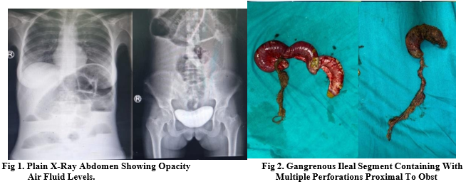
A 17 years old girl presented to the emergency room (ER) with history of pain abdomen, vomiting (greenish) for last 10 days. She had previous history of passage of foreign body per rectally. She had history of eating her own hairs since last 10 years. On examination, signs of dehydration were present, pallor present, her pulse rate was 102/ minute, blood pressure- 100/60 mm Hg, abdominal distension present, tender to touch, guarding present. Her haemoglobin (Hb) was 7g/dl with microcytic hypochromic picture, biochemical investigations were normal. On plain X-ray abdomen, an large opacity was seen in the pelvic region with distended small bowel along with multiple air fluid levels (fig 1).
After resuscitation, patient was taken for laparotomy and intra-operative findings were noted. There was hard intraluminal mass measuring 15×5×5 cm present in distal ileum causing bowel obstruction with its tail extending distally upto the mid transverse colon. There was a gangrenous segment along with multiple perforations present in the bowel proximal to obstruction. The gangrenous bowel containing perforations was resected and the distal part of trichobezoar was removed by milking movement and ileo-ileal anastomosis was done (fig 2). Psychiatrist opinion was taken. Patient was kept nil per oral for 3 days and then started oral feeds. On follow up patient is doing well and she is under treatment of psychiatrist.
CASE 2
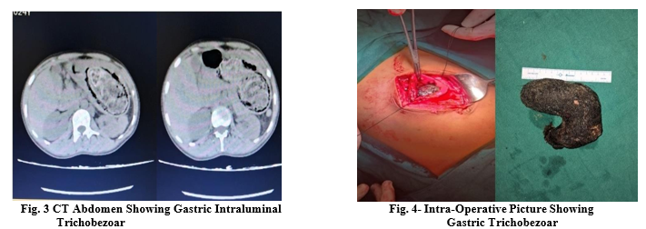
A 25 years old female presented to emergency with history of pain abdomen, vomiting and abdominal lump since last one year. On examination her vitals were stable, on per abdomen examination a palpable lump of size ~10cm×6cm was present in epigastrium and left hypochondrium extending upto umbilicus. Her haemoglobin was 6.9g/dl with microcytic hypochromic picture and biochemical investigations were normal. On ultrasonography there was a curvy linear hyperechoic area measuring 10cm in size in the stomach. On CECT abdomen there was a heterogeneous intraluminal mass 5×12×14cm extending from fundus to pyloric region of stomach (? Gastric bezoar) (fig 3). After resuscitation patient was taken for laparotomy under General anaesthesia. Intra-operatively the stomach was distended and hard mass was palpable. After opening the stomach, trichobezoar was present measuring 7×10×15cm completely occluding gastric lumen (fig. 4). The mass was removed and stomach closed in two layers with 3-0 vicryl suture.The postoperative period was uneventful and she was given oral feeds on 4th postoperative day. Psychiatrist opinion was taken and counselling was done. On follow up the patient is doing well.
CASE 3
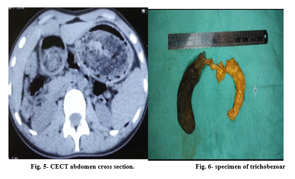
A 25-year-old female presented in ER with complaint of diffuse epigastric pain, postprandial nausea, persistent non-bilious vomiting, and significant unintended weight loss (9kg)over 6 months. Her symptoms progressively worsened over last 2 months. Her past history revealed a long-standing habit hair pulling (trichotillomania) and hair swallowing (trichophagia) since age 14, triggered by stress. A thin built young female in mild distress. Vital signs were stable. Abdominal examination demonstrated a prominent, firm, non-tender, mobile mass occupying the epigastrium and left upper quadrant. Patchy alopecia was noted on scalp examination. Her Haemoglobin was 8.2g/dL (Microcytic hypochromic anaemia), serum electrolytes and liver function parameters were within normal limits. Contrast-enhanced CT demonstrated a well-defined, heterogeneous, non-enhancing intraluminal mass in the stomach containing air pockets, extending through the pylorus into the second and third parts of the duodenum (figure 5). Based on imaging findings and clinical history, a primary diagnosis of Rapunzel Syndrome with partial gastric outlet obstruction was made. After preoperative workup patient was taken for exploratory laparotomy through an upper midline incision under general anaesthesia. Upon opening the peritoneal cavity, stomach was distended with a dense intraluminal mass. Anterior gastrostomy was done after placing stay sutures along the greater curvature to gain access to the mass. The main body of the trichobezoar was mobilized. Gentle traction was applied to carefully retract the long tail out of the pylorus, duodenum, and proximal jejunum. To prevent recurrence/obstruction, small intestine from the duodeno-jejunal junction to the ileo-cecal junction was palpated to rule out fragmented hairballs. The gastrotomy was closed in two layers using continuous absorbable sutures, followed by a standard abdominal closure. The extracted specimen was a complete, J-shaped hair mass (figure6). On postoperative day 4, patient was given oral liquids and she tolerated well and advanced to a full regular diet in next 2 days. She was discharged on day 7. Psychiatric assessment confirmed trichotillomania and underlying Obsessive-Compulsive Disorder (OCD). She was started on Cognitive Behavioural Therapy (CBT) and fluoxetine (20mg/day) prior to discharge. On follow-up, the abdominal wound had healed completely. She had gained weight and behavioural therapy continued.
DISCUSSION:
Trichobezoar is usually seen in adolescent girls, often with an underlying psychiatric or social problem. Swain first described trichobezoar while conducting an autopsy in 1854. 4 The formation of bezoars can occur in individuals with normal GI physiology and anatomy. However, patients with altered GI anatomy and/or motility are at an increased risk for the development of bezoars. Risk factors for bezoar formation include a partial gastrectomy with or without a vagotomy, diabetes mellitus complicated by gastroparesis, or other systemic illnesses that may affect GI motility. 5 Bezoars are typically grouped into 1 of 4 types according to their composition: phytobezoars (which are composed of indigestible food particles that are found in vegetable or fruit fibers), trichobezoars (which are composed of a conglomeration of hair and food particles), lactobezoars (which are composed of milk protein), or pharmacobezoars (which are concretions of various medications). 6 Trichobezoars occur because hairs are ineffectively moved by peristalsis due to its smooth surface and poorly digested keratinaceous substance. As a result, the hair becomes matted into a ball and is retained in the folds of gastric mucosa. The ball can reach sizes sufficient to cause stomach distension and inhibit gastric emptying. Trichobezoars occur more frequently in women, with only isolated cases reported in males. 3 Affected patients occasionally remain asymptomatic for many years. Symptoms develop as the bezoar increases in size to the point of obstruction. Not surprisingly, most of the cases have been reported in countries where women traditionally have long hair. The commonly presenting features are abdominal pain, nausea and vomiting, obstruction, and peritonitis. Less commonly, patients have presented with weight loss, anorexia, hematemesis and intussusception.⁷ In uncomplicated cases, a range of non-invasive and invasive radiological investigations can aid in the diagnosis. Upper gastrointestinal endoscopy serves both diagnostic and, in many cases, therapeutic roles, particularly for gastric trichobezoars. In patients presenting with complications, CECT of the abdomen is the investigation of choice, as it allows precise localization of the obstruction, identification of the transition point, and characterization of the intraluminal mass—typically demonstrating a mottled gas pattern that is characteristic of a bezoar. 8 Surgery is indicated when a very large or solid GB causes perforation or haemorrhage, or in the case of Rapunzel syndrome, when there is significant extension of the trichobezoar. Since the advent of minimally invasive surgery, the use of laparoscopic techniques for small to moderate-sized GBs has been proposed.1 Laparotomy is the most effective and therefore the most common technique in the medical literature. Due to the high success rate, the relatively low complication rate, the simple nature of the operation and the ability to carefully examine the entire gastrointestinal tract for satellites over a short period of time, laparotomy is still widely considered as the treatment of choice for complicated trichobezoar. ³
Trichobezoars may cause a range of complications such as bowel obstruction, perforation, intussusception, protein-losing enteropathy, obstructive jaundice, and pancreatitis, with bowel obstruction being the most commonly observed presentation. ⁹ Recently, a review of 108 cases of gastric trichobezoar showed that the most common complication is perforation of either the stomach or the intestine (occurring in 10.1% of cases), followed by intussusceptions (1.85%), pancreatitis (0.92%) and cholangitis (0.92%). 3 In our study, one patient presented with perforation peritonitis and the other two presented with gastric outlet obstruction, all were managed by exploratory laparotomy and trichobezoar removal.
CONCLUSION:
Gastric and intestinal trichobezoars, while rare, should be considered in patients—particularly young females—with chronic gastrointestinal symptoms, a palpable abdominal mass, and a history of trichotillomania or trichophagia. Timely diagnosis through imaging and clinical suspicion is essential to prevent serious complications such as obstruction, perforation, or bleeding. Surgical removal remains the mainstay of treatment for large or complicated bezoars, while long-term psychiatric evaluation and intervention are critical to address underlying behavioural disorders and prevent recurrence.
REFERENCES:
1. Gonuguntla V, Joshi DD: Rapunzel syndrome: a comprehensive review of an unusual case of trichobezoar. Clin Med Res 2009;7:99–102.
2. Yeo CJ, McFadden DW, Pemberton JH, Peters JH, Matthews JB. Shackelford’s surgery of the alimentary tract. New York: Elsevier Health Sciences; 2012.p805.
3. Gorter RR, Kneepkens CMF, Mattens ECJL, Aronson DC, Heij HA: Management of trichobezoar: case report and literature review. Pediatr Surg Int 2010;26:457–463.
4. Chisholm EM, Leong HT, Chung SC, Li AK. Phytobezoar: an uncommon cause of small bowel obstruction. Ann R Coll Surg Engl. 1992;74:342–4.
5. Hewitt AN, Levine MS, Rubesin SE, Laufer I. Gastric bezoars: reassessment of clinical and radiographic findings in 19 patients. Br J Radiol. 2009;82:901–907.
6. Klipfel AA, Kessler E, Schein M. Rapunzel syndrome causing gastric emphysema and small bowel obstruction. Surgery 2003;133:120-121.
7. Sanders M. Bezoars: from mystical charms to medical and nutritional management. Practical Gastroenterology. 2004;28:37–50.
8. Khattala K, Boujraf S, Rami M, Elmadi A, Afifi A, Sbai H, et al. Trichobezoar with small bowel obstruction in children: Two cases report. Afr J Paediatr Surg. 2008;5(1):48-51.
9. Goyal V, Goyal PK, Gupta M. A rare case of small bowel obstruction due to primary trichobezoar. J Clin Diagn Res. 2014;8(3):168-9.