Comparison of 128-Slice Multidetector Computed Tomography Versus 1.5-Tesla Magnetic Resonance Imaging in the Preoperative Local Staging of Rectal Carcinoma: A Prospective Cross-Sectional Study.
- Mohan Bhavikatti , Professor, Department of Radio-Diagnosis, KLE Jagadguru Gangadhar Mahaswamigalu Moorusavirmath Medical College and Hospital, Hubli, KLE Academy of Higher Education and Research, Deemed to be University, Belagavi, Karnataka, India- 590010.
- Karthikeya D. Hebbar , Department of Radio-Diagnosis, Diya Scans and Diagnostics, Surandai, Tamilnadu, India- 627859.
- Ambika S Gayad , Associate Professor, Department of Radio-Diagnosis, KLE Jagadguru Gangadhar Mahaswamigalu Moorusavirmath Medical College and Hospital, Hubli, KLE Academy of Higher Education and Research, Deemed to be University, Belagavi, Karnataka, India- 590010.
- Venkatesh H A , Assistant Professor, Department of Radio-Diagnosis, KLE Jagadguru Gangadhar Mahaswamigalu Moorusavirmath Medical College and Hospital, Hubli, KLE Academy of Higher Education and Research, Deemed to be University, Belagavi, Karnataka, India- 590010.
Article Information:
Abstract:
Background: Accurate preoperative local staging of rectal carcinoma is essential for selecting appropriate treatment strategies, predicting prognosis and minimizing local recurrence. Magnetic resonance imaging (MRI) is considered the preferred modality for local staging because of its superior soft-tissue contrast and assessment of the mesorectal fascia. However, advances in multidetector computed tomography (MDCT), particularly 128-slice scanners with multiplanar reconstruction, have considerably improved spatial resolution and may offer comparable diagnostic performance. Aim: To compare the diagnostic performance of 128-slice multidetector computed tomography (MDCT) and 1.5-Tesla magnetic resonance imaging (MRI) in the preoperative local staging of biopsy-proven rectal carcinoma. Materials and Methods: A prospective hospital-based cross-sectional study was conducted in the Department of Radiodiagnosis, SDM College of Medical Sciences and Hospital, Dharwad, India, between October 2016 and March 2018. Forty-six consecutive patients with biopsy-confirmed rectal carcinoma underwent both contrast-enhanced 128-slice MDCT and 1.5-T MRI before any therapeutic intervention. Tumour localization, T stage, nodal status, and adjacent organ involvement were evaluated. Histopathological findings were available for 15 surgically treated patients and served as the reference standard. Diagnostic performance was assessed using sensitivity, specificity, positive predictive value (PPV), negative predictive value (NPV), and overall accuracy. Results: Both MDCT and MRI achieved a tumour detection rate of 100%. The mean patient age was 54.6 years, with a male-to-female ratio of 1.5:1. Bleeding per rectum was the most common presenting symptom (46%), while the lower rectum was the most frequent tumour location (39%). For T staging, MDCT demonstrated a sensitivity of 82.4%, specificity of 84.6%, PPV of 87.5%, NPV of 78.6%, and an overall diagnostic accuracy of 83.3%. MRI demonstrated a sensitivity of 82.4%, specificity of 76.9%, PPV of 82.4%, NPV of 76.9%, and diagnostic accuracy of 80.0%. Both imaging modalities demonstrated comparable performance in nodal staging. Conclusion: Modern 128-slice MDCT demonstrated diagnostic performance comparable to 1.5-T MRI for preoperative local staging of rectal carcinoma. Although MRI remains the preferred modality for evaluating mesorectal fascia, sphincter complex, and pelvic soft-tissue anatomy, MDCT provides a reliable alternative in patients with MRI contraindications or limited access to MRI.
Keywords:
Article :
INTRODUCTION:
Rectal carcinoma is one of the most common gastrointestinal malignancies and accounts for approximately one-third of all colorectal cancers(1,2). Despite significant advances in surgical techniques and multimodality treatment, rectal cancer continues to be associated with substantial morbidity and mortality because of its propensity for local recurrence and distant metastases. Accurate preoperative staging using cross sectional imaging has therefore become a cornerstone of modern rectal cancer management and imaging studies thus play a crucial role in optimizing oncological outcomes.
The primary objective of local staging is to accurately determine the depth of tumour invasion, involvement of the mesorectal fascia, regional lymph node status, and invasion of adjacent pelvic organs. These parameters are critical in deciding whether patients should undergo surgery alone or receive neoadjuvant chemoradiotherapy followed by total mesorectal excision(3). Accurate imaging also facilitates prediction of circumferential resection margin involvement, which is one of the strongest predictors of local recurrence and long-term survival(3).
Magnetic resonance imaging (MRI)has emerged as the imaging modality of choice for local staging because of its excellent soft-tissue contrast and superior visualization of the rectal wall, mesorectal fat, mesorectal fascia, circumferential resection margin and sphincter complex. High-resolution T2-weighted sequences provide detailed anatomical information that assists surgeons in planning sphincter-preserving procedures and selecting patients who may benefit from neoadjuvant therapy(4,5).
Computed tomography(CT), on the other hand, is widely available, rapid, and routinely performed for staging colorectal malignancies. Advances in CT technology have emphasized the role of MDCT as a comprehensive imaging modality capable of providing both local and distant staging within a single examination(3).
Several studies have reported encouraging results regarding the diagnostic performance of modern MDCT in evaluating tumour invasion and regional nodal disease. Nevertheless, MRI continues to be regarded as the standard imaging modality, particularly for assessing the mesorectal fascia and predicting circumferential resection margin involvement. Comparative evidence between contemporary MDCT and MRI remains limited, especially in resource-constrained settings where MRI availability may be restricted.
This study is therefore proposed to compare the diagnostic performance of 128-slice MDCT and 1.5-T MRI in the preoperative local staging of biopsy-proven rectal carcinoma. The study also aims to evaluate the usefulness of each modality in treatment planning, and determination of the most appropriate surgical approach.
MATERIALS AND METHODS:
Study Design and Study Population
This is a prospective, hospital-based cross-sectional study conducted in the Department of Radiodiagnosis, SDM College of Medical Sciences and Hospital, Dharwad, Karnataka, India, over an 18-month period from October 2016 to March 2018. The study was conducted after obtaining approval from the Institutional Ethics Committee. Written informed consent was obtained from all participants. All patients referred to the Department of Radiodiagnosis with biopsy-proven rectal carcinoma following colonoscopic evaluation were screened for eligibility. Consecutive patients fulfilling the inclusion criteria were recruited. Patients aged more than or equal to 18 years , histopathologically confirmed primary rectal carcinoma located within 15 cm of the anal verge, patients who underwent both contrast-enhanced 128-slice MDCT and 1.5-T MRI before initiation of any treatment and those available for clinical follow-up or histopathological correlation were enrolled in the study. Patients with previous surgery for rectal carcinoma, prior neoadjuvant chemotherapy and/or radiotherapy, recurrent rectal carcinoma,those with contraindications to MRI and/or with contraindications to contrast CT study were excluded from the study.
A total of 46 consecutive patients were included in the study. Histopathological correlation following surgical resection was available in 15 patients, while the remaining patients were evaluated using imaging findings and clinical follow-up.
Image acquisition
All the patients enrolled for the study underwent both contrast-enhanced MDCT and pelvic MRI before initiation of surgery, chemotherapy, or radiotherapy.
All CT scans were performed on a 128-slice Somatom Definition AS scanner (Siemens Healthcare). All scans were performed on a 64-slice CT scanner (Toshiba Aquilion). Using a pressure injector, 60 to 70 mL of intravenous (IV) Iopamidol (350 mg iodine/mL) was injected at the rate of 4 to 4.5 mL/ s. Administration of contrast was followed by a flush of 40 mL normal saline at the same injection rate. A dual-phase CT scan with arterial phase(acquired after a delay of 15 to 20 seconds) and venous phase(acquired after a delay of 80 to 90 seconds) was performed after an unenhanced CT scan. Bolus tracking method was used for acquisition of arterial and venous phases. Images were acquired in axial plane in cranio-caudal direction from lung bases to 10 cm below pubic symphysis in a single breath hold.
Axial datasets were transferred to work station for reconstruction and review. High-resolution multiplanar reformations in all 3 planes were studied to facilitate local tumour assessment.
Magnetic Resonance Imaging(MRI) examinations were performed on a 1.5-T GE Signa MRI system equipped with a phased-array external body coil.The standard imaging protocol included:
- Axial T1-weighted spin-echo sequence
- Axial high-resolution T2-weighted fast spin-echo sequence
- Sagittal T2-weighted fast spin-echo sequence
- Coronal T2-weighted fast spin-echo sequence
- Sagittal T1-weighted spin-echo sequence
Sagittal T2-weighted images were initially obtained to identify tumour location and facilitate planning of subsequent axial and coronal images perpendicular and parallel to the tumour axis(Ref: Fig 1). High-resolution T2-weighted imaging served as the principal sequence for evaluating tumour extent, mesorectal fascia involvement, and adjacent pelvic structures.
Image Interpretation
All CT and MRI examinations were independently analysed by two experienced radiologists with 2 years and more than 20 years of experience independently.
The following imaging parameters were evaluated: Tumour location (upper, middle, lower rectum), morphological appearance, maximum mural thickness, T stage, regional lymph node involvement, mesorectal fascia involvement, adjacent organ invasion and presence of distant metastatic disease. Tumours were identified as irregular polypoidal or circumferential mural thickening demonstrating heterogeneous enhancement on CT and intermediate T2 signal intensity on MRI.
Tumour staging was performed according to the TNM classification applicable during the study period(Ref: Figure 2).
Reference Standard
Histopathological examination of the surgical specimen served as the reference standard in patients undergoing definitive surgery (n = 15). For patients who did not undergo surgery, imaging findings were interpreted in conjunction with clinical follow-up and multidisciplinary treatment decisions.
Statistical Analysis: Statistical analysis was performed using IBM SPSS Statistics Version 20.0 (IBM Corp., Chicago, IL, USA). Continuous variables are presented as mean ± standard deviation, whereas categorical variables are expressed as frequencies and percentages.
Diagnostic performance of MDCT and MRI was evaluated by calculating:Sensitivity, Specificity, Positive predictive value (PPV), Negative predictive value (NPV) and Overall diagnostic accuracy
Cross-tabulation analysis was performed using histopathological findings as the reference standard whenever available.
RESULTS:
All 46 patients enrolled during the study period successfully underwent both contrast-enhanced 128-slice MDCT and 1.5-T MRI examinations without procedure-related complications. Tumour detection was achieved in 100% of patients using both imaging modalities Study population included 28 males and 18 females. Mean age of study population was 54.6 years, ranged from 21 to 78 years. Most common presenting complaint in our study was bleeding per rectum (21/46) (46%). Circumferential irregular asymmetrical mural wall thickening was the most common morphological type of rectal cancer. Mucinous tumours were the most common histopathological type of adenocarcinoma, accounting for 53% of surgically treated cases.
Among the total 46 patients, MDCT classified 21 tumours (46%) as T2, 12 (26%) as T3, and 13 (28%) as T4. MRI staged 22 tumours (48%) as T2, 13 (28%) as T3, and 11 (24%) as T4.Compared with MRI, MDCT overestimated tumour stage in five patients and underestimated tumour stage in three patients. Histopathological confirmation was available in 15 surgically treated patients. One pathologically confirmed T1 tumour had been over staged as T2 by both imaging modalities. MDCT over staged one T2 tumour because of desmoplastic reaction mimicking mesorectal invasion. Both modalities underestimated two T3 tumours, while MDCT additionally over staged two T3 lesions as T4(Tables 1-5,Graphs 1-3, Figures 3-6)
TABLE 1: AGE DISTRIBUTION OF THE PATIENTS
|
Sex |
||||||
|
AGE IN YEARS |
MALE |
FEMALE |
TOTAL |
|||
|
|
N |
% |
N |
% |
N |
% |
|
21–30 |
2 |
7 |
4 |
22 |
6 |
13 |
|
31–40 |
1 |
4 |
2 |
11 |
3 |
7 |
|
41–50 |
4 |
14 |
2 |
11 |
6 |
13 |
|
51–60 |
8 |
29 |
3 |
17 |
11 |
24 |
|
61–70 |
9 |
32 |
5 |
28 |
14 |
30 |
|
71–80 |
4 |
14 |
2 |
11 |
6 |
13 |
|
TOTAL |
28 |
100 |
18 |
100 |
46 |
100 |
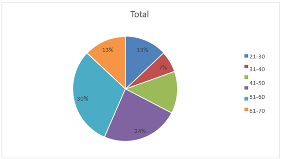
GRAPH 1: AGE DISTRIBUTION OF THE PATIENTS
TABLE 2: COMPARISON BETWEEN MDCT AND MRI IN ASSESSING TUMOR PENETRATION IN THE RECTAL WALL: IN ALL 46 PATIENTS
|
T stage |
CT |
% |
MRI |
% |
Over estimated by CT |
Underestimated by CT |
|
T0 |
0 |
0 |
0 |
0 |
0 |
0 |
|
T1 |
0 |
0 |
0 |
0 |
0 |
0 |
|
T2 |
21 |
46 |
22 |
48 |
4 |
0 |
|
T3 |
12 |
26 |
13 |
28 |
1 |
3 |
|
T4 |
13 |
28 |
11 |
24 |
0 |
0 |
|
TOTAL |
46 |
100 |
46 |
100 |
5 |
3 |
TABLE 3: COMPARISON BETWEEN CT, MRI AND HISTOPATHOLOGY IN ASSESSING TUMOUR PENETRATION IN THE RECTAL WALL: IN 15 PATIENTS
|
T STAGE |
CT |
% |
MRI |
% |
HP |
% |
Overestimate d by CT |
Overestimated by MRI |
Underestimated by CT |
Underestimated by MRI |
|
T0 |
0 |
0 |
0 |
0 |
0 |
0 |
0 |
0 |
0 |
0 |
|
T1 |
0 |
0 |
0 |
0 |
1 |
7 |
1 |
1 |
0 |
0 |
|
T2 |
8 |
53 |
9 |
60 |
6 |
40 |
1 |
0 |
0 |
0 |
|
T3 |
5 |
33 |
5 |
33 |
8 |
53 |
2 |
1 |
2 |
2 |
|
T4 |
2 |
14 |
1 |
7 |
0 |
0 |
0 |
0 |
0 |
0 |
|
TOTAL |
15 |
100 |
15 |
100 |
15 |
100 |
|
|
|
|
TABLE 4: SENSITIVITY AND SPECIFICITY OF MDCT FOR T STAGING
|
SENSITIVITY % |
82.4 |
|
SPECIFICITY % |
84.6 |
|
PPV % |
87.5 |
|
NPV % |
78.6 |
|
ACCURACY % |
83.3 |
GRAPH 2: SENSITIVITY AND SPECIFICITY OF MDCT FOR T STAGING
TABLE 5: SENSITIVITY AND SPECIFICITY OF MRI FOR T STAGING
|
SENSITIVITY % |
82.4 |
|
SPECIFICITY % |
76.9 |
|
PPV % |
82.4 |
|
NPV % |
76.9 |
|
ACCURACY % |
80 |
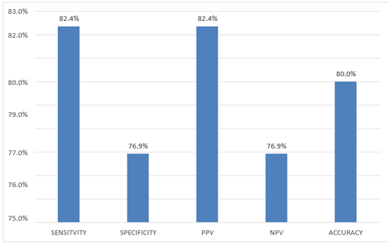
GRAPH 3: SENSITIVITY AND SPECIFICITY OF MRI FOR T STAGING
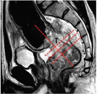
Figure 1: Planning MRI. (A) Sagittal T2W MRI. A bulky mid-rectal tumor (T); axial and coronal sequences are planned perpendicular and parallel to the tumor (lines).
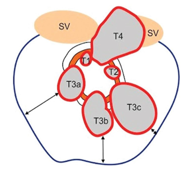
Figure 2: Drawing illustrates tumor staging in rectal cancer and CRM. T1 tumors are confined to the submucosa.T2 tumors extend to muscularispropria. T3 tumors extend into mesorectal fat. T4 tumors invade adjacent organs. Blue line represents the mesorectal fascia (MRF). Double ended arrows show the varying circumferential resection margin, which is the shortest distance between tumor edge and MRF.
CASE 1:
Figures 3a Figures 3b
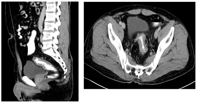
Figures 3a,3b: Post contrast sagittal and axial CT scan images show circimferential wall thickening involving the upper one-third of rectum and recto-sigmoid junction causing significant luminal narrowing. Mild perilesional fat stranding and few scattered perirectal nodes noted. Peritoneum is uninvolved.
Figures 3c
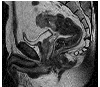
Fig 3c: Sagittal T2W images show stricturous growth involving the upper one-third of rectum and recto-sigmoid junction. Peritoneum is uninvolved.
CASE 2:
Figures 4a Figures 4b
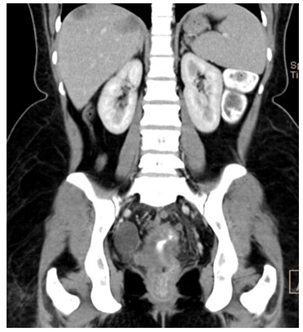
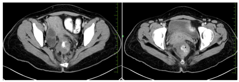
Fig 4c: Post contrast axial and coronal CT images showing heterogeneously enhancing polypoidal growth involving the middle and lower one-third of rectum with extension into the mesorectal fat and loss of fat planes with the uterus. Multiple enlarged mesorectal lymph nodes noted. Few heterogeneously enhancing metastatic lesions noted in the liver.
CASE 3
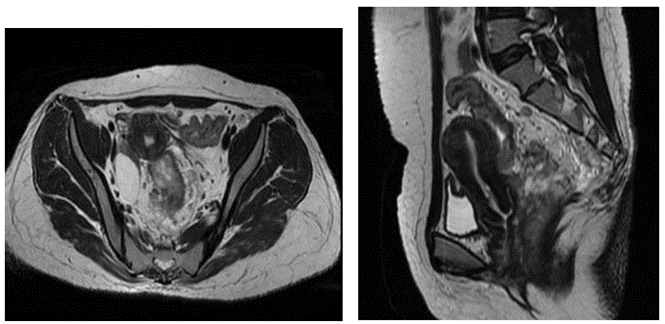
Figures 5a Figure 5b
Figures 5a and 5b: Axial and sagittal T2W images showing mixed intensity polypoidal growth involving the middle and lower one-third of rectum with extension into the mesorectal fat and with invasion into the uterus.
Multiple enlarged mesorectal lymph nodes noted.
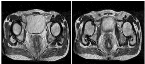
Figures 5c Figures 5d
Figures 5c, 5d: Post contrast axial CT images show circumferential growth involving the rectum with infiltration into the mesorectal fascia posteriorly and enlarged mesrorectal lymph nodes.
CASE 4
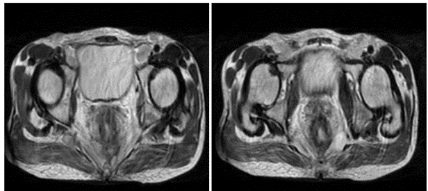
Figures 6a Figures 6b
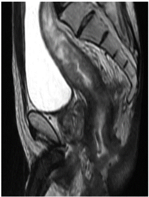
Figures 6c
Figures 6a,6b and 6c: Axial and sagittal T2W images show long segment circumferential growth involving the rectum and anal canal with infiltration into the mesorectal fascia posteriorly, possible invasion into the prostate and enlarged mesrorectal lymph nodes.
Histopathological assessment identified metastatic regional lymph nodes in 11 of 15 surgically treated patients. Both MDCT and MRI demonstrated comparable performance for nodal assessment. Overall diagnostic indices for nodal staging were identical for both modalities, with a sensitivity of 77.2%, specificity of 80.1%, and diagnostic accuracy of 82.8%.
DISCUSSION:
Accurate preoperative local staging is fundamental to the management of rectal carcinoma because treatment decisions depend on precise assessment of tumour depth, regional lymph node involvement, mesorectal fascia status, and invasion of adjacent pelvic organs. Reliable imaging enables appropriate selection of patients for primary surgery, neoadjuvant chemoradiotherapy, or sphincter-preserving procedures, thereby improving oncological outcomes while reducing local recurrence rates(3-6). The present prospective study compared the diagnostic performance of 128-slice multidetector computed tomography (MDCT) and 1.5-T magnetic resonance imaging (MRI) in the preoperative local staging of rectal carcinoma.
In the present study, both imaging modalities detected all biopsy-proven rectal tumours, resulting in a tumour detection rate of 100%. This finding confirms that both contrast-enhanced MDCT and MRI are highly reliable for identifying primary rectal malignancies. Although MRI is generally regarded as the reference standard for local staging because of its superior soft-tissue contrast resolution, the present study demonstrated that modern 128-slice MDCT achieved comparable and slightly higher diagnostic performance. These findings suggest that technological improvements have narrowed the gap between CT and MRI for assessment of rectal tumour extension. New generation CT scanner allows high-resolution, thinner collimation with improved MPR images and has been used to stage rectal cancer with a reported accuracy of over 90%(7,8). A recent meta-analysis reported an accuracy of 85%, sensitivity of 87%, and specificity of 75% for high resolution MRI in T staging of rectal cancer(9).
Dar et al in his study has concluded thar MDCT is a reliable radiological tool for local staging of rectal cancer with excellent accuracy rates for T and N-staging of rectal cancer(3). Similar results are observed in our study. Our study results are similar to that conducted by H. Matsuoka et al which has concluded that MDCT is equal to MRI in the preoperative local staging of rectal carcinoma(11).
However ,few discrepancies are observed between imaging findings and histopathology primarily involved over staging and under staging of intermediate-stage tumours . Difficulty in differentiating peri-tumoral inflammation, significant fat stranding, desmoplastic reaction and congestive changes probably would have falsely lead to under staging and over staging of the disease.
In our study, overall diagnostic indices for lymph nodal staging were identical for both modalities, with a sensitivity of 77.2%, specificity of 80.1%, and diagnostic accuracy of 82.8%. This is similar to study by Sinha et al who used a 5 mm cut-off value and reported an 84.8% overall accuracy rate in the N-staging(10).
MRI demonstrated superior accuracy in evaluating pelvic soft tissues and adjacent organ invasion. Accurate identification of these structures is essential because involvement of the circumferential resection margin is one of the strongest predictors of local recurrence following surgery. Although MDCT successfully identified advanced disease in many patients, MRI remained superior in demonstrating complex pelvic anatomy and predicting the extent of local tumour invasion. This is in agreement with other studies conducted by Shihab OC et al and How C et al.(12-15).
CONCLUSION:
The present prospective study demonstrated that both 128-slice multidetector computed tomography (MDCT) and 1.5-Tesla magnetic resonance imaging (MRI) provide excellent tumour detection and satisfactory diagnostic performance for the preoperative local staging of rectal carcinoma. While MRI remained advantageous for evaluating pelvic anatomical relationships and adjacent organ invasion, 128-slice MDCT exhibited comparable overall performance for local T staging and nodal assessment. The slightly higher specificity and diagnostic accuracy observed with MDCT suggest that modern multidetector CT may serve as a reliable alternative when MRI is unavailable or contraindicated.
Further multicentre prospective studies involving larger patient populations and complete histopathological validation are required to validate these findings and refine imaging-based treatment algorithms.
REFERENCES:
1. Jhaveri KS, Hosseini-Nik H. MRI of rectal cancer: an overview and update on recent advances. AJR Am J Roentgenol. 2015;205(1):42–55.
2. Teama AH, Alarabawy RA, Mohamed HA, Eissa HH. Role of magnetic resonance imaging in assessment of rectal neoplasms. Egypt J Radiol Nucl Med. 2015;46(3):833–846.
3. Dar RA, Chowdri NA, Parray FQ, Shaheen F, Wani SH, Mushtaque M. Pre-operative staging of rectal cancer using multi-detector row computed tomography with multiplanar reformations: single center experience. Indian J Cancer. 2014;51(2):170–175.
4. Iafrate F, Laghi A, Paolantonio P, Rengo M, Mercantini P, Ferri M, et al. Preoperative staging of rectal cancer with MR imaging: correlation with surgical and histopathologic findings. Radiographics. 2006;26(3):701–714.
5. Arya S, Das D, Engineer R, Saklani A. Imaging in rectal cancer with emphasis on local staging with MRI. Indian J Radiol Imaging. 2015;25(2):148–161.
6. Chun HK, Choi D, Kim MJ, et al. Preoperative staging of rectal cancer: comparison of 3-T high-field MRI and endorectal sonography. AJR Am J Roentgenol. 2006;187(6):1557–1562.
7. Sinha R, Verma R, Rajesh A, Richards CJ. Diagnostic value of multidetector row CT in rectal cancer staging: comparison of multiplanar and axial images with histopathology. Clin Radiol. 2006;61(11):924–931.
8. Matsuoka H, Nakamura A, Masaki T, Sugiyama M, Takahara T, Hachiya J, et al. A prospective comparison between multidetector row computed tomography and magnetic resonance imaging in the preoperative evaluation of rectal carcinoma. Am J Surg. 2003;185(6):556–559.
9. Al-Sukhni E, Milot L, Fruitman M, Beyene J, Victor JC, Schmocker S, et al. Diagnostic accuracy of MRI for assessment of T category, lymph node metastases, and circumferential resection margin involvement in patients with rectal cancer: a systematic review and meta-analysis. Ann Surg Oncol. 2012;19(7):2212–2223.
10. Sinha R, Verma R, Rajesh A, Richards CJ. Diagnostic value of multidetector row CT in rectal cancer staging: comparison of multiplanar and axial images with histopathology. Clin Radiol. 2006;61(11):924–931.
11. Matsuoka H, Nakamura A, Masaki T, Sugiyama M, Takahara T, Hachiya J, et al. A prospective comparison between multidetector row computed tomography and magnetic resonance imaging in the preoperative evaluation of rectal carcinoma. Am J Surg. 2003;185(6):556–559.
12. Shihab OC, Moran BJ, Heald RJ, Quirke P, Brown G. MRI staging of low rectal cancer. Eur Radiol. 2009;19(3):643–650.
13. Shihab OC, How P, West NP, George C, Patel U, Quirke P, et al. Can a novel MRI staging system for low rectal cancer aid surgical planning? Dis Colon Rectum. 2011;54(10):1260–1264.
14. Smith NJ, Shihab O, Arnaout A, Swift RI, Brown G. MRI for detection of extramural vascular invasion in rectal cancer. AJR Am J Roentgenol. 2008;191(5):1517–1522.
15. Koh DM, Smith NJ, Swift RI, Brown G. The relationship between MR demonstration of extramural venous invasion and nodal disease in rectal cancer. Clin Med Oncol. 2008;2:267–273.