Integrating Digital Labelling Technologies into Anatomy Museums for Contemporary Medical Education.
- Sharadkumar Pralhad Sawant , Department of Anatomy, Prince Medical College, Sikar, Rajasthan University of Health Sciences, Rajasthan, India.
- Amit Manchanda , Department of Anatomy, Prince Medical College, Sikar, Rajasthan University of Health Sciences, Rajasthan, India.
- Shaheen Rizvi , Department of Anatomy, Geetanjali Medical College, Udaipur, Rajasthan, India.
- Nikita Jakhar , Assistant Professor, Department of Pharmacology, KM Medical college and Hospital, Mathura (UP)
- Priyatama Sharadkumar Sawant , Department of Ayush, Prince Hospital, Sikar, Rajasthan University of Health Sciences, Rajasthan, India.
- Viren Sharadkumar Sawant , Mahaveer Prasad Institute of Technology and Paramedical Ghaziabad, Uttar Pradesh
- Kunal Sharadkumar Sawant , Mahaveer Prasad Institute of Technology and Paramedical Ghaziabad, Uttar Pradesh.
Article Information:
Abstract:
Background: Aim and Objectives: This review aimed to explore the development and educational importance of specimen labeling in anatomy museums and to assess how contemporary digital technologies can improve the identification, interpretation, accessibility, and educational use of anatomical specimens. Particular attention was given to QR codes, barcodes, three-dimensional visualization, augmented reality, virtual museum platforms, and artificial intelligence as emerging tools for supporting student-centered and competency-based anatomical education. Material and Method: A narrative review was undertaken using published literature, educational resources, and contemporary practices concerning anatomy museums, anatomical specimen labeling, museum-based teaching, digital cataloguing, and technology-assisted medical education. Traditional labeling approaches were compared with emerging digital systems with regard to their educational usefulness, ease of access, clinical integration, opportunities for independent learning, and practical applicability. The review also considered the use of QR-linked resources, digital databases, multimedia content, three-dimensional models, augmented and virtual reality, artificial intelligence-supported identification, and multilingual digital interfaces. Ethical considerations related to the respectful presentation and digital documentation of human anatomical material were also examined. Results: The review indicates that specimen labeling has evolved considerably from simple handwritten descriptions and basic printed tags to structured, standardized, and digitally connected information systems. Contemporary labels can provide specimen numbers, anatomical terminology, regional relationships, developmental information, pathological findings, clinical correlations, preservation details, and references to supplementary learning resources. QR codes and barcodes enable students to obtain additional information through mobile devices, including radiological images, histological preparations, clinical photographs, videos, and three-dimensional anatomical resources. Augmented and virtual reality technologies can extend the learning experience beyond the physical museum by providing interactive visualization and remote access to selected collections. Artificial intelligence offers further possibilities for specimen recognition, automated cataloguing, translation, information retrieval, and personalized educational support. Collectively, these approaches can promote active learning, improve information accessibility, and strengthen connections between basic anatomical knowledge and clinical practice. Conclusion: Digital specimen labeling has considerable potential to modernize anatomy museums and enhance their contribution to medical education. When appropriately designed, digitally enhanced labels can convert conventional museum displays into interactive learning resources while maintaining the educational value of real anatomical specimens. Integration of QR codes, barcodes, three-dimensional resources, augmented and virtual reality, and artificial intelligence can facilitate independent learning, clinical correlation, and competency development. Nevertheless, technological adoption should be supported by standardized nomenclature, appropriate infrastructure, regular system maintenance, faculty involvement, and ethical principles governing the display and digital representation of human remains. A balanced combination of direct specimen-based learning and carefully implemented digital technologies may provide a sustainable and educationally effective model for the anatomy museum of the modern medical institution.
Keywords:
Article :
INTRODUCTION:
Anatomy museums have historically occupied a central position in medical education because they provide preserved anatomical specimens for observation, demonstration, research, and self-directed learning. Since the early development of anatomical sciences, museums have functioned as important repositories of human anatomical knowledge and have significantly contributed to understanding normal anatomy, developmental anomalies, pathological changes, and surgical correlations. (1)
Museum specimens remain indispensable educational tools because they enable medical students to visualize three-dimensional anatomical relationships that are often difficult to appreciate through textbooks alone. Anatomy museums also facilitate clinical correlation by demonstrating pathological variations, congenital anomalies, surgical specimens, embryological stages, and radiological associations. (2)
Among the many components of museum organization, specimen labeling is one of the most critical educational elements. A properly labeled specimen provides essential scientific information including:
i. Name of anatomical structure
ii. Anatomical orientation
iii. Embryological significance
iv. Pathological findings
v. Clinical importance
vi. Preservation method
vii. Museum catalog number (3)
Accurate specimen labeling enhances self-directed learning, improves anatomical retention, supports competency-based medical education, and assists students in correlating theoretical and practical knowledge.
Historically, anatomy museum labeling systems were simple and manually written. Early anatomists used handwritten tags containing brief anatomical descriptions attached directly to specimens preserved in glass jars or dry preparations. These early labels were often vulnerable to fading, moisture damage, poor readability, and lack of scientific standardization.
The advancement of anatomical nomenclature systems such as Basle Nomina Anatomica, Nomina Anatomica, and later Terminologia Anatomica significantly improved consistency and scientific accuracy in specimen labeling worldwide.
Printed labels, color-coded systems, laminated charts, and multilingual educational displays gradually improved museum organization and educational quality. (4)
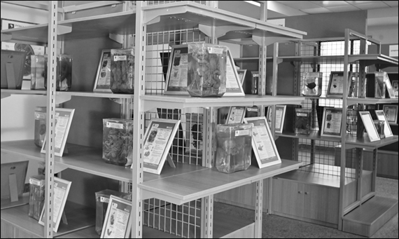
Figure 1: Photograph showing Modern Anatomy Museum with preserved anatomical specimens in glass jars, accompanied by professional specimen labels
The emergence of modern digital technologies has revolutionized anatomy museum pedagogy. Contemporary museum labeling systems now incorporate:
i. QR-code-enabled labels
ii. Barcode identification systems
iii. Interactive touch-screen displays
iv. Virtual museum access
v. Three-dimensional visualization
vi. Augmented reality applications
vii. Artificial Intelligence-assisted identification systems
Artificial Intelligence further enhances anatomy museum learning through automated specimen recognition, adaptive learning platforms, virtual educational assistance, and intelligent digital catalog management.
Modern competency-based medical education strongly emphasizes interactive, clinically oriented, and self-directed learning approaches. Consequently, anatomy museums are evolving from static display centers into dynamic educational environments integrating traditional preservation with advanced digital innovation. (5)
The present article discusses the historical evolution, educational significance, technological advancement, challenges, and future perspectives of specimen labeling systems in anatomy museums
Aim and Objectives
This review aimed to explore the development and educational importance of specimen labeling in anatomy museums and to assess how contemporary digital technologies can improve the identification, interpretation, accessibility, and educational use of anatomical specimens. Particular attention was given to QR codes, barcodes, three-dimensional visualization, augmented reality, virtual museum platforms, and artificial intelligence as emerging tools for supporting student-centered and competency-based anatomical education.
MATERIALS AND METHODS:
The present article is a narrative review and conceptual analysis based on published literature related to anatomy museums, anatomical preservation, museum pedagogy, specimen labeling systems, educational technologies, Artificial Intelligence, anatomical nomenclature, and competency-based medical education.
Relevant information was collected from:
1. Standard anatomy textbooks
2. Anatomical museum literature
3. Medical education journals
4. Museum pedagogy publications
5. Digital anatomy education research
6. Artificial Intelligence healthcare publications
7. International anatomical nomenclature guidelines
8. Recent educational technology studies
The collected literature was critically analyzed to evaluate the evolution, educational applications, technological innovations, advantages, limitations, and future perspectives of specimen labeling systems in anatomy museums.
Historical Evolution of Specimen Labeling
Early Handwritten Labeling Systems
The earliest anatomy museums used handwritten paper labels tied directly to preserved specimens. These labels generally included:
i. Name of organ
ii. Date of preservation
iii. Name of collector
iv. Basic anatomical description
However, such labels lacked durability and standardization.
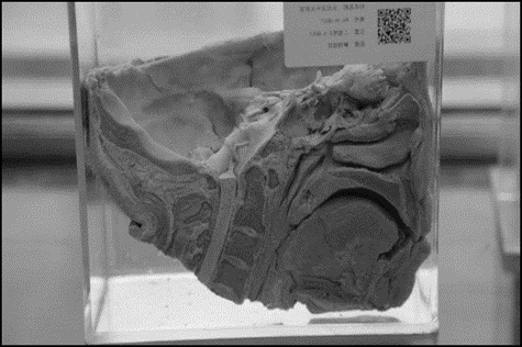
Figure 2: Photograph showing Evolution of anatomy museum labeling: handwritten tag → printed label → laminated label → QR/digital label
Development of Scientific Museum Labeling
As anatomical sciences evolved, labels became more detailed and educationally oriented.
Additional information included:
i Anatomical terminology
ii Developmental anatomy
iii Pathological diagnosis
iv. Clinical significance
v. Preservation techniques (6)
Introduction of Printed and Laminated Labels
Printing technologies improved readability, scientific presentation, and durability.
Advantages included:
i. Uniformity
ii. Better organization
iii. Enhanced visual appeal
iv. Long-term preservation (7)
Standardization through Anatomical Nomenclature
International anatomical nomenclature systems standardized specimen labeling globally.
Important systems included:
i. Basle Nomina Anatomica
ii. Nomina Anatomica
iii. Terminologia Anatomica
These systems improved consistency in anatomical communication and education. (8)
Educational Importance of Specimen Labeling
i. Enhancement of Anatomical Understanding
ii. Proper labels facilitate accurate identification of anatomical structures.
iii. Clinical Correlation
iv. Clinically oriented labels improve application of anatomy in patient care.
v. Promotion of Self-Directed Learning
vi. Students independently study museum specimens using detailed labels.
vii. Improvement of Spatial Orientation
viii. Labels help students appreciate anatomical relationships and orientation.
ix. Support for Competency-Based Medical Education
x. Interactive museum learning aligns with modern CBME objectives. (9)
Modern Components of Specimen Labeling
Modern anatomy museum labels commonly include:
1. Anatomical name
2. Region and orientation
3. Embryological details
4. Histological information
5. Clinical relevance
6. Preservation method
7. Pathological significance
8. Specimen numbering system
9. QR code integration
10. Radiological correlation (10)
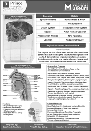
Figure 3: Photograph showing Anatomical specimen with a modern standardized museum label.
Digital Evolution of Specimen Labeling
QR-Code-Based Labeling Systems
QR codes provide direct access to:
i. Virtual dissection videos
ii. Radiological images
iii. Histological slides
iv. Clinical discussions
v. Interactive educational modules (11)
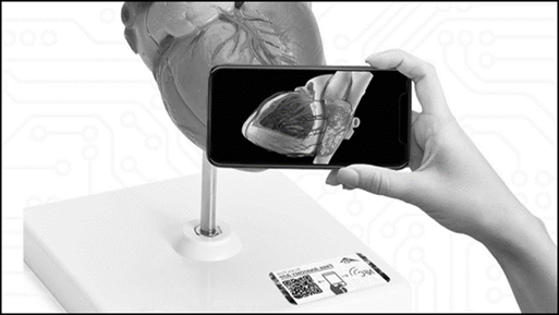
Figure 4: Photograph showing Barcode-based digital specimen catalog/inventory system showing specimen identification and tracking.
Barcode and Digital Catalog Systems
Facilitate museum inventory management and specimen tracking.
Augmented Reality Applications
Allow dynamic three-dimensional visualization of anatomical structures.
Virtual Anatomy Museums
Enable remote access to museum collections and educational materials.
Interactive Touch-Screen Displays
Provide multimedia educational content integrated with specimens. (12)
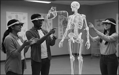
Figure 5: Photograph showing Augmented Reality anatomy museum: student viewing a preserved specimen with 3-D anatomical structures overlaid digitally.
Role of Artificial Intelligence in Specimen Labeling
Artificial Intelligence is increasingly transforming anatomy museum education through:
Automated Anatomical Identification
AI systems identify structures rapidly and accurately.
Intelligent Digital Cataloging
Large museum databases can be efficiently organized and searched.
Adaptive Learning Systems
Educational content is personalized according to learner requirements.
Multilingual Translation Support
AI improves accessibility for international learners.
Voice-Assisted Learning
Students can receive interactive explanations through smart systems.
Virtual Educational Assistance
AI chat-based educational systems support anatomy learning remotely. (13)
Advantages of Modern Labeling Systems
1. Improved Student Engagement
Interactive labels enhance learning interest and motivation.
2. Better Retention of Anatomical Knowledge
Visual and digital integration improves memory retention.
3. Enhanced Clinical Application
Clinical correlation improves practical understanding.
4. Accessibility and Remote Learning
Virtual access supports distance education.
5. Efficient Museum Organization
Digital cataloging improves administrative management.
6. Integration with Modern Technologies
Supports technologically advanced medical education. (14)
Challenges of Modern Specimen Labeling
1. Financial Limitations
Advanced digital systems require substantial investment.
2. Technological Maintenance
Regular updates and technical support are necessary.
3. Ethical Concerns
Respectful handling and display of human remains are essential.
4. Standardization Difficulties
Labeling systems vary among institutions.
5. Infrastructure Constraints
Resource-limited institutions may face implementation barriers.
6. Preservation Issues
Environmental damage may affect both specimens and labels. (15)
Future Perspectives
Future anatomy museums may incorporate:
i. AI-powered smart museums
ii. Holographic anatomy displays
iii. Fully virtual museum environments
iv. Interactive multilingual learning systems
v. Cloud-based anatomy databases
vi. Wearable augmented reality devices
vii. AI-driven competency assessment tools
These innovations may significantly transform anatomy education globally. (16).
DISCUSSION:
Specimen labeling has evolved from a simple method of anatomical identification into a highly sophisticated educational and technological system that significantly contributes to modern anatomical sciences and medical education. The progression of labeling systems reflects continuous advancements in anatomical research, preservation techniques, educational philosophy, and digital innovation. (17) Historically, anatomy museums primarily functioned as repositories of preserved human specimens intended for demonstration and reference purposes. Early specimen labels were handwritten and often contained only minimal information regarding the anatomical structure or pathological condition. Although these primitive labeling systems served basic identification purposes, they lacked scientific uniformity, educational depth, durability, and clinical orientation. Variability in terminology and handwriting additionally created confusion among learners and educators. (18)
The emergence of international anatomical nomenclature systems such as Basle Nomina Anatomica and later Terminologia Anatomica revolutionized specimen labeling by introducing globally accepted scientific terminology. Standardization significantly improved communication among anatomists, clinicians, educators, and students. Uniform terminology enhanced clarity in medical education and reduced ambiguity in anatomical identification. (19) With the advancement of museum pedagogy and educational psychology, specimen labeling gradually shifted from simple identification toward comprehensive educational interpretation. Modern labels now include embryological details, developmental anatomy, pathological findings, radiological correlation, histological information, and clinical significance. This multidimensional educational approach supports competency-based medical education by encouraging integration of anatomy with pathology, surgery, radiology, embryology, and clinical medicine. (10)
The incorporation of color coding, structured formatting, laminated educational charts, and multilingual labeling systems has further improved readability, organization, and accessibility of museum learning. Color-coded labels help students rapidly identify systems such as nervous, cardiovascular, musculoskeletal, gastrointestinal, and reproductive anatomy. Multilingual educational support is particularly important in countries with diverse linguistic backgrounds because it improves inclusiveness and comprehension among learners. (11)
One of the most significant transformations in anatomy museum labeling has occurred through integration of digital technology. QR-code-based labeling systems allow students to instantly access detailed educational resources using smartphones and digital devices. These resources may include:
1. Virtual dissection videos
2. Histological images
3. Radiological scans
4. Clinical case discussions
5. Surgical demonstrations
6. Three-dimensional anatomical models
This integration of physical museum specimens with digital educational platforms has greatly enhanced self-directed learning and independent exploration. (13,16) Artificial Intelligence has introduced another revolutionary dimension in specimen labeling and anatomy education. AI-assisted identification systems can recognize anatomical structures, provide automated explanations, and assist in digital catalog management. Intelligent educational platforms can personalize learning experiences according to student performance and educational needs. AI additionally facilitates multilingual translation, voice-assisted learning, and virtual educational support, thereby increasing accessibility and global educational connectivity.
The importance of digital anatomy museum systems became particularly evident during the COVID-19 pandemic, when physical access to anatomy museums and dissection halls was restricted worldwide. Virtual museum platforms, AI-assisted teaching systems, and QR-code-enabled digital learning resources played a critical role in maintaining continuity of anatomy education during this period. Many medical institutions rapidly adopted hybrid museum learning models combining physical specimens with online educational platforms. (14,15) Despite remarkable technological progress, several important concerns remain. Financial limitations represent a major challenge because advanced digital museum infrastructure, Artificial Intelligence systems, augmented reality devices, and virtual learning platforms require substantial institutional investment. Many resource-limited medical colleges may face difficulty implementing such technologies. (17)
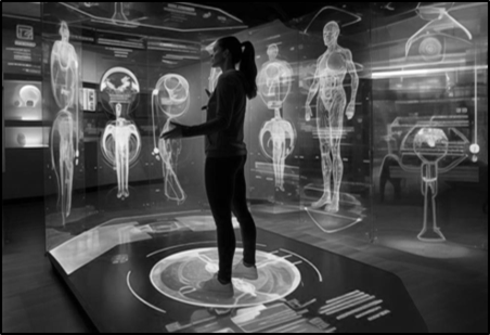
Figure 5: Photograph showing Conceptual model of an AI-enabled anatomy museum integrating automated specimen identification, digital cataloguing, adaptive learning, multilingual support, and virtual educational assistance.
Ethical considerations also remain extremely important in anatomy museum practice. Respectful preservation, display, and labeling of human remains are fundamental ethical responsibilities in anatomical sciences. Labels must maintain scientific dignity and avoid sensationalism while ensuring educational value. (18). Another concern involves overdependence on digital technology. While virtual learning systems provide numerous educational advantages, they should complement rather than completely replace direct observation of physical specimens. Physical specimens provide tactile appreciation, spatial orientation, texture perception, and emotional educational experiences that remain irreplaceable in anatomy education. (19)
Future anatomy museums are expected to evolve into fully interactive educational environments integrating Artificial Intelligence, virtual reality, augmented reality, holographic anatomy, cloud-based educational databases, and intelligent competency assessment systems. Such advancements may transform anatomy learning into a more personalized, immersive, and globally accessible educational experience. (20). Thus, the evolution of specimen labeling represents not merely a technical advancement but a major educational transformation that continues to redefine anatomy museum pedagogy and medical education worldwide.
CONCLUSION:
The evolution of specimen labeling in anatomy museums has significantly transformed anatomical education from traditional static learning into a technologically advanced, interactive, and competency-based educational process. From handwritten descriptive tags to AI-assisted digital systems, specimen labeling has continuously evolved to improve anatomical understanding, clinical correlation, educational accessibility, and museum organization. Modern labeling systems integrating QR codes, Artificial Intelligence, augmented reality, virtual museum technologies, and digital educational platforms have enhanced self-directed learning, student engagement, and multidisciplinary clinical integration. These innovations support competency-based medical education and facilitate global accessibility to anatomical learning resources.
Despite challenges related to ethics, standardization, infrastructure, financial investment, and technological maintenance, modern museum labeling systems continue to advance rapidly. Importantly, digital technologies should complement rather than replace direct interaction with preserved anatomical specimens because physical observation remains fundamental in anatomical sciences. The future of anatomy museums lies in harmonious integration of traditional anatomical preservation with intelligent digital educational systems to create immersive, ethical, student-centered, and clinically relevant learning environments for future healthcare professionals.
REFERENCES:
1. Standring S, editor. Gray’s Anatomy: The Anatomical Basis of Clinical Practice. 42nd ed. London: Elsevier; 2021.
2. Moore KL, Dalley AF, Agur AMR. Clinically Oriented Anatomy. 9th ed. Philadelphia: Wolters Kluwer; 2023.
3. Snell RS. Clinical Anatomy by Regions. 10th ed. Philadelphia: Wolters Kluwer; 2019.
4. Riva A, Conti G, Solinas P, Loy F. The evolution of anatomical museums and medical education. Clin Anat. 2010;23(5):546-556.
5. International Federation of Associations of Anatomists. Terminologia Anatomica. 2nd ed. Stuttgart: Thieme; 2019.
6. Estai M, Bunt S. Best teaching practices in anatomy education: A critical review. Ann Anat. 2016;208:151-157.
7. Moro C, Štromberga Z, Raikos A, Stirling A. The effectiveness of virtual and augmented reality in anatomy and medical education. Anat Sci Educ. 2017;10(6):549-559.
8. Azer SA, Azer S. 3D anatomy models and virtual learning in anatomy education. Adv Med Educ Pract. 2016;7:1-9.
9. Topol EJ. Deep Medicine: How Artificial Intelligence Can Make Healthcare Human Again. New York: Basic Books; 2019.
10. National Medical Commission. Competency Based Undergraduate Curriculum for the Indian Medical Graduate. New Delhi: NMC; 2023.
11. Turney BW. Anatomy in a modern medical curriculum. Ann R Coll Surg Engl. 2007;89(2):104-107.
12. Ghosh SK. Human cadaveric dissection: A historical account from ancient Greece to the modern era. Anat Cell Biol. 2015;48(3):153-169.
13. Sugand K, Abrahams P, Khurana A. The anatomy of anatomy: A review for its modernization. Anat Sci Educ. 2010;3(2):83-93.
14. McLachlan JC, Patten D. Anatomy teaching: Ghosts of the past, present and future. Med Educ. 2006;40(3):243-253.
15. Papa V, Vaccarezza M. Teaching anatomy in the XXI century: New aspects and pitfalls. ScientificWorldJournal. 2013;2013:310348.
16. Nicholson DT, Chalk C, Funnell WRJ, Daniel SJ. Can virtual reality improve anatomy education? Clin Anat. 2006;19(2):120-127.
17. COVID-19 and digital transformation of anatomy education. Anat Sci Educ. 2021;14(5):683-695.
18. Brassett C, Cosker T, Davies DC, et al. COVID-19 and anatomy: Stimulus for innovation. Clin Anat. 2020;33(8):1088-1092.
19. Langlois J, Wells GA, Lecourtois M, et al. Spatial abilities and anatomy learning performance. Anat Sci Educ. 2020;13(1):71-79.
20. Peterson DC, Mlynarczyk GS. Analysis of traditional versus digital anatomy museum learning methods. J Med Educ Curric Dev. 2016;3:39-45.