Comparison of Propofol and Etomidate for Induction of General Anaesthesia in Patients with Cardiovascular Risk.
- Veereshkumar Gobburkar , Senior Resident Dept of Anesthesiology ESIC Medical college Gulbarga Karnataka.
- J Vennela , Senior Resident Dept of Anesthesiology ESIC Medical College Gulbarga Karnataka.
Article Information:
Abstract:
Background: Haemodynamic stability during induction of general anaesthesia is particularly important in patients with cardiovascular risk. This study compared propofol and etomidate for their haemodynamic effects and adverse events during induction. Methods: In this prospective randomized study, 100 patients with cardiovascular risk undergoing elective surgery were allocated to propofol (Group P, n=50; 2 mg/kg) or etomidate (Group E, n=50; 0.3 mg/kg). Heart rate, blood pressure, and mean arterial pressure (MAP) were assessed before and after induction and intubation. Haemodynamic events, vasopressor requirement, and adverse effects were recorded. Results: Baseline characteristics were comparable between groups. Maximum reduction in MAP was significantly greater with propofol than etomidate (22.14 ± 7.62% vs 9.82 ± 5.46%; p<0.001). Hypotension (36.0% vs 10.0%; p=0.002) and vasopressor requirement (28.0% vs 8.0%; p=0.009) were significantly higher with propofol. Pain on injection was more frequent with propofol (30.0% vs 8.0%; p=0.005), whereas myoclonus was more frequent with etomidate (26.0% vs 4.0%; p=0.002). Conclusion: Etomidate provided superior haemodynamic stability with less hypotension and vasopressor requirement than propofol, making it a suitable induction agent in patients with cardiovascular risk.
Keywords:
Article :
INTRODUCTION:
Induction of general anaesthesia is a critical phase of perioperative management and is frequently associated with significant alterations in cardiovascular function[1]. Intravenous induction agents can influence systemic vascular resistance, myocardial contractility, sympathetic activity, heart rate, and arterial blood pressure.[2] Laryngoscopy and endotracheal intubation may further provoke cardiovascular responses, making haemodynamic stability particularly important in patients with underlying cardiovascular risk or limited cardiovascular reserve.[3]Propofol is one of the most commonly used intravenous agents for induction of general anaesthesia because of its rapid onset, short duration of action, predictable recovery profile, and favourable conditions for airway manipulation. However, propofol frequently causes a reduction in arterial blood pressure through venodilatation, increased vascular capacitance, inhibition of sympathetic vasoconstrictor activity, and reduction in systemic vascular resistance.[4,5] Muzi et al. demonstrated that venodilatation contributes significantly to propofol-mediated hypotension,[4] while Hoka et al. showed that propofol-induced increases in vascular capacitance are related to inhibition of sympathetic vasoconstrictor activity.[5] These effects may be clinically significant in elderly patients and in those with coronary artery disease, impaired ventricular function, hypertension, hypovolaemia, or other cardiovascular comorbidities.Etomidate is a rapidly acting intravenous hypnotic agent characterized by relatively minimal cardiovascular depression and is therefore particularly useful when haemodynamic stability is important.[3,6] It produces comparatively smaller changes in arterial pressure and cardiovascular function during induction.[6,7] Comparative studies have demonstrated better preservation of haemodynamic parameters with etomidate than with propofol.[7,8] In patients with coronary heart disease undergoing major non-cardiac surgery, etomidate has also been associated with fewer haemodynamic disturbances and reduced vasopressor requirements compared with propofol.[8]
The haemodynamic advantages of etomidate, however, must be balanced against its adverse-effect profile. Etomidate may cause myoclonus, postoperative nausea and vomiting, and transient suppression of adrenal steroidogenesis.[9,10] Myoclonic movements are particularly characteristic of etomidate induction,[10] although lipid formulations of etomidate may produce considerably less injection pain than propofol.[11] Thus, the clinical selection of an induction agent requires consideration of both haemodynamic stability and drug-related adverse effects.The selection of an appropriate induction agent is particularly relevant in patients with cardiovascular risk, where the principal objective is to achieve adequate hypnosis while minimizing haemodynamic instability and maintaining myocardial oxygen balance. Previous comparative studies have generally demonstrated a greater reduction in arterial pressure with propofol and better haemodynamic stability with etomidate.[7,8,12] Nevertheless, the choice of induction agent should be individualized according to the patient's cardiovascular reserve, associated comorbidities, anticipated haemodynamic response, and adverse-effect profile.Therefore, the present study was undertaken to compare propofol and etomidate for induction of general anaesthesia in patients with cardiovascular risk, with particular emphasis on their haemodynamic effects during induction and following endotracheal intubation.
MATERIALS AND METHODS:
This prospective, randomized, comparative study was conducted in the Department of Anaesthesiology A total of 100 patients with cardiovascular risk, scheduled to undergo elective surgery under general anaesthesia with endotracheal intubation, were enrolled. Written informed consent was obtained from all participants before inclusion in the study.
Study Population
Adult patients aged 18–70 years, of either sex, belonging to American Society of Anesthesiologists (ASA) physical status II or III, and having at least one cardiovascular risk factor were included. Cardiovascular risk factors included hypertension, diabetes mellitus, dyslipidaemia, obesity, smoking, or documented stable coronary artery disease.
Inclusion Criteria
Patients aged 18–70 years, of either sex, with ASA physical status II–III, having at least one cardiovascular risk factor, scheduled for elective surgery under general anaesthesia with endotracheal intubation, and willing to provide written informed consent were included.
Exclusion Criteria
Patients with known hypersensitivity to propofol or etomidate, haemodynamic instability, severe or decompensated heart failure, significant arrhythmias, severe valvular heart disease, adrenal insufficiency, chronic corticosteroid therapy, severe hepatic or renal dysfunction, anticipated difficult airway, pregnancy, and those undergoing emergency surgery were excluded.
Sample Size and Randomization
A total of 100 patients were enrolled and randomly allocated into two groups of 50 patients each using a computer-generated randomization sequence. Allocation was concealed using sequentially numbered, opaque, sealed envelopes.Group P (n=50) received intravenous propofol 2 mg/kg for induction, while Group E (n=50) received intravenous etomidate 0.3 mg/kg for induction.
Pre-anaesthetic Assessment
All patients underwent detailed pre-anaesthetic evaluation, including demographic characteristics, medical history, cardiovascular risk factors, current medications, general and systemic examination, airway assessment, and relevant laboratory investigations. Electrocardiography and other cardiovascular investigations were performed as clinically indicated. Patients were kept fasting according to standard institutional guidelines.
Anaesthetic Technique
On arrival in the operating room, standard monitoring consisting of electrocardiography, non-invasive blood pressure, pulse oximetry, and capnography was instituted. Intravenous access was secured, and patients were allowed to rest for approximately 5 minutes before recording baseline parameters.Baseline heart rate (HR), systolic blood pressure (SBP), diastolic blood pressure (DBP), mean arterial pressure (MAP), and peripheral oxygen saturation (SpO₂) were recorded.
All patients were preoxygenated with 100% oxygen for 3 minutes and received a standardized intravenous opioid. Anaesthesia was then induced according to group allocation. Patients in Group P received propofol 2 mg/kg intravenously, while those in Group E received etomidate 0.3 mg/kg intravenously.
Following loss of consciousness, an appropriate neuromuscular blocking agent was administered. After adequate neuromuscular relaxation, direct laryngoscopy and endotracheal intubation were performed by an experienced anaesthesiologist. Anaesthesia was subsequently maintained using a standardized anaesthetic technique in both groups.
Haemodynamic Assessment
Haemodynamic parameters, including HR, SBP, DBP, MAP, and SpO₂, were recorded at predefined intervals:
|
Time point |
Assessment |
|
T0 |
Baseline, before induction |
|
T1 |
1 minute after induction |
|
T2 |
Immediately after endotracheal intubation |
|
T3 |
3 minutes after intubation |
|
T4 |
5 minutes after intubation |
|
T5 |
10 minutes after intubation |
The percentage change in haemodynamic parameters from baseline was also calculated and compared between the two groups.
Haemodynamic Events
Hypotension was defined as a reduction in MAP of more than 20% from baseline or MAP <65 mmHg. Hypotension was managed with intravenous fluids and vasopressors when clinically indicated. Bradycardia was defined as HR <50 beats/min and treated with intravenous atropine when required. Tachycardia, hypertension, arrhythmias, and the requirement for vasopressor therapy were also recorded.
Adverse Effects
Patients were monitored for induction-related adverse effects, including pain on injection, myoclonus, apnoea, nausea, vomiting, bradycardia, hypotension, and arrhythmias. Any additional adverse event occurring during the study period was documented and managed according to standard clinical practice.
Outcome Measures
The primary outcome was the difference in haemodynamic stability between propofol and etomidate, assessed primarily by changes in MAP following induction and endotracheal intubation.
The secondary outcomes included changes in HR, SBP, and DBP; incidence of hypotension, bradycardia, tachycardia, and hypertension; requirement for vasopressor therapy; and incidence of induction-related adverse effects.
Statistical Analysis
Data were entered into Microsoft Excel and analysed using IBM SPSS Statistics version 26.0. Continuous variables were expressed as mean ± standard deviation (SD) or median (interquartile range [IQR]), depending on data distribution. Categorical variables were expressed as frequency and percentage.
Normality was assessed using the Shapiro–Wilk test. Continuous variables between the two groups were compared using the independent-samples t-test or Mann–Whitney U test, as appropriate. Categorical variables were compared using the Chi-square test or Fisher's exact test. Changes in haemodynamic parameters over different time points were analysed using repeated-measures ANOVA, with between-group comparisons performed as appropriate. A p-value <0.05 was considered statistically significant.
RESULTS:
A total of 100 patients were included in the study, with 50 patients each in Group P and Group E. The two groups were comparable with respect to baseline demographic and clinical characteristics. The mean age was 57.16 ± 8.74 years in Group P and 56.42 ± 9.12 years in Group E (p=0.680), while the mean BMI was 26.48 ± 3.21 kg/m² and 26.12 ± 3.08 kg/m², respectively (p=0.569). Males constituted 60.0% of Group P and 58.0% of Group E. The distribution of ASA physical status and cardiovascular risk factors, including hypertension, diabetes mellitus, dyslipidaemia, smoking, obesity, and stable coronary artery disease, was also comparable between the groups, with no statistically significant differences (all p>0.05) (Table 1).
Baseline heart rate was comparable between Group P and Group E (78.24 ± 9.16 vs 77.68 ± 8.72 beats/min; p=0.755). Immediately after intubation (T2), heart rate was significantly higher in Group P than in Group E (89.36 ± 10.24 vs 84.18 ± 9.32 beats/min; p=0.009). This difference remained significant at 3 minutes (85.42 ± 9.62 vs 81.26 ± 8.91 beats/min; p=0.027) and 5 minutes after intubation (81.56 ± 8.94 vs 78.12 ± 8.21 beats/min; p=0.047). By 10 minutes, the difference was no longer statistically significant (p=0.340). Repeated-measures ANOVA demonstrated a significant time × group interaction (p<0.001), indicating different heart-rate trends between the two groups over the observation period (Table 2).
Baseline systolic and diastolic blood pressures were comparable between the groups. At 1 minute after induction (T1), Group P showed a substantially greater reduction in both SBP and DBP compared with Group E. Mean SBP was 105.42 ± 12.18 mmHg in Group P compared with 121.36 ± 11.42 mmHg in Group E (p<0.001), while corresponding DBP values were 62.18 ± 8.14 mmHg and 71.92 ± 7.68 mmHg, respectively (p<0.001). Significantly lower SBP and DBP values in Group P persisted immediately after intubation and at 3 and 5 minutes after intubation (all p≤0.010). By 10 minutes, the differences in SBP (128.36 ± 11.52 vs 132.18 ± 10.94 mmHg; p=0.092) and DBP (71.42 ± 7.28 vs 74.18 ± 7.04 mmHg; p=0.056) were no longer statistically significant (Table 3).
A similar pattern was observed for mean arterial pressure (MAP). Baseline MAP was comparable between Group P and Group E (96.28 ± 8.46 vs 95.74 ± 8.12 mmHg; p=0.745). At 1 minute after induction, MAP decreased markedly to 76.32 ± 9.18 mmHg in Group P compared with 88.46 ± 8.34 mmHg in Group E (p<0.001). MAP remained significantly lower in Group P immediately after intubation (87.46 ± 9.74 vs 93.82 ± 8.65 mmHg; p=0.001), at 3 minutes (82.64 ± 8.86 vs 91.16 ± 8.12 mmHg; p<0.001), and at 5 minutes (85.78 ± 8.42 vs 92.34 ± 7.84 mmHg; p<0.001). At 10 minutes, the difference was no longer statistically significant (p=0.056). The maximum percentage reduction in MAP from baseline was significantly greater in Group P than in Group E (22.14 ± 7.62% vs 9.82 ± 5.46%; p<0.001) (Table 4).
Haemodynamic adverse events were more frequent in Group P. Hypotension occurred in 18 (36.0%) patients in Group P compared with 5 (10.0%) patients in Group E (p=0.002). Similarly, vasopressor support was required significantly more frequently in Group P than in Group E (28.0% vs 8.0%; p=0.009). Bradycardia, tachycardia, hypertension, and arrhythmias did not differ significantly between the groups (Table 5, Figure 1).
The induction-related adverse-effect profile differed between the two groups. Pain on injection was significantly more frequent in Group P (30.0%) than in Group E (8.0%; p=0.005). In contrast, myoclonus was significantly more frequent in Group E (26.0%) compared with Group P (4.0%; p=0.002). Apnoea occurred in 22.0% of patients in Group P and 10.0% in Group E, although this difference was not statistically significant (p=0.102). The incidences of nausea and vomiting were also comparable between the groups (Table 6, Figure 2).
Correlation analysis demonstrated a weak but statistically significant positive correlation between age and maximum percentage reduction in MAP (r/ρ=0.286, p=0.004), indicating that the magnitude of MAP reduction tended to increase with advancing age. BMI, baseline heart rate, baseline DBP, and baseline MAP showed no statistically significant correlations with maximum MAP reduction. Baseline SBP showed a weak positive correlation that approached but did not reach statistical significance (r/ρ=0.194, p=0.053) (Table 7, Figure 3).
Overall, Group E demonstrated better haemodynamic stability than Group P, as evidenced by a smaller reduction in MAP, better preservation of arterial blood pressure during the early post-induction period, and significantly lower incidences of hypotension and vasopressor requirement. However, the adverse-effect profiles differed, with pain on injection being more frequent in Group P and myoclonus being more frequent in Group E.
Table 1. Baseline demographic and clinical characteristics of the study participants
|
Variable |
Group P (n=50) |
Group E (n=50) |
p-value |
|
Age (years), mean ± SD |
57.16 ± 8.74 |
56.42 ± 9.12 |
0.680 |
|
Male, n (%) |
30 (60.0) |
29 (58.0) |
0.839 |
|
Female, n (%) |
20 (40.0) |
21 (42.0) |
|
|
BMI (kg/m²), mean ± SD |
26.48 ± 3.21 |
26.12 ± 3.08 |
0.569 |
|
ASA II, n (%) |
31 (62.0) |
29 (58.0) |
0.683 |
|
ASA III, n (%) |
19 (38.0) |
21 (42.0) |
|
|
Hypertension, n (%) |
35 (70.0) |
37 (74.0) |
0.656 |
|
Diabetes mellitus, n (%) |
19 (38.0) |
21 (42.0) |
0.683 |
|
Dyslipidaemia, n (%) |
16 (32.0) |
18 (36.0) |
0.673 |
|
Smoking, n (%) |
14 (28.0) |
13 (26.0) |
0.822 |
|
Obesity, n (%) |
12 (24.0) |
11 (22.0) |
0.812 |
|
Stable coronary artery disease, n (%) |
10 (20.0) |
12 (24.0) |
0.629 |
Table 2. Comparison of heart rate at different time points
|
Time point |
Group P (n=50), mean ± SD |
Group E (n=50), mean ± SD |
p-value |
|
Baseline (T0) |
78.24 ± 9.16 |
77.68 ± 8.72 |
0.755 |
|
1 min after induction (T1) |
75.18 ± 8.84 |
76.42 ± 8.35 |
0.472 |
|
Immediately after intubation (T2) |
89.36 ± 10.24 |
84.18 ± 9.32 |
0.009 |
|
3 min after intubation (T3) |
85.42 ± 9.62 |
81.26 ± 8.91 |
0.027 |
|
5 min after intubation (T4) |
81.56 ± 8.94 |
78.12 ± 8.21 |
0.047 |
|
10 min after intubation (T5) |
78.42 ± 8.51 |
76.84 ± 7.96 |
0.340 |
Table 3. Comparison of systolic and diastolic blood pressure at different time points
|
Time point |
SBP Group P |
SBP Group E |
p-value |
DBP Group P |
DBP Group E |
p-value |
|
T0 |
139.12 ± 12.64 |
138.46 ± 12.18 |
0.791 |
74.86 ± 8.12 |
74.38 ± 7.94 |
0.765 |
|
T1 |
105.42 ± 12.18 |
121.36 ± 11.42 |
<0.001 |
62.18 ± 8.14 |
71.92 ± 7.68 |
<0.001 |
|
T2 |
126.38 ± 13.24 |
133.16 ± 12.36 |
0.010 |
68.12 ± 8.46 |
74.16 ± 7.92 |
<0.001 |
|
T3 |
117.46 ± 12.68 |
129.24 ± 11.84 |
<0.001 |
65.18 ± 7.92 |
72.12 ± 7.46 |
<0.001 |
|
T4 |
121.18 ± 11.96 |
130.16 ± 11.38 |
<0.001 |
67.24 ± 7.64 |
73.42 ± 7.18 |
<0.001 |
|
T5 |
128.36 ± 11.52 |
132.18 ± 10.94 |
0.092 |
71.42 ± 7.28 |
74.18 ± 7.04 |
0.056 |
Table 4. Comparison of mean arterial pressure at different time points
|
Time point |
Group P (n=50), mean ± SD |
Group E (n=50), mean ± SD |
p-value |
|
Baseline (T0) |
96.28 ± 8.46 |
95.74 ± 8.12 |
0.745 |
|
1 min after induction (T1) |
76.32 ± 9.18 |
88.46 ± 8.34 |
<0.001 |
|
Immediately after intubation (T2) |
87.46 ± 9.74 |
93.82 ± 8.65 |
0.001 |
|
3 min after intubation (T3) |
82.64 ± 8.86 |
91.16 ± 8.12 |
<0.001 |
|
5 min after intubation (T4) |
85.78 ± 8.42 |
92.34 ± 7.84 |
<0.001 |
|
10 min after intubation (T5) |
90.46 ± 8.15 |
93.52 ± 7.62 |
0.056 |
|
Maximum reduction from baseline, % |
22.14 ± 7.62 |
9.82 ± 5.46 |
<0.001 |
Table 5. Comparison of haemodynamic events and requirement for intervention
|
Outcome |
Group P (n=50), n (%) |
Group E (n=50), n (%) |
p-value |
|
Hypotension |
18 (36.0) |
5 (10.0) |
0.002 |
|
Bradycardia |
5 (10.0) |
2 (4.0) |
0.436 |
|
Tachycardia |
10 (20.0) |
5 (10.0) |
0.161 |
|
Hypertension |
6 (12.0) |
4 (8.0) |
0.505 |
|
Arrhythmia |
3 (6.0) |
2 (4.0) |
>0.999 |
|
Vasopressor requirement |
14 (28.0) |
4 (8.0) |
0.009 |
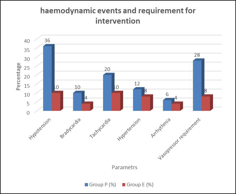
Figure 1. Comparison of haemodynamic events and requirement for intervention
Table 6. Comparison of induction-related adverse effects
|
Adverse effect |
Group P (n=50), n (%) |
Group E (n=50), n (%) |
p-value |
|
Pain on injection |
15 (30.0) |
4 (8.0) |
0.005 |
|
Myoclonus |
2 (4.0) |
13 (26.0) |
0.002 |
|
Apnoea |
11 (22.0) |
5 (10.0) |
0.102 |
|
Nausea |
3 (6.0) |
5 (10.0) |
0.715 |
|
Vomiting |
2 (4.0) |
3 (6.0) |
>0.999 |
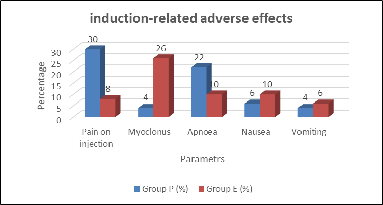
Figure 2. Comparison of induction-related adverse effects
Table 7. Correlation of clinical variables with maximum percentage reduction in MAP
|
Variable |
Correlation coefficient (r/ρ) |
p-value |
|
Age |
0.286 |
0.004 |
|
BMI |
0.102 |
0.313 |
|
Baseline HR |
0.084 |
0.406 |
|
Baseline SBP |
0.194 |
0.053 |
|
Baseline DBP |
0.121 |
0.231 |
|
Baseline MAP |
0.173 |
0.085 |
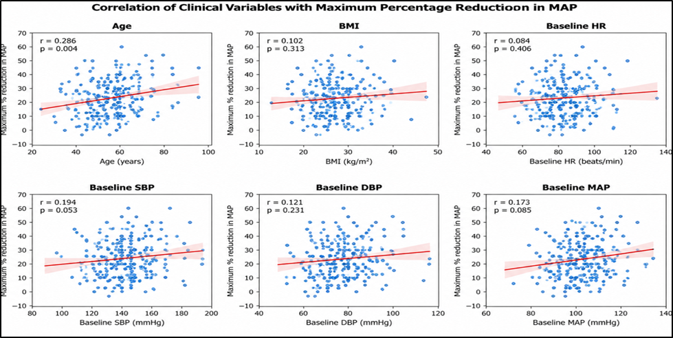
Figure 3. Correlation of clinical variables with maximum percentage reduction in MAP
DISCUSSION:
The two groups were comparable at baseline. Mean age was 57.16 ± 8.74 years in the propofol group and 56.42 ± 9.12 years in the etomidate group (p=0.680), with males comprising 60.0% and 58.0%, respectively. BMI, ASA status, hypertension, diabetes, dyslipidaemia, smoking, obesity, and stable coronary artery disease were also comparable (all p>0.05). Similar baseline comparability was maintained in the comparative studies by Saricaoglu et al. [13] and Miner et al. [14], supporting an unbiased assessment of drug-related haemodynamic differences.
Baseline HR was comparable between propofol and etomidate (78.24 ± 9.16 vs 77.68 ± 8.72 beats/min; p=0.755). After intubation, HR was significantly higher with propofol (89.36 ± 10.24 vs 84.18 ± 9.32 beats/min; p=0.009), with significant differences persisting at 3 and 5 minutes. Saricaoglu et al. [13] similarly demonstrated significant differences in haemodynamic responses among etomidate, propofol, and their admixture, with the admixture showing the least overall alteration. Boysen et al. [15] also reported differing cardiovascular responses among propofol, thiopental, and etomidate, supporting the relatively stable cardiovascular profile of etomidate.
Propofol produced a greater fall in arterial pressure. At 1 minute after induction, SBP was 105.42 ± 12.18 mmHg with propofol versus 121.36 ± 11.42 mmHg with etomidate (p<0.001), while DBP was 62.18 ± 8.14 versus 71.92 ± 7.68 mmHg (p<0.001). Miner et al. [14] similarly reported a greater maximum reduction in SBP with propofol than etomidate (7.9% vs 3.8%). Boysen et al. [15] also observed a greater fall in arterial pressure following propofol. These findings confirm better preservation of arterial pressure with etomidate.
Baseline MAP was comparable (96.28 ± 8.46 vs 95.74 ± 8.12 mmHg; p=0.745), but at 1 minute after induction it decreased to 76.32 ± 9.18 mmHg with propofol compared with 88.46 ± 8.34 mmHg with etomidate (p<0.001). The maximum reduction in MAP was 22.14 ± 7.62% versus 9.82 ± 5.46%, respectively (p<0.001).
Zausig et al. [16] provided experimental support for this finding, reporting a maximum reduction in cardiac work of 50 ± 6% with propofol compared with 17 ± 6% with etomidate in septic rat hearts; propofol also reduced contractility by 38% and lusitropy by 44%. Ray and McKeown [17] likewise emphasized the haemodynamic stability of etomidate in critically ill patients despite concerns regarding transient adrenal suppression. These findings support the substantially better preservation of MAP with etomidate observed in our study.
Hypotension was significantly more frequent with propofol (36.0% vs 10.0%; p=0.002), and vasopressor requirement was also higher (28.0% vs 8.0%; p=0.009). Ray and McKeown [17], in 159 patients with septic shock, including 74 receiving etomidate and 25 receiving propofol, found that etomidate was not associated with increased subsequent vasopressor or inotrope requirements and was associated with less cardiovascular intervention around induction. Together with our findings, these observations favour etomidate when avoidance of post-induction hypotension is particularly important.
Bradycardia (10.0% vs 4.0%), tachycardia (20.0% vs 10.0%), hypertension (12.0% vs 8.0%), and arrhythmias (6.0% vs 4.0%) were numerically more frequent with propofol, although differences were not statistically significant. The overall pattern nevertheless favoured etomidate and is consistent with the cardiovascular stability described by Ray and McKeown[17] and Saricaoglu et al. [13].
Pain on Injection and Myoclonus
Pain on injection was significantly more frequent with propofol (30.0% vs 8.0%; p=0.005), whereas myoclonus was significantly more frequent with etomidate (26.0% vs 4.0%; p=0.002). Saricaoglu et al. [13] also identified injection pain and myoclonus as important differences between propofol- and etomidate-based induction. Boysen et al. [15] reported more involuntary muscular movements with etomidate. Similarly, Miner et al. [14] observed myoclonus in 20.0% of etomidate-treated patients compared with 1.8% receiving propofol, closely corresponding to our findings.Apnoea was numerically more frequent with propofol (22.0% vs 10.0%; p=0.102), while nausea (6.0% vs 10.0%) and vomiting (4.0% vs 6.0%) were comparable. Miner et al. [14] similarly reported subclinical respiratory depression in 42.2% of propofol-treated and 34.3% of etomidate-treated patients, although clinically important respiratory interventions were comparable. Thus, respiratory depression may occur with either agent, warranting appropriate airway monitoring.
CONCLUSION:
Etomidate provided better haemodynamic stability than propofol during induction of general anaesthesia in patients with cardiovascular risk, with significantly less reduction in MAP, hypotension, and vasopressor requirement. Propofol was associated with more pain on injection, whereas myoclonus was more frequent with etomidate. Thus, etomidate may be preferred when maintenance of cardiovascular stability during induction is a major concern.
LIMITATIONS
The study was limited by its relatively small sample size and single-centre design, which may limit the generalizability of the findings. In addition, only short-term peri-induction haemodynamic changes were evaluated, without assessment of long-term cardiovascular outcomes or adrenal function.
REFERENCES:
1. Aono H, Hirakawa M, Unruh GK, Kindscher JD, Goto H. Anesthetic induction agents, sympathetic nerve activity and baroreflex sensitivity: a study in rabbits comparing thiopental, propofol and etomidate. Acta Med Okayama. 2001;55(4):197-203.
2. Paris A, Philipp M, Tonner PH, et al. Activation of alpha 2B-adrenoceptors mediates the cardiovascular effects of etomidate. Anesthesiology. 2003;99:889-95.
3. Sarkar M, Laussen PC, Zurakowski D, et al. Hemodynamic responses to etomidate on induction of anesthesia in pediatric patients. Anesth Analg. 2005;101:645-50.
4. Muzi M, Berens RA, Kampine JP, Ebert TJ. Venodilation contributes to propofol-mediated hypotension in humans. Anesth Analg. 1992;74:877-83.
5. Hoka S, Yamaura K, Takenaka T, Takahashi S. Propofol-induced increase in vascular capacitance is due to inhibition of sympathetic vasoconstrictive activity. Anesthesiology. 1998;89:1495-500.
6. Morel J, Salard M, Castelain C, et al. Haemodynamic consequences of etomidate administration in elective cardiac surgery: a randomized double-blinded study. Br J Anaesth. 2011;107:503-9.
7. Shah SB, Chowdhury I, Bhargava AK, Sabbharwal B. Comparison of hemodynamic effects of intravenous etomidate versus propofol during induction and intubation using entropy guided hypnosis levels. J Anaesthesiol Clin Pharmacol. 2015;31:180-5.
8. Dai ZL, Cai XT, Gao WL, Lin M, Lin J, Jiang YX, et al. Etomidate vs propofol in coronary heart disease patients undergoing major noncardiac surgery: a randomized clinical trial. World J Clin Cases. 2021;9:1293-303.
9. Forman SA. Clinical and molecular pharmacology of etomidate. Anesthesiology. 2011;114:695-707.
10. Nyman Y, von Hofsten K, Ritzmo C, et al. Effect of a small priming dose on myoclonic movements after intravenous anaesthesia induction with Etomidate-Lipuro in children. Br J Anaesth. 2011;107:225-8.
11. Nyman Y, von Hofsten K, Palm C, et al. Etomidate-Lipuro is associated with considerably less injection pain in children compared with propofol with added lidocaine. Br J Anaesth. 2006;97:536-9.
12. Meena K, Meena R, Nayak SS, Prakash S, Kumar A. A comparative study between propofol and etomidate in patients under general anesthesia. Braz J Anesthesiol. 2016;66:237-41.
13. Saricaoglu F, Uzun S, Arun O, Arun F, Aypar U. A clinical comparison of Etomidate-Lipuro, propofol and admixture at induction. Saudi J Anaesth. 2011;5:62-6.
14. Miner JR, Danahy M, Moch A, Biros M. Randomized clinical trial of etomidate versus propofol for procedural sedation in the emergency department. Ann Emerg Med. 2007;49:15-22.
15. Boysen K, Sanchez R, Krintel JJ, Hansen M, Haar PM, Dyrberg V. Induction and recovery characteristics of propofol, thiopental and etomidate. Acta Anaesthesiol Scand. 1989;33:689-92.
16. Zausig YA, Busse H, Lunz D, Sinner B, Zink W, Graf BM. Cardiac effects of induction agents in the septic rat heart. Crit Care. 2009;13:R144.
17. Ray DC, McKeown DW. Etomidate for critically ill patients. Pro: yes we can use it. Eur J Anaesthesiol. 2012;29:506-10.