Intracranial Nail Penetration Through the Parietal Bone with Minimal Neurological Deficit: A Rare Case Report.
- Sawan Dahiya , Junior Resident Department of Forensic Medicine and Toxicology Jawarharlal Nehru Medical College, Aligarh Muslim University, Aligarh, Uttar Pradesh, India
Article Information:
Abstract:
Background: Penetrating cranial injuries are rare but highly significant traumatic brain injuries associated with considerable morbidity and mortality. These injuries involve disruption of the skull and intracranial structures by an external object and may result in cerebral tissue damage, intracranial hemorrhage, vascular complications, infection, and delayed neurological deterioration. Intracranial foreign body injuries caused by metallic objects, particularly nails, represent an uncommon subset of penetrating brain trauma. Although these injuries may appear clinically less severe in some patients, the potential for catastrophic complications remains high, and neurological stability at presentation does not necessarily exclude significant intracranial injury. Case Presentation: A 15-year-old male presented to the emergency department with a history of penetrating cranial injury following an alleged assault approximately two days prior to admission. The patient complained of severe headache but had no history of loss of consciousness, vomiting, seizures, or focal neurological deficits. On examination, two metallic nail-like foreign bodies were identified embedded vertically in the left parietal scalp region, approximately 3 cm apart. The patient was conscious, oriented, and neurologically intact with a Glasgow Coma Scale (GCS) score of 15/15. Skull radiography demonstrated metallic foreign bodies penetrating the cranial vault. Computed tomography (CT) of the brain confirmed intracranial penetration of two metallic nails through the parietal bone, showing extension into the cranial cavity without evidence of major intracranial hemorrhage or significant midline shift. Conclusion: Intracranial nail penetration is a rare but potentially fatal form of penetrating brain injury. This case emphasizes that minimal neurological symptoms do not exclude severe intracranial trauma. Early CT-based evaluation is essential for defining foreign body trajectory, identifying associated complications, and planning safe surgical intervention.
Keywords:
Article :
INTRODUCTION:
Penetrating cranial injuries (PCIs) are a distinct and severe category of traumatic brain injuries characterized by the disruption of the skull and dura mater by an external object, resulting in direct communication between the extracranial environment and intracranial structures. Unlike closed head injuries, penetrating injuries cause direct mechanical damage to cerebral tissue and may involve intracranial hemorrhage, vascular disruption, cerebrospinal fluid leakage, infection, and secondary neurological deterioration. Although they represent a relatively small proportion of traumatic brain injuries, PCIs are associated with considerable morbidity and mortality because of the potential involvement of critical brain regions and intracranial vascular structures. The outcome depends on multiple factors, including the velocity and nature of the penetrating object, anatomical pathway, depth of penetration, associated vascular injury, and development of infectious complications [1,2]. Despite advances in trauma care, penetrating brain injuries continue to represent challenging neurosurgical emergencies requiring rapid diagnosis, multidisciplinary management, and individualized treatment strategies [3].
Metallic foreign body penetration, particularly injuries caused by nails, represents an uncommon but important subset of penetrating cranial trauma. Nail-related cranial injuries may occur through multiple mechanisms, including accidental discharge from industrial nail guns, occupational mishaps among construction workers, assault-related trauma, and intentional self-inflicted injuries. Nail gun injuries are generally considered low-velocity penetrating injuries; however, their clinical impact can be devastating because the narrow trajectory of the nail may allow deep intracranial penetration with limited external evidence of trauma [4]. Occupational injuries involving nail guns have been increasingly recognized due to the widespread use of pneumatic tools in construction and manufacturing environments. Assault-related nail injuries are rare but carry significant medico-legal importance because they may result from deliberate attempts to cause fatal cranial trauma. Similarly, self-inflicted nail gun injuries, although extremely uncommon, have been reported and may involve multiple intracranial foreign bodies with variable neurological outcomes [5,6].
The prognosis of penetrating cranial injuries is primarily determined by the characteristics of the injury pathway rather than the size or appearance of the external wound. The depth of penetration is a major determinant because deeper trajectories increase the likelihood of injury to eloquent cortical areas, deep nuclei, ventricles, and major vascular structures. The trajectory of the foreign body is particularly important because injuries crossing the midline, involving the skull base, or traversing vascular territories are associated with increased risk of catastrophic hemorrhage and neurological impairment [1,3]. The anatomical region involved also influences clinical outcome; penetration of non-eloquent cortical areas may result in minimal neurological findings despite significant radiological abnormalities, whereas involvement of the brainstem, basal ganglia, or major vessels can be rapidly fatal. Vascular complications, including arterial laceration, pseudoaneurysm formation, venous sinus injury, and delayed hemorrhage, remain important causes of morbidity following penetrating brain trauma [7]. Furthermore, contamination of penetrating objects introduces a significant risk of intracranial infection, including meningitis, cerebral abscess formation, and osteomyelitis, necessitating appropriate antimicrobial therapy and meticulous surgical debridement [8].
Radiological assessment plays a fundamental role in the evaluation and management of intracranial foreign body injuries. Plain skull radiography remains useful for rapid identification of radiopaque foreign materials and assessment of skull fractures, particularly in emergency settings where immediate imaging is required. However, computed tomography (CT) of the brain is considered the imaging modality of choice because it provides detailed information regarding the trajectory of penetration, intracranial extension, hemorrhage, pneumocephalus, bone fragments, and associated structural damage [2,3]. CT imaging is essential for surgical planning because removal of penetrating objects without adequate anatomical assessment may result in uncontrolled hemorrhage, particularly when the foreign body is tamponading an injured vessel. In cases where vascular involvement is suspected, CT angiography (CTA) provides additional information regarding arterial injury, pseudoaneurysm formation, and vascular relationships with the penetrating object, thereby assisting in operative decision-making and reducing procedural risks [7,9].
The present case is unique because it demonstrates intracranial nail penetration through the parietal bone with surprisingly minimal neurological impairment despite significant structural injury. Many penetrating cranial injuries present with severe neurological deterioration; however, selected cases may exhibit preserved consciousness and normal neurological examination when the foreign body trajectory avoids critical cortical and vascular structures. Such cases highlight the importance of maintaining a high index of suspicion, as apparently stable patients may still harbor life-threatening intracranial injuries. Early radiological evaluation, careful surgical planning, and multidisciplinary management are essential to optimize outcomes. The present case highlights the importance of recognizing intracranial nail penetration as a potentially severe traumatic injury despite minimal neurological deficit and emphasizes the role of prompt imaging-based assessment and controlled neurosurgical intervention in achieving favorable outcomes.
CASE PRESENTATION:
A 15-year-old male patient was brought to the Emergency Department with a history of penetrating cranial injury caused by metallic nail penetration through the parietal region. The patient presented with complaints of severe headache following an alleged assault. According to the history provided by the patient, the injury occurred approximately two days prior to hospital presentation following an assault related to a gambling-associated monetary dispute.
The patient had no documented history of loss of consciousness, vomiting, seizures, or focal neurological symptoms after the injury. At the time of admission, he was conscious, oriented, and hemodynamically stable. Neurological examination revealed a Glasgow Coma Scale (GCS) score of 15/15, with intact cranial nerve function and no motor or sensory deficits.
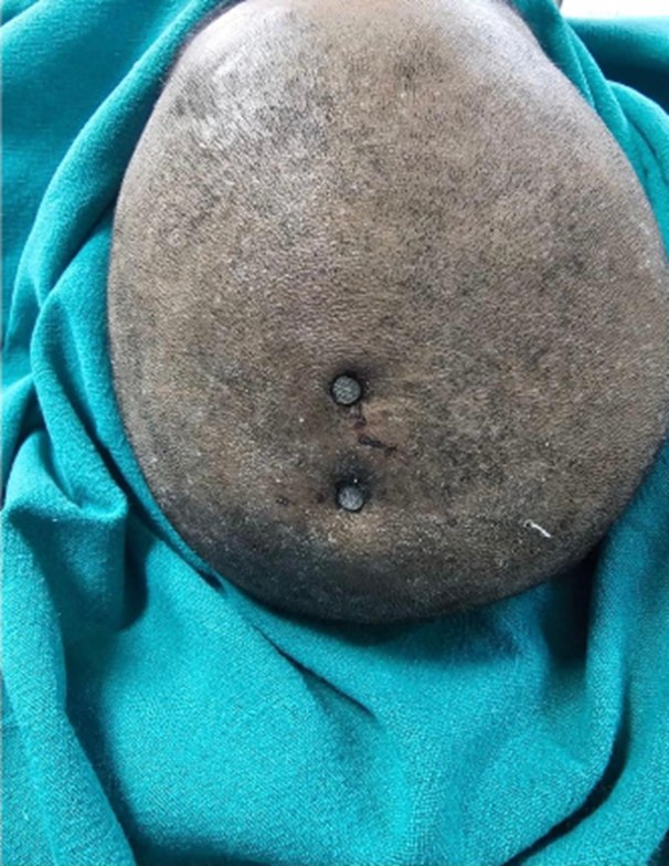
Local examination of the scalp demonstrated two metallic button-like foreign bodies embedded vertically in the left parietal region, approximately 3 cm apart. Mild local tenderness was noted around the injury site, but there was no active bleeding. The unusual combination of intracranial metallic penetration with preserved neurological function prompted urgent radiological evaluation and neurosurgical assessment.
Timeline of Clinical Events
|
Time |
Clinical Event |
|
Day 0 (Approximately 2 days after injury) |
Patient presented to the Emergency Department with severe headache following alleged assault resulting in penetrating cranial injury. |
|
Day 0 |
Initial clinical assessment performed. Patient was conscious, oriented, hemodynamically stable, and had a GCS score of 15/15 without focal neurological deficit. |
|
Day 0 |
Local scalp examination revealed two metallic foreign bodies embedded vertically in the left parietal region with mild tenderness and no active bleeding. |
|
Day 0 |
Plain skull radiography performed, demonstrating two metallic foreign bodies penetrating the cranial vault. |
|
Day 0 |
CT brain evaluation confirmed intracranial penetration of two metallic nails through the parietal bone. The foreign bodies extended into the cranial cavity without major intracranial hemorrhage or significant midline shift. |
|
Day 0 |
Patient admitted under neurosurgical care. Surgical exploration with craniectomy and controlled removal of the penetrating foreign bodies was performed under general anaesthesia. |
|
Postoperative period |
The patient underwent postoperative neurological monitoring with assessment for complications including infection, hemorrhage, and neurological deterioration. |
|
Follow-up period |
Clinical outcome assessment demonstrated favourable recovery following removal of the intracranial foreign bodies. The case emphasized the possibility of survival despite delayed presentation and minimal neurological symptoms. |
Clinical Findings
On local examination, the scalp revealed two metallic foreign bodies embedded vertically within the left parietal region. The two metallic objects were positioned approximately 3 cm apart, suggesting two separate penetrating points within the parietal scalp and underlying cranial vault. Mild tenderness was present around the injury site; however, there were no features suggestive of active external bleeding or significant soft tissue disruption.
The entry wound characteristics were consistent with a penetrating cranial injury, with the metallic objects firmly retained within the scalp and skull region. No active hemorrhage was observed externally at the time of examination. The retained foreign bodies represented the visible external component of a deeper intracranial penetrating injury requiring radiological assessment before attempted removal.
The location of the foreign bodies was identified in the left parietal region of the scalp. Subsequent radiological evaluation demonstrated that the metallic objects had penetrated the cranial vault and extended intracranially through the parietal bone.
Neurological assessment demonstrated a surprisingly preserved clinical status despite the presence of intracranial metallic penetration. The patient was conscious, alert, and oriented at presentation. His Glasgow Coma Scale (GCS) score was 15/15, indicating normal consciousness without evidence of impaired neurological function.
Cranial nerve examination was normal, with no detectable cranial nerve dysfunction. There were no clinical signs suggestive of brainstem involvement, raised intracranial pressure, or meningeal irritation.
Motor examination revealed no weakness or focal motor deficit, with preserved limb movements. Sensory examination was also unremarkable, with no reported sensory impairment. The absence of neurological deficit despite intracranial foreign body penetration represented an important clinical feature of this case and emphasized the discrepancy that may occur between radiological severity and neurological presentation in penetrating cranial injuries.
Summary of Clinical Findings
|
Clinical Parameter |
Findings |
|
Scalp examination |
Two metallic foreign bodies embedded vertically in the left parietal scalp region |
|
Entry wound characteristics |
Penetrating injury with retained metallic objects; mild local tenderness |
|
Foreign body location |
Left parietal region, approximately 3 cm apart |
|
External bleeding |
No active bleeding observed |
|
Neurological status |
Conscious, oriented, and neurologically intact |
|
Glasgow Coma Scale |
15/15 |
|
Cranial nerve examination |
Normal |
|
Motor examination |
No motor deficit |
|
Sensory examination |
No sensory deficit |
|
Meningeal signs |
Absent |
Diagnostic Assessment
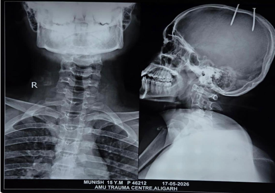
Plain skull radiography was performed as an initial imaging investigation to identify the location and extent of the retained metallic foreign bodies. The radiograph demonstrated two radiopaque metallic foreign bodies within the left parietal region, consistent with retained metallic nail-like objects. The foreign bodies were visualized as penetrating structures involving the cranial vault.
The radiographic findings confirmed the presence of metallic foreign material crossing the skull bone, indicating a penetrating cranial injury rather than an isolated superficial scalp injury. The location of the foreign bodies corresponded with the clinically identified embedded metallic objects in the left parietal scalp region.
The skull radiograph was important for rapid identification of the retained metallic objects and provided initial confirmation of cranial vault penetration, prompting further evaluation with computed tomography for accurate assessment of intracranial extension and surgical planning.
CT Brain
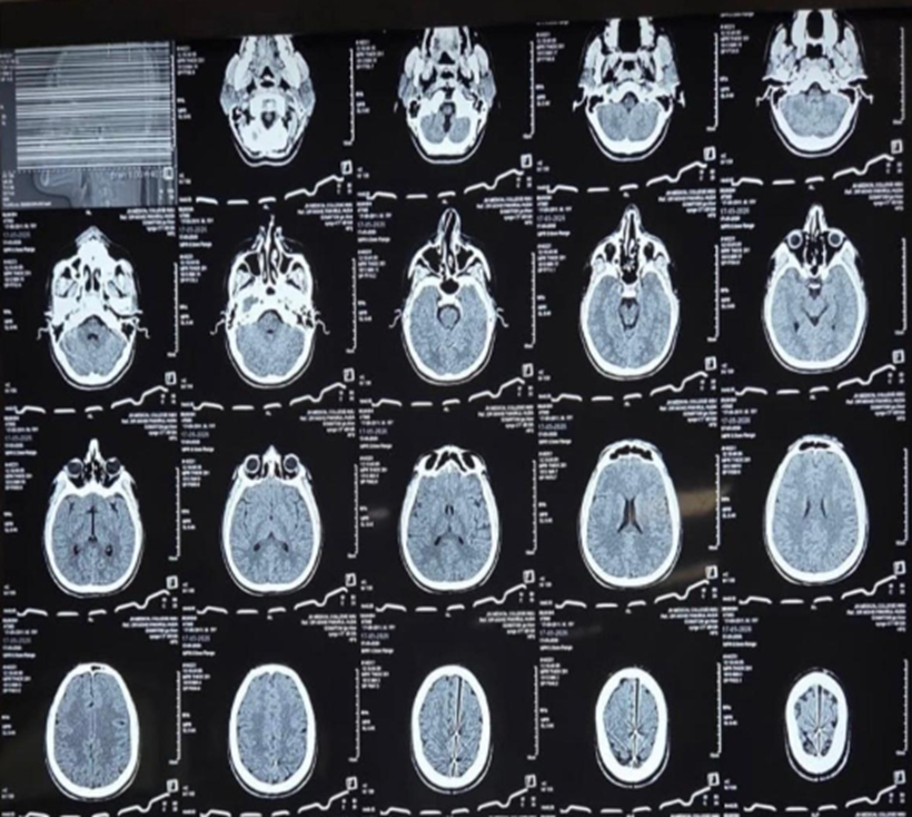
Computed tomography (CT) of the brain was subsequently performed to define the exact trajectory of the penetrating foreign bodies and assess associated intracranial injury. CT imaging confirmed intracranial penetration of two metallic nails through the left parietal bone.
Foreign Body Trajectory
The CT scan demonstrated that the metallic foreign bodies followed a trajectory through the parietal cranial vault, extending from the external scalp region into the cranial cavity. The intracranial pathway of the objects was clearly visualized, confirming a true penetrating cranial injury rather than superficial impalement.
Parietal Bone Breach
The imaging demonstrated a breach of the left parietal bone, with the metallic objects traversing the cranial vault. The bony penetration represented the entry pathway for intracranial extension of the foreign bodies.
Intracranial Extension
Both metallic objects extended beyond the parietal bone into the cranial cavity. The presence of intracranial foreign bodies confirmed a penetrating traumatic brain injury requiring neurosurgical management.
Intracranial Hemorrhage Assessment
CT evaluation did not demonstrate evidence of major intracranial hemorrhage associated with the penetrating objects. Despite intracranial penetration, there was no reported significant hemorrhagic complication at the time of imaging.
Midline Shift and Mass Effect
There was no significant midline shift identified on CT imaging. The absence of substantial mass effect correlated with the patient's preserved neurological status at presentation.
Brain Injury Assessment
Although the metallic foreign bodies penetrated intracranially, the patient did not demonstrate major radiological complications such as extensive hemorrhage or significant structural displacement. The imaging findings illustrated the unusual nature of the case, where considerable penetrating injury occurred with minimal neurological impairment.
Surgical Implications
The CT findings were crucial for operative planning by defining the location, depth, and trajectory of the penetrating foreign bodies. Because blind extraction of intracranial foreign objects may result in catastrophic hemorrhage or additional neurological injury, controlled surgical removal after adequate imaging assessment was required. The patient was subsequently managed under neurosurgical care with surgical exploration and removal of the foreign bodies under general anaesthesia.
Radiological Summary Table
|
Imaging Parameter |
Findings |
|
Imaging modality |
Skull radiograph followed by CT brain |
|
Foreign body |
Two metallic nail-like foreign bodies |
|
Location |
Left parietal region |
|
Bone involvement |
Penetration through parietal cranial vault |
|
Trajectory |
External scalp → parietal bone → intracranial cavity |
|
Intracranial extension |
Present |
|
Major intracranial hemorrhage |
Not identified |
|
Midline shift |
Absent |
|
Mass effect |
No significant mass effect reported |
|
Clinical significance |
Required planned neurosurgical removal |
Therapeutic Intervention
The patient was initially managed in the emergency department following the principles of trauma stabilization. On arrival, the patient was assessed for airway, breathing, circulation, and neurological status. Despite the presence of intracranial penetrating foreign bodies, the patient remained conscious, oriented, and hemodynamically stable, with a Glasgow Coma Scale (GCS) score of 15/15 and no evidence of focal neurological deficit.
Immediate attention was directed toward preventing secondary complications, including intracranial hemorrhage, infection, and neurological deterioration. The retained metallic objects were not manipulated or removed in the emergency setting because premature extraction of penetrating intracranial foreign bodies may increase the risk of uncontrolled bleeding, particularly in cases involving occult vascular injury.
The patient was stabilized and transferred for definitive neurosurgical evaluation.
Neurosurgical Consultation
Following radiological confirmation of intracranial penetration, the patient was admitted under neurosurgical care for further management. The decision for operative intervention was based on the presence of retained intracranial metallic foreign bodies, the risk of delayed complications, and the potential for infection and vascular injury.
Although the patient demonstrated minimal neurological symptoms, the presence of intracranial foreign material represented a high-risk condition requiring specialist management. The clinical decision-making emphasized that neurological stability does not exclude significant structural injury in penetrating cranial trauma.
Antibiotic Prophylaxis
Because penetrating cranial injuries involve direct communication between the external environment and intracranial structures, there is a substantial risk of contamination and subsequent infectious complications, including meningitis, cerebral abscess formation, and wound infection.
Appropriate antimicrobial prophylaxis was considered an essential component of management to reduce infection risk associated with retained metallic foreign bodies and contaminated penetrating injuries.
Surgical Planning
Detailed preoperative planning was performed based on the clinical examination and radiological findings. CT imaging demonstrated two metallic nails penetrating through the parietal bone and extending into the cranial cavity without major intracranial hemorrhage or significant midline shift.
The objectives of surgery were:
- Safe removal of the intracranial foreign bodies
- Prevention of secondary infection
- Assessment and control of potential bleeding sources
- Management of contaminated tissue and bone injury
- Prevention of delayed neurological complications
The foreign bodies were planned for controlled extraction rather than emergency blind removal because the objects could potentially be associated with vascular structures or provide temporary tamponade against bleeding.
Anaesthesia
Surgical removal of the penetrating foreign bodies was performed under general anaesthesia to ensure adequate patient immobilization, optimal surgical exposure, and controlled extraction conditions.
General anaesthesia also allowed appropriate airway protection and intraoperative neurological and haemodynamic monitoring during the procedure.
Foreign Body Removal Technique
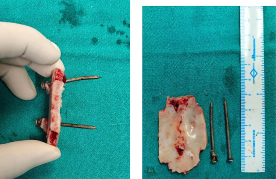
Surgical exploration with craniectomy and removal of the retained metallic foreign bodies was performed under neurosurgical supervision.
The metallic objects were carefully extracted after adequate surgical exposure. The procedure was performed cautiously to minimize the risk of:
- Secondary intracranial hemorrhage
- Additional brain injury
- Vascular disruption
- Contamination of intracranial structures
The extracted foreign bodies consisted of two metallic nails measuring approximately 4 cm each, which were removed along with the involved skull fragment.
Wound Management
Following removal of the foreign bodies, the operative wound was managed with appropriate surgical debridement and cleaning to minimize the risk of infection. Contaminated tissue and injured bone fragments were addressed during the procedure.
The principles of wound management included:
- Removal of contaminated material
- Adequate irrigation
- Prevention of infectious complications
- Secure closure following neurosurgical assessment
Postoperative Care
After surgery, the patient was monitored closely for potential complications associated with penetrating cranial injuries. Postoperative care focused on:
- Serial neurological assessment
- Monitoring for intracranial hemorrhage
- Observation for seizures
- Infection surveillance
- Assessment of wound healing
The patient's favourable neurological presentation despite delayed presentation and intracranial penetration highlighted the importance of timely imaging assessment and appropriate surgical intervention. The reported outcome demonstrated that survival with minimal neurological deficit is possible when penetrating cranial injuries are managed through coordinated neurosurgical care.
Rationale for Management Decisions
The management strategy was guided by the principle that intracranial penetrating foreign bodies require careful evaluation and controlled removal rather than immediate extraction. The absence of severe neurological symptoms did not eliminate the possibility of serious intracranial injury. Radiological assessment was therefore essential to determine the trajectory of penetration and guide safe surgical intervention.
Early neurosurgical involvement, controlled removal under general anaesthesia, infection prevention measures, and postoperative neurological monitoring were critical components contributing to the favourable outcome in this rare case.
DISCUSSION:
Penetrating brain injuries (PBI) represent a rare but devastating form of traumatic brain injury characterized by disruption of the scalp, skull, dura mater, and underlying brain tissue by an external object. Although they account for a relatively small proportion of traumatic brain injuries, they are associated with substantial morbidity and mortality due to the possibility of direct cerebral destruction, intracranial hemorrhage, vascular injury, infection, and delayed neurological complications. The clinical outcome of penetrating injuries depends primarily on the trajectory of the penetrating object, depth of penetration, velocity, anatomical region involved, and associated vascular damage rather than the external appearance of the wound alone [10]. The management of these injuries remains challenging because patients may present with variable neurological findings ranging from complete neurological preservation to profound neurological deterioration [11].
Metallic foreign body penetration, particularly injuries caused by nails, represents an uncommon subgroup of penetrating cranial trauma. Intracranial nail injuries are most frequently associated with nail guns used in occupational environments, especially construction-related activities. Although nail guns generally produce low-velocity injuries compared with firearms, the narrow diameter and pointed structure of nails allow them to penetrate the skull and travel deeply into intracranial structures. The severity of injury is determined by the nail trajectory and the structures encountered along its pathway rather than the apparent simplicity of the external wound [12]. In addition to accidental occupational injuries, intracranial nail penetration has been reported following assault, homicidal attempts, and self-inflicted mechanisms, highlighting the diverse circumstances associated with this rare injury pattern [13].
Previous reports have demonstrated considerable variability in clinical presentation and outcome following intracranial nail penetration. Miller et al. described the potential lethality of intracranial nail injuries and emphasized the risk of catastrophic vascular complications, particularly when the penetrating object involves major intracranial vessels [14]. Al-Mefty et al. highlighted the importance of angiographic evaluation in selected cases of nail gun injuries because vascular injury may remain clinically silent initially but can result in delayed complications such as pseudoaneurysm formation and secondary hemorrhage [15]. Similarly, Beaver and Cheatham reported life-threatening nail gun injuries and emphasized that early recognition, appropriate imaging, and coordinated surgical management are essential for reducing morbidity [16].
The present case demonstrates an unusual clinical scenario in which the patient survived a significant penetrating cranial injury with minimal neurological impairment. This phenomenon has been described in previous reports where patients maintained preserved neurological function despite radiologically confirmed intracranial foreign body penetration. Such outcomes are largely attributed to favourable trajectories that avoid critical cortical regions, major vascular structures, and eloquent brain areas. However, a normal neurological examination should not be interpreted as evidence of minor injury because delayed complications, including infection, vascular events, and seizures, may occur despite initial stability [10,11].
A major challenge in the management of intracranial nail injuries is determining the safest approach for foreign body removal. Blind extraction is discouraged because the penetrating object may be tamponading an injured vessel, and sudden removal can precipitate uncontrolled intracranial hemorrhage. Advanced imaging, particularly computed tomography (CT), plays a central role in defining the trajectory of the foreign body, identifying associated skull fractures, evaluating intracranial hemorrhage, and assisting surgical planning [11,15]. In cases where vascular injury is suspected, additional vascular imaging such as CT angiography or conventional angiography may be required to minimize operative risks.
Several rare cases have expanded understanding of intracranial nail injuries. Albuali et al. reported a unique self-inflicted nail-gun injury involving intracranial and intracardiac nail penetration, demonstrating that even extensive foreign body injuries may have variable outcomes depending on anatomical involvement [17]. Aggrawal et al. described a fatal nail injury case with forensic implications, emphasizing the importance of careful documentation and evaluation of injury circumstances in medico-legal cases [18]. These reports collectively highlight that intracranial nail penetration requires individualized assessment based on clinical presentation, imaging findings, and mechanism of injury.
The current case contributes to the existing literature because of the combination of intracranial nail penetration through the parietal bone, delayed presentation, and preservation of neurological function despite significant structural injury. It reinforces the principle that penetrating cranial injuries require aggressive diagnostic evaluation even when clinical findings appear reassuring. Early radiological assessment, multidisciplinary neurosurgical management, controlled removal of the foreign body, and vigilant postoperative monitoring remain essential for achieving favourable outcomes.
CONCLUSION:
Intracranial nail penetration through the parietal bone is an exceptionally rare but potentially life-threatening form of penetrating cranial injury. The main learning point from this case is that a preserved neurological examination and minimal clinical symptoms do not exclude significant intracranial trauma. Patients with retained intracranial foreign bodies may appear neurologically stable despite the risk of delayed hemorrhage, infection, vascular complications, and neurological deterioration.
The clinical implication of this case is that penetrating cranial injuries require a high index of suspicion, meticulous neurological assessment, and comprehensive radiological evaluation even when the initial presentation appears reassuring. Computed tomography plays a critical role in defining the foreign body trajectory, assessing associated intracranial injury, and guiding safe surgical planning.
REFERENCES:
1. Aarabi B. Management of penetrating brain injury. Neurosurg Focus. 2001;10(1):1-7.
2. Kazim SF, Shamim MS, Tahir MZ, Enam SA, Waheed S. Management of penetrating brain injury. J Emerg Trauma Shock. 2011;4(3):395-402.
3. Helling TS, McNabney WK, Whittaker CK, Schultz CC, Watkins M. The role of early surgical intervention in penetrating brain injuries. J Trauma. 1992;32(3):398-400.
4. Beaver AC, Cheatham ML. Life-threatening nail gun injuries. Am Surg. 1999;65(11):1113-1116.
5. Albuali A, Reimann A, Nicolaou S. Self-inflicted nail-gun injury with intracranial and intracardiac nails. Radiol Case Rep. 2015;6(3):525.
6. Aggrawal A, Pradhan M, Sreenivas M. Nail injury to the brain obfuscated by a fall from height: homicide or suicide? A case report. Med Sci Law. 2015;55(1):68-71.
7. Al-Mefty O, Holoubi A, Fox JL. Value of angiography in cerebral nail-gun injuries. AJNR Am J Neuroradiol. 1986;7:164-165.
8. Bayston R, de Louvois J, Brown EM, Johnston RA, Lees P, Pople IK. Use of antibiotics in penetrating craniocerebral injuries. Lancet. 2000;355(9217):1813-1817.
9. Blankenship BA, Baxter AB, McKahn GM. Delayed cerebral artery pseudoaneurysm after nail gun injury. AJR Am J Roentgenol. 1999;172(2):541-542.
10. Aarabi B. Management of penetrating brain injury. Neurosurg Focus. 2001;10(1):1-7.
11. Kazim SF, Shamim MS, Tahir MZ, Enam SA, Waheed S. Management of penetrating brain injury. J Emerg Trauma Shock. 2011;4(3):395-402.
12. Beaver AC, Cheatham ML. Life-threatening nail gun injuries. Am Surg. 1999;65(11):1113-1116.
13. Blankenship BA, Baxter AB, McKahn GM. Delayed cerebral artery pseudoaneurysm after nail gun injury. AJR Am J Roentgenol. 1999;172(2):541-542.
14. Miller CF, Brodkey JS, Colombi BJ. The danger of intracranial nail injuries. Neurosurgery. 1977;1(2):173-177.
15. Al-Mefty O, Holoubi A, Fox JL. Value of angiography in cerebral nail-gun injuries. AJNR Am J Neuroradiol. 1986;7:164-165.
16. Beaver AC, Cheatham ML. Life-threatening nail gun injuries. Am Surg. 1999;65:1113-1116.
17. Albuali A, Reimann A, Nicolaou S. Self-inflicted nail-gun injury with intracranial and intracardiac nails. Radiol Case Rep. 2015;6(3):525.
18. Aggrawal A, Pradhan M, Sreenivas M. Nail injury to the brain obfuscated by a fall from height: homicide or suicide? A case report. Med Sci Law. 2015;55(1):68-71.