Study Of Serum Albumin Levels In Acute Ischemic Stroke And It’s Association With Clinical Outcome.
- Ankesh Rajesh Amle , Junior Resident, General Medicine, K.S Hegde Medical Academy, Mangalore, Karnataka
- Shama Prakash K , Professor, General Medicine, K.S Hegde Medical Academy, Mangalore Karnataka
Article Information:
Abstract:
Background: Acute Ischaemic stroke (AIS) is one of the leading causes of morbidity and mortality due to non-communicable diseases globally. Due to the rapid onset of symptoms in AIS, immediate medical intervention is necessary to restore perfusion to the affected area and thus minimize neuronal damage. Serum albumin is one of the most abundant protein in the human plasma. It plays a crucial role in maintaining oncotic pressure. Recent studies have highlighted the neuroprotective benefits of normal serum albumin levels in the context of AIS. Several knowledge gaps and challenges remain in the understanding of the role of albumin in AIS. Addressing these challenges is necessary to establish the role of albumin in stroke management and improving patient outcomes. Our study aims to find out association between serum albumin and clinical outcome in acute ischemic stroke. Methods: A hospital based cross sectional study was conducted during 2023-2024 among 65 participants. Serum Albumin level on admission is used as predictor of clinical outcome in Acute ischemic stroke and it was compared with NIHSS and mRS score. Statistical analysis of the data was performed using SPSS version 20.0. Results: The mean age of the participants of 63.4 years (SD=11.4 years). A total of 38 patients (58.46%) were males while 27 (41.54%) were female. The mean serum albumin levels of the study population were 4.07 grams/dL (SD=0.6 gm/dL). Further, we performed linear logistic regression analyses to determine the relationship between serum albumin levels and NIHSS and mRS scores. The R2 value (Serum albumin vs NIHSS score) was found to be 0.217 indicating that around 21.7% of the variability in NIHSS scores can be explained by the serum albumin levels of the patients (p<0.0001). The R2 value (Serum albumin vs mRS score) was found to be 0.179 indicating that around 18% of the variability in mRS scores can be explained by the serum albumin levels of the patients (p=0.0004). Conclusion: This study adds to the growing body of evidence that serum albumin is a reliable, cost-effective biomarker associated with both stroke severity and short-term functional outcomes. Lower serum albumin levels are associated with poorer outcomes in stroke patients, including higher mortality rates and increased risk of complications. Despite the short-follow up period in our study, the potential inclusion of serum albumin assessment in composite prognostic models and tools warrants further exploration.
Keywords:
Article :
INTRODUCTION:
Acute Ischemic stroke (AIS) is one of the leading causes of morbidity and mortality due to non-communicable diseases globally1. It is a medical emergency characterized by the sudden loss of circulation to the territory of the occluded artery. This leads to tissue damage and loss of function to the brain tissue. AIS accounts for approximately 87% of all stroke cases and is primarily caused by the occlusion of a cerebral artery, usually due to thrombus or embolus formation.2, 3 Due to the rapid onset of symptoms in AIS, immediate medical intervention is necessary to restore perfusion to the affected area and thus minimize neuronal damage. Recent studies have given more insight into the pathophysiology of AIS, which have helped refine treatment strategies. However, the challenges in management still remain and prognosis is limited, especially in older age groups.4 Therefore, it is necessary to establish the prognosis early and guide interventions accordingly.
Due to occlusion of the involved artery, blood flow is restricted. This begins an ischemic cascade due to deprivation of glucose and oxygen supply. These are vital for maintaining cellular metabolism, therefore, their absence triggers energy failure, enhanced excitotoxicity caused by excessive glutamate release, formation of reactive oxygen species (ROS), and inflammation.5 These processes eventually lead to cerebral oedema and raised intracranial tension (ICT). Raised ICT, in turn, further exacerbates the ischemia in both healthy and affected tissue. The above sequence of events ultimately leads to neuronal and glial cell death. This further releases toxic products which exacerbate the injury. Thus, a vicious cycle ensues which spreads the damage to adjoining healthy tissue and worsens neurological outcomes. Thus, the extent of damage is influenced by the duration of ischemia, the size of the affected vascular territory, and the presence of collateral circulation.6
Serum albumin is one of the most abundant protein in the human plasma. It plays a crucial role in maintaining oncotic pressure. It also helps to transport various endogenous and exogenous substances, and exhibits antioxidant properties.7 Structurally, it is a globular protein with a molecular weight of approximately 66.5 kDa, composed of a single polypeptide chain folded into multiple domains. Thus created domains, in turn, create hydrophobic pockets which allow the binding and transport of fatty acids such as Omega-3 along with hormones and drugs.8 Thus, Albumin has multiple functions, including its capacity to maintain plasma oncotic pressure, scavenge free radicals and modulate inflammatory responses. Thus, it may play an important role in predicting the prognosis of ischemic stroke9. Although, it is to be noted that Albumin extravasates to the brain tissue in the case of hemorrhagic stroke and thus may exacerbate cerebral oedema.
Recent studies have highlighted the neuroprotective benefits of normal serum albumin levels in the context of AIS.9 Lower serum albumin levels at baseline have been shown to be associated with poorer outcomes10-13. Albumin has the ability to bind to and neutralize harmful substances, reduce oxidative stress, and stabilize the endothelial barrier function.14 These properties help confer it neuroprotective properties. Additionally, albumin has been shown to inhibit apoptosis and support neuronal survival during ischemic events.15
Serum albumin levels have been identified as a potential prognostic marker for AIS. Hypoalbuminemia, or low serum albumin levels, is associated with poorer outcomes in stroke patients, including higher mortality rates and increased risk of complications.16 The prognostic value of albumin in AIS may be linked to its role in maintaining vascular integrity, modulating immune responses, and mitigating oxidative damage.17
Monitoring serum albumin levels in stroke patients could provide valuable insights into disease progression and outcomes, aiding in the identification of individuals at higher risk of adverse events.18 Due to the above neuroprotective properties, albumin infusions have also been explored for management of AIS. Multiple preclinical and clinical studies have indicated that albumin administration during acute ischemic stroke can reduce infarct size, improve neurological outcomes, and may enhance overall recovery.19 The proposed mechanisms of action include restoration of plasma oncotic pressure, reduction of cerebral edema, and attenuation of the ischemic cascade. However, despite promising results in animal models and early-phase clinical trials, large-scale studies have yielded mixed results, necessitating further research to determine the efficacy and safety of albumin therapy in stroke patients.20
Several knowledge gaps and challenges remain in the understanding of the role of albumin in AIS. The timing, dosage, and route of administration have yet to be clearly defined.21 There is also a lack of large scale randomized clinical trials (RCTs) investigating the efficacy of Albumin infusions in AIS22. Additionally, the mechanisms underlying the neuroprotective effects of albumin are not fully understood, and further research is needed to completely understand these pathways.23Addressing these challenges is necessary to establish the role of albumin in stroke management and improving patient outcomes.24
METHODOLOGY:
A prospective cross-sectional study was conducted at Justice K. S. Hegde Charitable Hospital, which is attached to K. S. Hegde Medical Academy, a constituent unit of Nitte (Deemed to be University), Mangalore-575018. The study included cases diagnosed with acute stroke who were 18 years of age and above. The sample size was a minimum of 65. Convenience sampling was used to select participants. All the participants were selected according to the inclusion and exclusion criteria. The approval from the Institute Ethics Committee was obtained before conducting the study.
Inclusion Criteria
All consenting patients above 18 years of age, all patients with acute stroke identified through clinical, laboratory, and radiological evaluation (including CT/MRI) admitted within 72 hours of symptom onset, with the neurological deficit persisting for more than 24 hours.
Exclusion Criteria
Patients with a previous history of stroke, patients presenting more than 72 hours after symptom onset, patients with known renal or hepatic disease, patients with sepsis, celiac disease, inflammatory bowel disease, cirrhosis, or malnutrition, stroke due to tuberculoma, tumour, or trauma.
RESULTS :
A total of 65 patients were diagnosed with acute ischemic stroke and were included in the study. The mean age of the participants of 63.4 years (SD=11.4 years). A total of 38 patients (58.46%) were males while 27 (41.54%) were female (Figure 5). The middle cerebral artery was the most common territory involved and was seen in 47 (72.31%) patients (Figure 6). This was followed by posterior cerebral artery in 10 patients (15.38%), multivessel infarct in 7 patients (10.77%), and a single case of Lacunar infarct (1.54%).
As far as comorbidities are concerned, hypertension was the most prevalent comorbidity and was seen in 53 (81.54%) patients (Figure 7). This was followed by diabetes mellitus in 28 (43.08%) patients and coronary artery disease in 10 (15.38%) patients. A total of 27 (41.53%) patients from the above had more than one comorbidity and 4 (6.15%) patients had no comorbidities.
Regarding personal habits which were risk factors for stroke development, 24 (36.92%) patients had a history of smoking while 18 (27.69%) had a history of alcohol consumption (Figure 8). Further, 13 (20%) patients included in above distribution had co-occurring smoking and alcohol consumption.
Table 1. Baseline characteristics of the participants included in the study.
|
Parameter |
Value |
|
Age (Mean ± SD) |
63.4 ± 11.4 |
|
Males (n,%) |
38, 58.46% |
|
Females (n,%) |
27, 41.54% |
|
Vessel(s) involved (n,%) |
|
|
Middle Cerebral Artery |
47, 72.31% |
|
Multivessel infarct |
7, 10.77% |
|
Posterior Cerebral Artery |
10, 15.38% |
|
Lacunar |
1, 1.54% |
|
Co-morbidities (n,%) |
|
|
Hypertension |
53, 81.54% |
|
Diabetes Mellitus |
28, 43.08% |
|
Coronary Artery Disease |
10, 15.38% |
|
History of addiction (n,%) |
|
|
Smoking |
24, 36.92% |
|
Alcohol |
18, 27.69% |
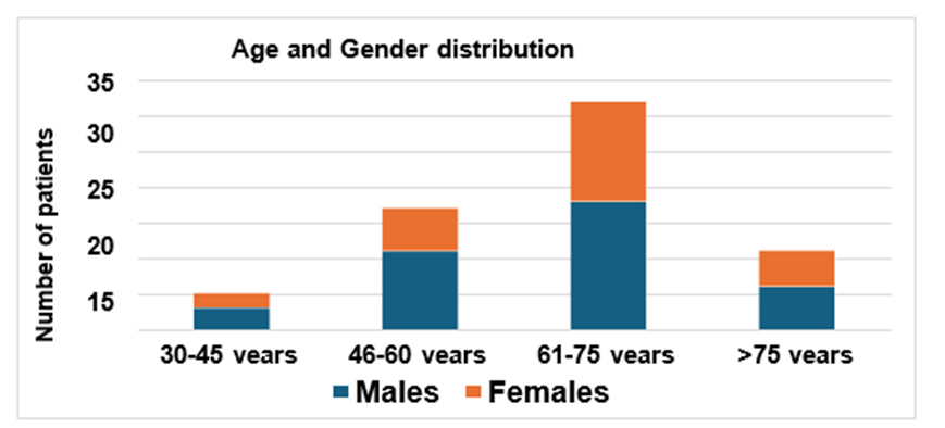
Figure 1. Age and gender distribution of the participants included in the study.
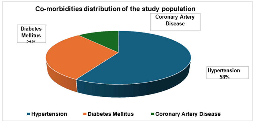
Figure 2. Distribution of vessels involved in stroke diagnosis of the patients included in the study. All percentages are calculated from the total number of patients involved in the study i.e. n=65.
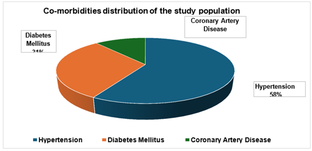
Figure 3. Comorbidities distribution of the patients included in the study. All percentages are calculated from the total number of patients involved in the study i.e. n=65.
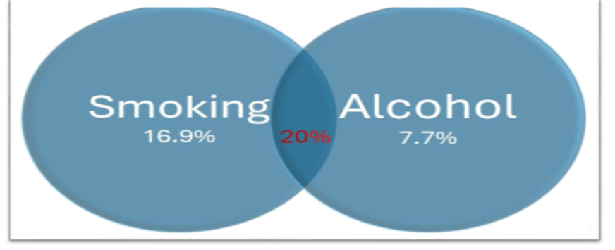
Figure 4. Distribution of smoking and alcohol habits of the patients included in the study. All percentages are calculated from the total number of patients involved in the study i.e. n=65.
TABLE 2: Serum Albumin and Clinical Scores
|
|
S. albumin
(grams/dL) |
NIHSS score |
mRS score |
|
Mean |
4.07 |
4.94 |
2.77 |
|
SD |
0.60 |
3.42 |
1.28 |
The mean serum albumin levels of the study population was 4.07 grams/dL (SD=0.6 gm/dL). The mean NIHSS score at admission was 4.94 (SD=3.42). The mean modified Rankin Scale (mRS) score at day 7 was 2.77 (SD=1.28). Further, we performed linear logistic regression analyses to determine the relationship between serum albumin levels and NIHSS and mRS scores. Regression Analyses
Serum Albumin vs NIHSS Score
We performed a linear regression to evaluate the relationship between serum albumin levels and NIHSS scores at admission (Figure 9, Table 2). The R2 value was found to be 0.217 indicating that around 21.7% of the variability in NIHSS scores can be explained by the serum albumin levels of the patients (p<0.0001). The slope of the regression line was -2.64 (95% confidence interval -3.91 to - 1.38), while the Y-intercept (NIHSS) was found to be 15.72 (95% confidence interval 10.51 to 20.91). The derived regression equation was as follows.
NIHSS = 15.72 – 2.64 × Albumin.
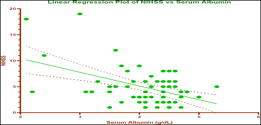
Figure 5. Linear regression plot of NIHSS versus serum albumin. The green line represents the regression line while the red dotted lines indicate the 95% confidence interval values.
Table 3. Estimates of NIHSS intercept and slope of the linear regression along with their 95% confidence interval values
|
|
Coefficient |
Estimate |
95% CI (Lower – Upper) |
|
NIHSS |
Intercept |
15.23 |
10.51 – 20.91 |
|
Albumin (slope) |
-2.65 |
-3.91 – -1.38 (significant) |
Serum Albumin vs mRS Score
We also performed a linear regression to evaluate the relationship between serum albumin levels and mRS scores at day 7 (Figure 10, Table 3). The R2 value was found to be 0.179 indicating that around 18% of the variability in mRS scores can be explained by the serum albumin levels of the patients (p=0.0004). The slope of the regression line was -0.90 (95% confidence interval -1.39 to -0.42), while the Y-intercept (mRS) was found to be 6.44 (95% confidence interval 4.44 to 8.44). The derived regression equation was as follows.
mRS = 6.442 – 0.90 × Albumin
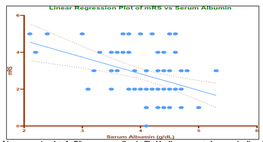
Figure 6. Linear regression plot of mRS versus serum albumin. The blue line represents the regression line while the red dotted lines indicate the 95% confidence interval values.
Table 4. Estimates of mRS intercept and slope of the linear regression along with their 95% confidence interval values
|
|
Coefficient |
Estimate |
95% CI (Lower – Upper) |
|
mRS |
Intercept |
6.44 |
4.44 – 8.44 |
|
Albumin (slope) |
-0.90 |
-1.39 – -0.42 (significant) |
DISCUSSION:
Acute ischemic stroke (AIS) is among the most disabling neurological conditions globally. Stroke is the second most common cause of death among non-communicable diseases in the world and third most common overall for death and disabilities1, 25. Ischemic stroke accounts for around 62% of this burden25. This is true especially in developing countries like India where its risk factors such as hypertension, diabetes mellitus, and metabolic syndrome are on the rise26-28. Therefore, it is important to identify the patients at risk of stroke and focus on preventive strategies. However, this is not often feasible, especially in large countries like India. After the occurrence of stroke, it is important to determine which patients are at a higher risk of poor outcomes and adjust interventions accordingly. While imaging is central to stroke identification, scales such as the NIHSS and mRS are useful in tracking the progress of the patient and predicting outcomes serially during the hospitalization. Serum biomarkers such as Serum albumin can also be conveniently repeated in the patient to assess the prognosis. Albumin is known to play neuroprotective, antioxidant, and endothelial-stabilizing roles in the patients of stroke29. These properties may help modulate the cascade of ischemic brain injury. Therefore, we designed this study to evaluate the association between serum albumin measured at admission and both stroke severity (via NIHSS) and short-term functional outcome (via mRS on day 7). Stroke incidence increases with age due to cumulative vascular changes and comorbidity burden. In our study, the mean age of the cohort was 63.4 years, with a slight male predominance (58.46%) which is consistent with the age-related risk factors of stroke. Further, older age may be associated with worse outcomes post-stroke due to higher risk of complications, especially beyond the age of 65 years29. Male predominance in incidence of stroke is commonly reported in the age group of 55-75 years and may also reflect differential exposure to modifiable risk factors such as smoking and hypertension. However, in our study, there was only a slight predominance of male patients.
We found that the most commonly affected territory was that of the middle cerebral artery (72.31%), followed by posterior cerebral artery (15.38%), multivessel infarcts (10.77%), and lacunar infarcts (1.54%). This distribution is consistent with the present knowledge that MCA territory is most commonly affected in acute ischemic stroke globally31. MCA strokes are also more likely to produce high NIHSS scores due to the broader neurological territories involved in this stroke type32. Hypertension (81.54%) and diabetes mellitus (43.08%) were the leading co-morbidities in our study with overlaps in some cases. Both of the above are well-established risk factors for ischemic stroke. They contribute to atherosclerosis, endothelial dysfunction, and microvascular damage thereby increasing the chances of thromboembolism and consequent stroke33. The presence of these comorbidities has also been linked to worse outcomes post-stroke due to impaired collateral perfusion, delayed recovery, and increased risk of complications33. Smoking and alcohol consumption were observed in 36.92% and 27.69% of patients, respectively in our study. Smoking enhances the risk of stroke with higher consumption relating to higher risks33, 25. This is primarily due to its role in vascular inflammation and thromboembolism. Alcohol, especially binge drinking, may also exacerbate hypertension and atrial fibrillation33. Both of these are known to be contributors to ischemic stroke. A substantial fraction of the study population ( 64.61%) reported consumption of one or both of the above which could have contributed to the development of stroke in these patients.
Primary findings of the study
Our study found a significant inverse association between serum albumin levels and NIHSS scores at admission (R² = 0.217, p < 0.0001). Therefore, patients with higher albumin levels had milder neurological deficits and those with lower albumin levels had more severe neurological deficits. This pattern was consistently seen in the study population (Figure 9) with a high level of significance indicating that the strength of the correlation was strong. These findings are consistent with previous studies. A multicentre study by Abubakar S, Sabir A, Ndakotsu M et al 10 showed that low admission serum albumin independently predicted more severe stroke and higher early mortality. Similarly, Idicula TT, Waje-Andreassen U, Brogger J et al11 findings from the Bergen Stroke Study emphasized that higher albumin levels were consistently associated with less severe strokes and fewer early complications11. Another 2024 study by Jena PK, Padhy T.el al 12 supported this, identifying serum albumin as a strong independent predictor of baseline stroke severity. Thus, our study substantiates their findings in Indian population.
Albumin is known to have several protective effects. It has a central role in maintaining oncotic pressure of the plasma and thus may contribute in reducing cerebral oedema in ischemic stroke patients28. Cerebral oedema is a known exacerbating factor of neuronal injury in stroke and is associated with poorer outcomes34. Therefore, patients with higher baseline albumin levels may be less inclined to develop severe cerebral oedema early. This may help in its protective role. Further, due to its antioxidant properties, it helps in scavenging reactive oxygen species (ROS)29. Reactive oxygen species generated by ischemia are responsible for neuronal damage of the brain tissue. Thus, study of van der Vusse GJ et al 35 albumin may also help by reducing ROS at the site of injury and conferring a neuroprotective role. Further, being an important carrier molecule in the plasma, it helps in transporting neuroprotective fatty acids and hormones1. Omega-3 fatty acids have an established neuroprotective role in stroke29. Therefore, transport of neuroprotective fatty acids such as Omega-3 by Albumin helps in preserving the blood-brain barrier, thereby limiting secondary injury to the brain tissue post-ischemia. These cumulative effects of Albumin may explain the observed inverse relationship between albumin and NIHSS in ischemic stroke. However, this relationship may not be observed in hemorrhagic stroke. This could be owing to the oncotic properties of the extravasated albumin within the brain tissue in this stroke type36. Albumin present in the brain tissue after haemorrhagic stroke would lead to increased cerebral oedema rather than decreasing it. Therefore, it is important to note that the findings of the present study should be limited to ischemic stroke.
Our study also found a significant negative association between serum albumin levels at admission and mRS score at 7 days (R² = 0.179, p = 0.0004). Thus, we found that higher albumin levels at admission predicted better short-term functional recovery in patients of ischaemic stroke (Figure 10). A study by Li C, Yang C, Zhu J el al13 reported that albumin and related ratios (globulin-to-prealbumin) were independently predictive of 3-month functional outcomes post-stroke, which is consistent with our observations13. Another study by Bc PK, Somannavar VG et al37 found similar trends in their Indian cohort, where hypoalbuminemia was associated with higher mRS scores and longer hospital stays37. Our study, through a more robust regression analysis has confirmed these findings. While many of the above studies have used longer-term mRS scores, short-term mRS may help to predict the length of hospital stay along with planning of early discharge and rehabilitation.
Other than its direct neuroprotective role as discussed above, serum albumin levels at admission may also indicate better baseline nutritional status and lower baseline systemic inflammation38, 39. This is especially important in elderly patients as lower serum albumin levels may indicate frailty. Interestingly, a study by Rael LT, Leonard J, Salottolo K el al 40 from 2019 noted that even oxidized forms of albumin may exert protective effects by neutralizing oxidative stress during ischemia-reperfusion injury. However, this is an isolated finding and may need to be substantiated by other studies. The findings of our study are aligned with multiple large-scale studies demonstrating that hypoalbuminemia is a poor prognostic factor in AIS13, 41. The direction and magnitude of the regression coefficients in this study (e.g., slope = –2.64 for NIHSS) are within the ranges reported in these studies. The differences in the values can be owed to the variations in sample sizes and population included. In our study we used similar methods of albumin estimation and timing (within 24 hours of admission), validated stroke scales (NIHSS and mRS), and similar patient demographics (age, risk factors) across Asian stroke populations. However, some of these studies report stronger associations over longer follow-up periods (e.g., 90-day mRS), whereas this study focused on a 7-day window. Therefore, the findings of our study may need to be substantiated in longer follow-up periods in South-east Asian population. There may also be differences in the baseline nutritional status, prevalence of chronic liver disease, or timing of measurement which may affect albumin levels.
In our study, we did not consider the effects of confounders such as CRP and ferritin in order to mimic a more real-world scenario. Lack of adjustment for markers like CRP, ferritin, and interleukins in some studies could either mask or exaggerate the albumin-outcome relationship. Further, Albumin is a negative acute phase reactant. Therefore, its levels may decline in inflammatory states, confounding associations unless adjusted for. However, we have undertaken baseline measurements at admission which disregards future albumin levels and focuses on the initial injury-prevention role of Albumin during the early hours of ischemic stroke. Further, it is to be noted that serum albumin levels alone cannot replace clinical assessment and established biomarkers. It should rather be seen as an additional tool.
CONCLUSION:
This study adds to the growing body of evidence that serum albumin is a reliable, cost-effective biomarker associated with both stroke severity and short-term functional outcomes. We found that lower serum albumin levels at admission were significantly associated with poorer neurological and short-term functional outcomes. However, it should not be considered a replacement of existing biomarkers and scales, rather an addition to them. Measuring albumin at the time of hospital admission is a simple inexpensive procedure which can assist clinicians in early risk stratification. It can further help to guide patient-family discussions and optimize resource allocation during the patient’s hospitalization. Despite the short-follow up period in our study, the potential inclusion of serum albumin assessment in composite prognostic models and tools warrants further exploration.
REFERENCES :
1. Feigin VL, Brainin M, Norrving B, Martins SO, Pandian J, Lindsay P, et al. World Stroke Organization: Global Stroke Fact Sheet 2025. International journal of stroke : official journal of the International Stroke Society. 2025;20(2):132-44.
2. O’Collins VE, Donnan GA, Macleod MR, Howells DW. Animal Models for the Study of Human Disease: Chapter 23. Animal Models of Stroke Versus Clinical Stroke: Comparison of Infarct Size, Cause, Location, Study Design, and Efficacy of Experimental Therapies: Elsevier Inc. Chapters; 2013.
3. Agarwal A, Devarajan R, Balbale S, Chopra A, Prabhakaran D, Huffman MD, et al. Heart Failure With Reduced Ejection Fraction Polypill Implementation Strategy in India: A Convergent Parallel Mixed Methods Study. Global heart. 2024;19(1):69.
4. Lakhan SE, Kirchgessner A, Hofer MJJotm. Inflammatory mechanisms in ischemic stroke: therapeutic approaches. 2009;7:1-11.
5. Belayev L, Pinard E, Nallet H, Seylaz J, Liu Y, Riyamongkol P, et al. Albumin therapy of transient focal cerebral ischemia: in vivo analysis of dynamic microvascular responses. Stroke. 2002;33(4):1077-84.
6. Wiedermann CJ, Joannidis MJTNEjom. Albumin replacement in severe sepsis or septic shock. 2014;371(1):83.
7. Ginsberg MD, Palesch YY, Hill MD, Martin RH, Moy CS, Barsan WG, et al. High-dose albumin treatment for acute ischaemic stroke (ALIAS) Part 2: a randomised, double-blind, phase 3, placebo-controlled trial. 2013;12(11):1049-58. Dziedzic T, Slowik A, Szczudlik A. Serum albumin level as a predictor of ischemic stroke outcome. Stroke. 2004;35(6):e156-8.
8. Nair R, Radhakrishnan K, Chatterjee A, Gorthi SP, Prabhu VA. Serum Albumin as a Predictor of Functional Outcomes Following Acute Ischemic Stroke. Journal of vascular and interventional neurology. 2018;10(2):65-8.
9. Abubakar S, Sabir A, Ndakotsu M, Imam M, Tasiu M. Low admission serum albumin as prognostic determinant of 30-day case fatality and adverse functional outcome following acute ischemic stroke. The Pan African medical journal. 2013;14:53.
10. Idicula TT, Waje-Andreassen U, Brogger J, Naess H, Thomassen L. Serum albumin in ischemic stroke patients: the higher the better. The Bergen Stroke Study. Cerebrovascular diseases (Basel, Switzerland). 2009;28(1):13-7.
11. Jena PK, Padhy T. Ischemic Stroke Outcome Predicted by Serum Albumin Levels. Cureus. 2024;16(5):e59816.
12. Li C, Yang C, Zhu J, Huang H, Zheng J, Hu X, et al. Predictive Value of Globulin to Prealbumin Ratio for 3-Month Functional Outcomes in Acute Ischemic Stroke Patients. Disease markers. 2022;2022:1120192.
13. Khatri P, Tayama D, Cohen G, Lindley RI, Wardlaw JM, Yeatts SD, et al. Effect of intravenous recombinant tissue-type plasminogen activator in patients with mild stroke in the third international stroke trial-3: post hoc analysis. 2015;46(8):2325-7.
14. Belayev L, Liu Y, Zhao W, Busto R, Ginsberg MDJS. Human albumin therapy of acute ischemic stroke: marked neuroprotective efficacy at moderate doses and with a broad therapeutic window. 2001;32(2):553-60.
15. Babu MS, Kaul S, Dadheech S, Rajeshwar K, Jyothy A, Munshi A. Serum albumin levels in ischemic stroke and its subtypes: correlation with clinical outcome. Nutrition (Burbank, Los Angeles County, Calif). 2013;29(6):872-5.
16. Kuller LH, Eichner JE, Orchard TJ, Grandits GA, McCallum L, Tracy RP. The relation between serum albumin levels and risk of coronary heart disease in the Multiple Risk Factor Intervention Trial. American journal of epidemiology. 1991;134(11):1266-77.
17. Li M, Chen Y, Chen Z, Wang L, Xie W, Zhang Y, et al. Dose-response relationship between C-reactive protein/albumin ratio and in-hospital mortality in elderly patients with acute ischemic stroke. 2024;70(2):125-33.
18. Elkind MS, Tai W, Coates K, Paik MC, Sacco RLJAoim. High-sensitivity C-reactive protein, lipoprotein-associated phospholipase A2, and outcome after ischemic stroke. 2006;166(19):2073-80.
19. de Liyis BG, Ardhaputra GYB, Liyis S, Wihandani DM, Siahaan YMT, Pinatih KJPJWN. High C-Reactive Protein/Albumin Ratio Predicts Mortality and Hemorrhage in Stroke Patients Undergoing Mechanical Thrombectomy: A Systematic Review and Meta-Analysis. 2024.
20. Alexandrova ML, Bochev PGJFRB, Medicine. Oxidative stress during the chronic phase after stroke. 2005;39(3):297-316.
21. Larsson M. Extracorporeal Membrane Oxygenation in Trauma Patients with Hypovolaemic Shock: Karolinska Institutet (Sweden); 2015.
22. Di Napoli M, Papa FJS. Inflammation, hemostatic markers, and antithrombotic agents in relation to long-term risk of new cardiovascular events in first-ever ischemic stroke patients. 2002;33(7):1763-71.
23. Shisler RJ, Baylis GC, Frank EMJA. Pharmacological approaches to the treatment and prevention of aphasia. 2000;14(12):1163-86.
24. Global, regional, and national burden of stroke and its risk factors, 1990-2019: a systematic analysis for the Global Burden of Disease Study 2019. The Lancet Neurology. 2021;20(10):795-820.
25. Khatri R, Afzal MR, Rodriguez GJ, Maud A, Miran MS, Qureshi MA, et al. Albumin-Induced Neuroprotection in Focal Cerebral Ischemia in the ALIAS Trial: Does Severity, Mechanism, and Time of Infusion Matter? Neurocritical care. 2018;28(1):60-4.
26. Koch S, Concha M, Wazzan T, Romano JG, Forteza A. High dose human serum albumin for the treatment of acute ischemic stroke: a safety study. Neurocritical care. 2004;1(3):335-41.
27. Huang Y, Xiao Z. Albumin therapy for acute ischemic stroke: a meta-analysis. Neurological sciences : official journal of the Italian Neurological Society and of the Italian Society of Clinical Neurophysiology. 2021;42(7):2713-9.
28. Lalkovičová M, Danielisová V. Neuroprotection and antioxidants. Neural regeneration research. 2016;11(6):865-74.
29. Mohammad R, Bansod DW. Hypertension in India: a gender-based study of prevalence and associated risk factors. BMC Public Health. 2024;24(1):2681
30. Nogles TE, Galuska MA. Middle cerebral artery stroke. StatPearls [Internet]: StatPearls Publishing; 2023.
31. Pradeepa R, Mohan V. Epidemiology of type 2 diabetes in India. Indian journal of ophthalmology. 2021;69(11):2932-8.
32. Boehme AK, Esenwa C, Elkind MS. Stroke Risk Factors, Genetics, and Prevention. Circulation research. 2017;120(3):472-95.
33. Gu Y, Zhou C, Piao Z, Yuan H, Jiang H, Wei H, et al. Cerebral edema after ischemic stroke: Pathophysiology and underlying mechanisms. Frontiers in neuroscience. 2022;16:988283.
34. van der Vusse GJ. Albumin as fatty acid transporter. Drug metabolism and pharmacokinetics. 2009;24(4):300-7.
35. Wang R, Ashwal S, Tone B, Tian HR, Badaut J, Rasmussen A, et al. Albumin Reduces Blood-Brain Barrier Permeability But Does Not Alter Infarct Size in a Rat Model of Neonatal Stroke. Pediatric Research. 2007;62(3):261
36. Bhalwar R. Metabolic syndrome: The Indian public health perspective. Medical journal, Armed Forces India. 2020;76(1):8-16.
37. Yousufuddin M, Young N. Aging and ischemic stroke. Aging. 2019;11(9):2542-4.
38. Yaghi S, Herber C, Boehme AK, Andrews H, Willey JZ, Rostanski SK, et al.The Association between Diffusion MRI-Defined Infarct Volume and NIHSS Score in Patients with Minor Acute Stroke. Journal of neuroimaging : official journal of the American Society of Neuroimaging. 2017;27(4):388-91.
39. Bc PK, Somannavar VG. A Study on the Role of Serum Calcium, Serum Albumin and Serum Uric Acid as Markers of Initial Neurological Severity and Short Term Outcome Indicators in Acute Ischemic Stroke. The Journal of the Association of Physicians of India. 2022;70(4):11-2.
40. Keller U. Nutritional Laboratory Markers in Malnutrition. Journal of clinical medicine. 2019;8(6).