Clinical Profile Of Non-Cardiac Pulmonary Hypertension In Real Life Practice.
- Amit Sharma. , Senior Consultant, Department of Internal Medicine, Lifeline Mediclinic, Gurugram, Haryana, India.
Article Information:
Abstract:
Background: Pulmonary Hypertension (PH) results from diverse causes and cardiac diseases are the commonest seen in clinical practice. There is paucity of data regarding non-cardiac PH seen at Referral centres at tertiary care hospitals. Our study was conducted to describe the etiological profile of Non-Cardiac Pulmonary Hypertension and to assess severity of Pulmonary Hypertension and see its association with aetiology. Methodology: We screened 66 patients who presented with unexplained or disproportionate dyspnoea in those having diseases which can cause Pulmonary Hypertension (i.e. chronic respiratory diseases, connective tissue diseases, drugs, obstructive sleep apnoea and pulmonary thromboembolism). Results: 95 cases with PH due to heart disease were excluded from the study. 66 cases were subjected to testing based on clinical suspicion and 35 subjects were diagnosed as Pulmonary Hypertension based on echocardiography Doppler (n=33) and right heart catheterization (n=2) and these were analysed further for aetiology and severity. The mean age of patients with Pulmonary Hypertension was 59.54 ± 14.45 years. The prevalence of Pulmonary Hypertension was highest in patients of age group of 51-60 years. Chronic Obstructive Pulmonary Disease (COPD) was found to be commonest cause (n=16, 45.71%) followed by idiopathic Pulmonary Artery Hypertension (iPAH) (n=8, 22.86%), Interstitial Lung Disease (n=6, 17.14%), and OSA (n=02, 5.71%). Conclusion: Chronic respiratory diseases (COPD and ILD) and Obstructive sleep apnea syndrome were the major cause of Non- cardiac PH found in our patients. Screening of patients who have unexplained dyspnoea by Doppler echocardiography for PH is advisable for timely detection of P.
Keywords:
Article :
INTRODUCTION:
Pulmonary Hypertension (PH) is defined as a mean pulmonary artery pressure (mPAP) ≥ 25 mmHg with a pulmonary capillary wedge pressure ≤ 15 mmHg, as measured by cardiac catheterization(1). The aetiology of pulmonary hypertension is diverse and most of the underlying causes of Pulmonary Hypertension are prevalent in the developing world in a much larger magnitude as compared to the western world(2,3). Cardiac diseases constitute the major group and other etiologic such as drugs, connective tissue diseases and pulmonary thromboembolism (PTE) are seen less often in clinical practice(4). In all these diseases, underlying CTD and chronic respiratory diseases causing pulmonary hypertension indicates poor prognosis and higher mortality(5). There are few studies that have described the aetiology of Pulmonary Hypertension from India(6,7); and there is paucity of data on the non-cardiac causes of PH seen in our population. Further it is not clear whether there is an association between aetiology and severity of pulmonary Hypertension(7). To look at these aspects we carried out this observational cross-sectional study at our hospital from September 2014 to September 2016.
2D Echocardiography with Doppler (ED) is a non-invasive screening tool for detection of pulmonary hypertension(8). Doppler is used to measure the maximum velocity (V) of the Tricuspid regurgitant jet, the systolic pressure gradient (AP) between right ventricle and right atrium is calculated by the modified Bernoulli equation (AP = 4V2). Adding the trans-tricuspid gradient to the mean right atrial pressure (RAP = 3-5 mm of Hg in healthy adults) gives an estimate of right ventricular systolic pressure (RVSP). In most studies using ED, estimated right ventricular systolic pressure( RVSP) correlates well with invasive cardiac catheterization measurements (9). The right ventricular systolic pressure of ≥35 mm Hg measured by echocardiography is suggestive of Pulmonary Hypertension as this pressure correlates with mean pulmonary artery pressure of ≥ 25 mmHg measured during right heart catheterisation(10, 11).
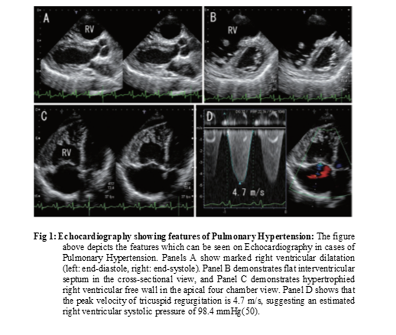
The earliest symptoms of Pulmonary Hypertension (PH) are effort related like exertional dyspnoea and cough. When right ventricular failure sets in, patients start having lower extremity oedema due to venous congestion. Orthopnoea and paroxysmal nocturnal dyspnoea (PND) may be seen in patients with PH due to underlying cardiac disease. Presence of dyspnoea which is disproportionate to the severity of the underlying cardio-pulmonary disease should be clue for evaluating for PH(12). The Physical examination in Patients of Pulmonary hypertension reveals prominent ‘a’ wave in jugular venous pressure (JVP), peripheral oedema, Left parasternal heave, accentuated pulmonary component of Second heart sound (S2), tricuspid regurgitation murmur, pulmonary early diastolic murmur, right ventricular third heart sound (S3), hepatomegaly, and ascites(4).
The severity of PH can be determined based on functional class by WHO classification of symptoms and limitation in activities. Exercise capacity assessed in PH clinics by the 6-min walk test(13).
The screening requires investigations that are able to detect presence of PH They include electrocardiogram (ECG), chest radiograph and transthoracic Doppler echocardiography (14). The underlying aetiology causing PH can be identified by tests such as pulmonary function tests, arterial blood gases, ventilation and perfusion lung scan, high resolution computed tomography (HRCT) of the chest and pulmonary angiography. Serological tests for HIV, hepatitis B or C serology should be performed to screen for cause of cirrhosis in porto-pulmonary hypertension. The thyroid hormone assays may reveal either hyperthyroid dysfunction or autoimmune thyroiditis, which are rarely associated with PH(14).
Despite certain limitations of echocardiography, it remains the most clinically useful non-invasive method allowing for multidimensional assessment of pulmonary circulation. However, in patients who have features suggestive of PH but no tricuspid regurgitation in echocardiography, RHC is warranted for diagnosis of PH (15).
The Etiological profile and prevalence of Pulmonary Hypertension in various non-cardiac disorders has not been evaluated in the real-world setting. This study was undertaken to find out the etiological and clinical profile of Pulmonary Hypertension due to non-cardiac causes. Many underlying diseases are associated with the development of PH which increases the morbidity and mortality of the disease. We decided to screen patients with disproportionate dyspnoea using echocardiography doppler and/or RHC to evaluate for Pulmonary Hypertension at our centre in this study to institute timely therapy to reduce the morbidity and mortality.
MATERIALS AND METHODS:
The study was a prospective cross-sectional observational study. The Patients coming to hospital outpatient or inpatient department who were diagnosed to have Pulmonary Hypertension based on Echocardiographic right ventricular systolic pressure (RVSP) or pulmonary artery systolic pressure (PASP) measured by right heart catheterisation ≥ 35 mmHg or mean Pulmonary Artery Pressure (mPAP) ≥ 25 mm of Hg on right heart catheterisation (RHC) with no structural or functional heart disease were included in the study for analysis. The study was carried out after approval from the institutional ethics committee and with fully informed written consent from the subjects. Patients with age more than 18 years and who had PH based on echo-doppler and/ or RHC were included in the study. Patients with Echocardiographic/ ECG evidence of underlying Heart diseases were excluded. A total of 35 Patients diagnosed with non-cardiac Pulmonary Hypertension were studied.
After obtaining written informed consent, each participant underwent a detailed clinical history and physical examination to identify features of PH and determine the likely underlying cause. Functional limitation was graded according to the WHO functional classification, based on symptoms and exercise capacity.
All patients initially underwent transthoracic echocardiographic study including two-dimensional, M-Mode, spectral Doppler. The echocardiographic mean pulmonary artery pressure was assessed by Tricuspid Regurgitation jet velocity for confirmation of the diagnosis of Pulmonary Hypertension (PASP was considered equivalent to RVSP in the absence of pulmonary outflow obstruction). The RVSP was approximated by measurement of the systolic regurgitant tricuspid flow velocity v and an estimate of right atrial pressure (2) applied in the formula: RVSP = 4v2 + RAP (RAP was assumed as 3-5 mmHg in this study). The patients were classified as suffering from mild, moderate or severe Pulmonary Hypertension as per calculated RVSP. The severity of Pulmonary Hypertension on the basis of measured Right Ventricular Systolic Pressure is classified as Mild for 35-50 mm of Hg, Moderate 51-65 mm of Hg and as Severe ≥ 65 mm of Hg (16).
All patients with PH were also tested for secondary causes by doing Complete Blood Count (CBC), Renal Function Test (RFT), Liver Function Test (LFT), Thyroid Function Test (TFT), Human Immunodeficiency Virus (HIV by ELISA), Anti-Nuclear Antibodies (ANA by ELISA), Chest radiograph PA view, Ultrasonography of Abdomen, and Pulmonary Function Testing (PFT) using spirometry. Arterial Blood Gas (ABG) analysis at room air was done in cases with hypoxemia (SpO2 < 90%). Mild hypoxemia was defined as a PaO2 of 60 to 79 mmHg; moderate hypoxemia as PaO2 40 to 59mm Hg; and severe hypoxemia with PaO2 less than 40 mm Hg (17).
Specialized tests such as High-Resolution Computed Tomography of the Chest (HRCT), CT Pulmonary Angiography, Diffusion Capacity for Carbon-Monoxide (DLCO), N Terminal pro- Brain Natriuretic Peptide (NT pro BNP) levels, Polysomnography, and Right Heart Cardiac Catheterization were carried out in selected cases and if consent was given.
Etiological diagnosis for PH was based on data from history, physical examination and results of various tests analysed together. Those where drugs history was excluded and no secondary cause was found after extensive testing including specialized tests were diagnosed as Idiopathic pulmonary arterial hypertension (iPAH). Genetic testing for mutations was not carried out in this study due to financial constraints.
The statistical analyses were performed by STATA 11.2 (College Station TX USA) and Microsoft Office Excel. Descriptive analysis was performed separately for Age, Gender, WHO functional class, RVSP values on echocardiography/ RHC, ABG analysis, Spirometry, GOLD classification for Obstructive pattern, and final Aetiology of Pulmonary Hypertension in all subjects.
RESULTS:
During the study period of 2 years (September 2014 to Sep 2016), the total OPD load was 1765. After excluding 95 cases with cardiac diseases related PH, 66 patients were screened for evidence of Pulmonary Hypertension by echo-doppler or RHC. Total 35 subjects with echocardiographic evidence or right heart catheterisation showing presence of Pulmonary Hypertension and with no underlying heart disease were included in the study. Out of these 35 patients, 33 patients were diagnosed based on Echo-doppler criteria (RVSP of ≥ 35 mm of Hg), and 02 patients were confirmed to have mean PAP ≥ 25 mm of Hg measured by Right Heart Catheterisation.
The age of all patients included in study ranged from 28 years to 90 years with mean of 59.54 years and standard deviation of 14.45 years. Most of the patients were in age group of 51-60 years (n=10, 28.57%), next common age group was 70-90 years (25.71%) followed by age group of 61-70 (22.86%). Cumulatively prevalence of Pulmonary Hypertension in patients of age >51 years was 77.14% of total patients diagnosed with disease. 17.14% of patients were in age group of 41-50 years. Subjects in the age group of 18-40 year constituted only 5.72% of total number of patients.
Out of total 35 subjects, male preponderance was noted with 69% of total patients and 31% of females. The male to female ratio was 2.1:1 this being a referral armed forces hospital. On clinical Examination 07 patients had Cyanosis, 11 had various grades of Clubbing, JVP was raised in 04 patients, 08 patients had loud pulmonary component of second heart sound (S2), 02 patients had left parasternal heave. 01 patient presented with right heart failure with RVS3. This patient was one of the 02 patients who underwent RHC for the diagnosis of PH.
Based on measured RVSP, 15 cases (43%) had mild Pulmonary Hypertension, 6 cases (17%) had moderate RVSP and 12 cases (34%) had severe Pulmonary Hypertension. The mean (+SD) estimated RVSP was 55.94 + 15.46. 02 (06%) cases underwent RHC during the study as Echo-doppler RVSP was < 35 mm of Hg, however the clinical suspicion of PH was strong.
Table1: Distribution of Severity of Pulmonary Hypertension on the basis of Right Ventricular Systolic Pressures (RVSP)
|
Severity on the basis of RVSP in Echo-doppler |
Number of Cases |
Percentage |
|
Mild (36-50) |
15 |
43% |
|
Moderate (51-65) |
6 |
17% |
|
Severe (> 65) |
12 |
34% |
|
Total |
33 |
94% |
19 cases (54.29%) were found to be in WHO class II. There were 10 cases (28.57%) in WHO Class III and 05 cases (14.29%) in WHO class IV. There was only 01 (2.86%) case in WHO class I.
The Spirometry results showed normal function in 11 cases (31%) and Restrictive pattern in 06 cases (17%). 18 cases (51%) showed obstructive pattern with FEV1 ranging from 26 to 68 % with a mean predicted FEV1 of 39% + 11.35%. ABG analysis of Partial Pressure of Oxygen (PaO2) revealed, normal PaO2 in 04 cases (11%), Mild Hypoxemia in 08 cases (23%), Moderate Hypoxemia in 10 cases (29%) and Severe Hypoxemia in 13 cases (37%).
The other investigations of all patients in this study like HIV test, USG Abdomen and Thyroid Assay were essentially normal/ negative. We arrived at the etiological diagnosis based on information from history, clinical exam along with laboratory and imaging tests in all our subjects. 18 patients (51.43%) were diagnosed to have Obstructive airway disease which included 16 patients of Chronic Obstructive Pulmonary Disease (COPD) (45.17%), 01 patient of Asthma COPD Overlap Syndrome (ACOS) and 01 with Allergic Broncho Pulmonary Aspergillosis (ABPA). 06 patients (17.14%) had Interstitial Lung Disease {03 patients had underlying connective tissue disease with 02 having ANA positivity (01 SLE and 01 Systemic Sclerosis) and 01 having RF positivity, 02 patient had drug induced (01 each due to Amiodarone and Dasatinib), and 01 patient was negative for CTD work up}. 02 patients (5.71%) Obstructive Sleep Apnoea and/or Obesity Hypoventilation Syndrome. One patient (2.86%) had Hereditary Haemorrhagic Telangiectasia (HHT) with Pulmonary Arterio-venous Malformation.
After detailed history, including drug and smoking history, thorough physical examination and lab tests, serological tests, spirometry and imaging studies including specialized tests; no underlying systemic disease could be identified in 08 (22.86%) patients and they were diagnosed as idiopathic Pulmonary Artery Hypertension (iPAH). The genetic testing for mutation analysis was not carried out in our study due to financial constraints.
Table 2: Etiological Diagnosis of cases with Pulmonary Hypertension
|
Aetiology |
Number of Cases |
Percentage (%) |
|
Obstructive Airway Disease |
18 |
51.43% |
|
Idiopathic PAH (iPAH) |
8 |
22.86% |
|
Interstitial Lung Disease |
6 |
17.14% |
|
Obstructive Sleep Apnoea (OSA) |
2 |
5.71% |
|
Pulmonary AV Malformation (HHT) |
1 |
2.86% |
|
Total |
35 |
100 |
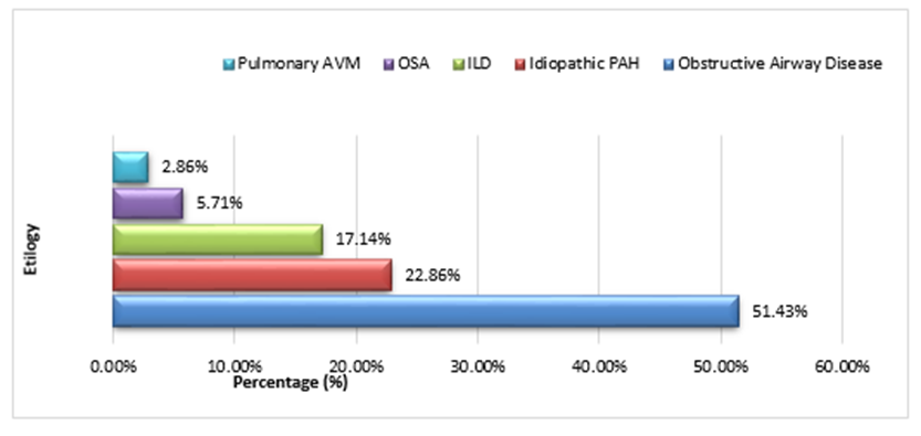
Chart 1: Etiological Diagnosis of cases with Pulmonary Hypertension
DISCUSSION:
Pulmonary Hypertension (PH) is a heterogeneous disease with different hemodynamic parameters and complex patho-physiology depending upon the underlying aetiology (18). The current updated classification of PH based on the underlying aetiology was given by WHO in 2013 comprising of five groups. Under this classification, group 2 includes causes of PH due to underlying left heart diseases (19).
Echo maybe normal in 20% cases with PH due to poor thoracic window or operator dependant factors and such cases would need RHC for the diagnosis of PH (20). In our study, out of 35 patients, 33 patients were diagnosed with PH by echocardiographic measurement of tricuspid jet velocity and RVSP. 02 patients underwent right heart catheterisation due to inability to detect tricuspid regurgitation for calculating the RVSP. These patients had clinical presentation and signs of PH, and were confirmed to have PH on RHC. One of these patients was finally diagnosed to have Hereditary Haemorrhagic Telangiectasia (HHT) and the other one with Idiopathic Pulmonary Artery Hypertension (iPAH).
Mean age of patients in our study was 59.54 ± 14.45 which is similar to the study conducted by Patel et al from Ahmedabad in India which shows the cumulative prevalence of disease to be 82% in age of > 41 years and maximum prevalence of 30% in age group of 51-60 years (14). However, the earlier studies have reported lower mean age of patients presenting with pulmonary hypertension. In a study by Hasan et al conducted at Aligarh, the mean age of the patients was 43.47±17.44 years (15). The western studies done by Shapiro et al, Burger et al and the REVEAL registry 2010 have reported a mean age of 49±18 years, 53±14 years and 50.4±16.8 years respectively (21-23). Thus, the mean age was relatively higher in our study, compared to earlier studies. This could be explained by the fact that, the most of the subjects in our study were retired soldiers of Indian Armed Forces who are healthier than corresponding general population when they are young due to their regular exercise regime and also because of mandatory Annual Medical Examination during their service tenure. Also, most of the previous studies cited above have included patients of underlying structural heart disease including valvular heart Diseases and Congenital Heart Diseases which is generally diagnosed in relatively younger age.
In the current study, males preponderance was noted with male to female ratio of 2.1 which was relatively comparable to study by Patel et al and Rich S et al which showed ratio of 1.5 and 1.7 respectively (15, 24).
In our study, 19 (54.29%) patients were found to be in WHO functional class II, 10 (28.57%) were found to be in WHO functional class III and 5 (14.29%) were found to be in WHO functional class IV. There was only one patient in WHO functional class I. Thus, the maximum number of patients was seen in the WHO functional class II. This contrasts with the other studies by Hasan et al, Burger et al, REVEAL registry 2010 and Korean registry 2011 in which they found maximum number of patients to be in functional class III (15, 22, 23, 25). This difference in finding is probably because of early diagnosis of PH in our patients who are either serving/ retired armed forces personnel or their dependants and have better access to healthcare facility.
In our study we found that mean arterial partial pressure of oxygen (PaO2) was 53.01 ± 18.05 mm of Hg. There were 13 (37%) patients who had severe hypoxemia (PaO2 < 40 mm of Hg). These finding are similar to the observations made by Keller et al (mean PaO2 of 59 ± 8 mmHg) and Kessler et al (60.3 ± 9.3 mmHg) in their patients with Pulmonary Hypertension (26, 27).
In present study, Chronic Obstructive Pulmonary Disease was found as the commonest cause (45.17%) of pulmonary hypertension which is comparable to the studies of Patel et al and Hasan et al which showed COPD as the most common cause of Pulmonary Hypertension in 66% and 36% of their study population respectively (14, 15). This finding contrasted with the western literature possibly due to higher incidence of Respiratory diseases specially COPD in Indian population due to increased use of biomass fuel for heating/ cooking in rural India, tobacco smoking and increasing air pollution in urban areas.
On further analysing the 18 patients with obstructive airway disease according to GOLD classification of COPD (28), we found the mean FEV1 percent of predicted to be 39 ± 11.35% and observed that out of these 18 patients, maximum number of patients i.e. 12 (66.67%) had moderate obstruction (GOLD III). This is comparable to the findings in the studies of Keller et al and Kessler et al which showed mean FEV1 of 33 ± 14% and 40.6 ± 12.3% respectively in patients of Pulmonary Hypertension (26, 27).
In our study we found mean Right Ventricular Systolic pressure (RVSP) to be 55.94 ± 15.46 mmHg. This is comparable to measured mean Pulmonary Artery Pressures of 50.7 ± 13.6, 50 ± 14, and 48 ± 16 in study by Burger et al, REVEAL registry 2010, and Korean registry 2011 respectively (22, 23, 25). Also, our study showed maximum patients to be suffering from mild Pulmonary Hypertension (43%) followed by Severe Pulmonary Hypertension (34%). The only Indian study which assessed severity of Pulmonary Hypertension by RVSP was by Hasan et al and they found higher severity of PH in Indian population with mean PAP of 69.77 ± 21.2.
This prospective cross-sectional study was designed to describe the aetiology of non-cardiac PH encountered in routine clinical practice. Several limitations should be acknowledged. The sample size was small, and genetic mutation analysis was not performed; consequently, some patients classified as having iPAH may have had an underlying heritable cause. The limited sample size also precluded a meaningful assessment of the relationship between PH aetiology and disease severity. In addition, systematic clinical and echocardiographic follow-up was not performed.
CONCLUSION:
The profile of non-cardiac PH in real life clinical practice in a tertiary care hospital showed Chronic Obstructive Pulmonary Disease (COPD) to be commonest cause. Other causes included Chronic Interstitial Lung Diseases and Obstructive Sleep Apnoea/ Hypopnea Syndrome.
Screening patients with Echo Doppler and or invasive right heart catheterization for diagnosis of Pulmonary Hypertension is recommended in those with risk factors for an early diagnosis and instituting appropriate treatment.
REFERENCES:
1. Hoeper MM. The new definition of pulmonary hypertension. Eur Respir J. 2009;34(4):790-1.
2. McCrory DC, Coeytaux RR, Schmit KM, Kraft B, Kosinski AS, Mingo A, et al. Pulmonary Arterial Hypertension: Screening, Management, and Treatment. Comparative Effectiveness Review. April 2013.
3. Simonneau G, Gatzoulis MA, Adatia I, Celermajer D, Denton C, Ghofrani A, et al. Updated clinical classification of pulmonary hypertension. J Am Coll Cardiol. 2013;62(25 Suppl): D34-41.
4. Gandharba Ray, Kumar LR. Pulmonary Arterial Hypertension. API Update. 2013; Chapter 27:126-30.
5. Cesar Machado, Ísis Brito, Denile Souza, Correia LC. Etiological Frequency of Pulmonary Hypertension in a Reference Outpatient Clinic in Bahia, Brazil. Arq Bras Cardiol. 2009;93(6):629-36.
6. Rahul Mehrotra, Manish Bansal, Ravi R Kasliwal, Trehan N. Epidemiological and Clinical Profile of Pulmonary Hypertension: Data from an Indian Registry. Journal of Clinical and Preventive Cardiology. April 2012; 2:51-7.
7. Natarajan R. Recent trends in pulmonary arterial hypertension. Lung India. 2011;28(1):39-48.
8. Paul G. Yock, Popp RL. Noninvasive estimation of right ventricular systolic pressure by Doppler ultrasound in patients with tricuspid regurgitation. Circulation. 1984;70(4):657-62.
9. Allemann Y. Echocardiographic and invasive measurements of pulmonary artery pressure correlate closely at high altitude. Am J Physiol Heart Circ Physiol. 2000;279:H2013–H6.
10. Chopra S, Badyal DK, Baby PC, Cherian D. Pulmonary arterial hypertension: advances in pathophysiology and management. Indian J Pharmacol. 2012;44(1):4-11.
11. Habib G, Torbicki A. The role of echocardiography in the diagnosis and management of patients with pulmonary hypertension. Eur Respir Rev. 2010;19(118):288-99.
12. Galie N, Torbicki A, Barst R, Dartevelle P, Haworth S, Higenbottam T, et al. Guidelines on diagnosis and treatment of pulmonary arterial hypertension. The Task Force on Diagnosis and Treatment of Pulmonary Arterial Hypertension of the European Society of Cardiology. Eur Heart J. 2004;25(24):2243-78.
13. Barst RJ, McGoon M, Torbicki A, Sitbon O, Krowka MJ, Olschewski H, et al. Diagnosis and differential assessment of pulmonary arterial hypertension. J Am Coll Cardiol. 2004;43(12 Suppl S):40S-7S.
14. Vaishali Patel, Kalpesh Khaped, Bhagirath Solanki, Amul Patel, Hitesh Rathod, Patel J. Profile of Pulmonary Hypertension Patients coming to Civil Hospital, Ahmedabad. Int J Res Med. 2013;2(1):94-7.
15. Asif Hasan, Muhammad Uwais Ashraf, Kumar S. A Study of Etio-Clinical and Echocardiographic Profile of Patients Presenting with Pulmonary Hypertension. International Journal of Enhanced Research in Medicines & Dental Care. April-2014;1(2):14-22.
16. Wrobel JP, Thompson BR, Snell GI, Williams TJ. Preoperative Echocardiographic-Defined Moderate–Severe Pulmonary Hypertension Predicts Prolonged Duration of Mechanical Ventilation Following Lung Transplantation for Patients with COPD. Lung. 2012;190(6):635-43.
17. William C. Pruitt, Jacobs M. Interpreting Arterial Blood Gases: Easy as ABC. Nursing. 2004;34(8):50-3.
18. Uma Kumar, R Ramteke, R Yadav, M Ramam, Rohini Handa, Kumar A. Prevalence and Predictors of Pulmonary Artery Hypertension in Systemic Sclerosis. Journal of Association of Physician in India. June 2008; 56:413-7.
19. Simonneau G, Gatzoulis MA, Adatia I, Celermajer D, Denton C, Ghofrani A, et al. Updated clinical classification of pulmonary hypertension. J Am Coll Cardiol. 2013;62(25 Suppl): D34-41.
20. Greiner S, Jud A, Aurich M, et al. Reliability of noninvasive assessment of systolic pulmonary artery pressure by Doppler echocardiography compared to right heart catheterization: analysis in a large patient population. J Am Heart Assoc. 2014;3(4): e001103. Published 2014 Aug 21. doi:10.1161/JAHA.114.001103
21. Shapiro S, Traiger GL, Turner M, McGoon MD, Wason P, Barst RJ. Sex differences in the diagnosis, treatment, and outcome of patients with pulmonary arterial hypertension enrolled in the registry to evaluate early and long-term pulmonary arterial hypertension disease management. Chest. 2012;141(2):363-73.
22. Burger CD, Foreman AJ, Miller DP, Safford RE, McGoon MD, Badesch DB. Comparison of body habitus in patients with pulmonary arterial hypertension enrolled in the Registry to Evaluate Early and Long-term PAH Disease Management with normative values from the National Health and Nutrition Examination Survey. Mayo Clinic proceedings. 2011;86(2):105-12.
23. Badesch DB, Raskob GE, Elliott CG, Krichman AM, Farber HW, Frost AE, et al. Pulmonary arterial hypertension: baseline characteristics from the REVEAL Registry. Chest. 2010;137(2):376-87.
24. Rich S. Primary pulmonary hypertension. A national prospective study. Ann Intern Med. 1987;107(2):16-23.
25. Chung WJ, Park YB, Jeon CH, Jung JW, Ko KP, Choi SJ, et al. Baseline Characteristics of the Korean Registry of Pulmonary Arterial Hypertension. J Korean Med Sci. 2015;30(10):1429-38.
26. Keller CA, Shepard JJW, Chun DS, Vasquez P, Dolan GF. Pulmonary hypertension in chronic obstructive pulmonary disease. multivariate analysis. Chest. 1986;90(2):185-92.
27. Romain Kessler, Michèle Faller, Emmanuel Weitzenblum, Ari Chaouat, Attila Aykut, Alain Ducoloné, et al. “Natural History” of Pulmonary Hypertension in a Series of 131 Patients with Chronic Obstructive Lung Disease. Am J Respir Crit Care Med. 2001; 164:219-24.
28. C. Volgelmeier, Chair, A. Agusti, A. Anzueto, L. Fabbri, F. Martinez, et al. Global Strategy for the Diagnosis, Management, and Prevention of Chronic Obstructive Pulmonary Disease (Updated 2016). 2016.