Correlation Of P16 And Elafin In Squamous Intraepithelial Lesions And Squamous Cell Carcinoma Of Cervix.
- Rashi Vaish , Senior Resident, Department of Pathology, Guru Teg Bahadur Hospital, Dilshad Garden, Delhi, India
- Noorin Zaidi , Professor, Department of Pathology, Era’s Lucknow Medical College and Hospital, Lucknow, Uttar Pradesh, India
- Astha Gupta , Assistant Professor, Department of Obstetrics and Gynecology, Blue Diamond Medical College, Darbhanga, Bihar, India
- Nirupma Lal , Professor and HOD, Department of Pathology, Era’s Lucknow Medical College and Hospital, Lucknow, Uttar Pradesh, India
- Aishwarya Gupta , Junior Resident, Department of Pathology, Hind Institute of Medical Sciences, Barabanki, Uttar Pradesh, India
- Ankita Dwivedi , Senior Resident, Department of Pathology, Rama Medical College, Kanpur, Uttar Pradesh, India.
Article Information:
Abstract:
Background: Aim: To evaluate the expression of p16 and Elafin in cervical SIL and SCC and assess the correlation between these biomarkers. Methods: This observational study was conducted in the Department of Pathology, Era’s Lucknow Medical College & Hospital, over a period of 24 months. A total of 55 histopathologically confirmed cases of cervical SIL and SCC were included. Immunohistochemical staining for p16 and Elafin was performed on formalin-fixed paraffin-embedded tissue sections. p16 expression was assessed using the Allred scoring system, while Elafin expression was evaluated in the plasma membrane, nucleus, and cytoplasm using a semi-quantitative scoring method. Statistical analysis was performed using SPSS version 26, and correlations were assessed using Spearman’s correlation coefficient. Results: p16 expression was significantly higher in SCC compared with SIL, with SCC cases predominantly exhibiting strong intensity scores and higher Allred scores (p<0.001). A significant association between p16 proportion score and CIN grade was observed (p=0.027). Elafin expression demonstrated distinct compartment-specific patterns. Membranous Elafin expression was significantly higher in SIL, whereas nuclear and cytoplasmic expression were significantly increased in SCC (p≤0.001). Among SIL grades, membranous expression progressively decreased, while nuclear and cytoplasmic expression increased with lesion severity (p<0.001). Significant correlations were identified between Elafin and p16 expression, with a negative correlation for plasma membrane Elafin (ρ=-0.618, p<0.001) and positive correlations for nuclear (ρ=0.619, p<0.001) and cytoplasmic Elafin (ρ=0.655, p<0.001). Conclusion: p16 is a reliable biomarker for distinguishing cervical precancerous lesions from invasive SCC. Elafin exhibits significant alterations in subcellular localization during cervical neoplastic progression, characterized by reduced membranous expression and increased nuclear and cytoplasmic expression. The significant correlation between p16 and Elafin suggests a potential synergistic role in cervical carcinogenesis. Combined assessment of p16 and Elafin may enhance diagnostic accuracy and provide additional prognostic information in cervical neoplastic lesions.
Keywords:
Article :
INTRODUCTION:
Cervical cancer remains a major public health concern worldwide and is one of the leading causes of cancer-related mortality among women, particularly in developing countries. In India, it is the second most common cancer among women and contributes substantially to the global cervical cancer burden1–3. Persistent infection with high-risk human papillomavirus (HPV), especially HPV types 16 and 18, is recognized as the principal etiological factor in the development of cervical squamous intraepithelial lesions (SILs) and invasive squamous cell carcinoma (SCC)2,4. The progression from low-grade squamous intraepithelial lesions (LSIL) to high-grade squamous intraepithelial lesions (HSIL) and ultimately invasive carcinoma involves a series of molecular and cellular alterations that can be identified using specific biomarkers.
Among the various biomarkers studied in cervical neoplasia, p16INK4a has emerged as a reliable surrogate marker of high-risk HPV infection.
p16 is a cyclin-dependent kinase inhibitor that regulates cell-cycle progression by inhibiting the transition from the G1 to S phase. In normal cervical epithelium, p16 expression is minimal; however, its expression becomes markedly increased following inactivation of the retinoblastoma protein (pRb) by the HPV E7 oncoprotein5,6. Consequently, diffuse overexpression of p16 is strongly associated with HPV-mediated dysplastic and malignant transformation. Several studies have demonstrated that p16 expression increases with the severity of cervical lesions, showing higher positivity in HSIL and SCC compared to LSIL7,8. Owing to its high diagnostic utility, p16 immunohistochemistry is increasingly used as an adjunct in the diagnosis and grading of cervical precancerous lesions and carcinoma9,10.
In recent years, Elafin (Peptidase Inhibitor 3; PI3) has attracted attention as a potential biomarker involved in tumor progression and aggressiveness. Elafin is an endogenous serine protease inhibitor that plays a protective role in epithelial tissues and is normally upregulated during inflammatory responses11. Aberrant expression of Elafin has been reported in several malignancies, including cancers of the lung, esophagus, bladder, and colorectum12. In cervical cancer, both nuclear and cytoplasmic expression of Elafin have been associated with tumor aggressiveness and disease progression, suggesting its potential role as a prognostic marker13.
The biological significance of Elafin may be linked to pathways involved in cellular proliferation and survival. The PI3K/AKT signaling pathway, which is frequently dysregulated in cervical carcinogenesis, promotes proliferation, invasion, and resistance to apoptosis, thereby contributing to progression from SIL to invasive SCC14. Since both p16 and Elafin are associated with distinct aspects of cervical carcinogenesis, evaluating their expression patterns may provide additional insight into disease progression and tumor behavior.
Therefore, the present study was undertaken to assess the immunohistochemical expression of p16 and Elafin in squamous intraepithelial lesions and squamous cell carcinoma of the cervix and to determine the correlation between these biomarkers, thereby evaluating their potential diagnostic and prognostic significance.
MATERIALS AND METHODS:
This was an observational study conducted over a period of 24 months at the Department of Pathology, Era’s Lucknow Medical College & Hospital. The study involved histopathologically confirmed cases of Squamous Intraepithelial Lesions (SIL) and Squamous Cell Carcinoma (SCC) of the cervix. The samples were obtained from newly diagnosed patients who had not previously been treated for cervical carcinoma. Specimens were processed and examined in the pathology laboratory using standard histological and immunohistochemical (IHC) techniques.
Inclusion Criteria: Histopathologically confirmed cases of Squamous Intraepithelial Lesions (SIL) and Squamous Cell Carcinoma (SCC) of the cervix were included in the study.
Exclusion Criteria
● Patients with any other malignancies were excluded.
● Cases already treated or on treatment for cervical carcinoma were also excluded from the study.
Sample size calculation: Sample size was calculated on the basis of proportion of SCC cases containing 2+ or higher grades and lower grades of Elafin expression using the formula:
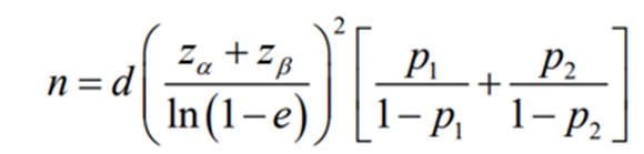
• p1 = 0.442 (44.2%) proportion of SCC cases containing 2+ or higher grades of Elafin.
• p2 = 0.558 (55.8%) proportion of SCC cases containing lower grades of Elafin15.
• e = 0.5, the proportion ratio considers to be clinically significant.
• d = 1.0, the design effect.
• Type I error α=5 %, for the significance level of 95%.
• Type II error β=10 %, for detecting the results with 90% power of study.
• The minimum sample size required comes out to be n = 55.
Sample Collection and Tissue Preparation: Upon receiving the cervical tissue samples, the following steps were performed:
1. Fixation: All tissue specimens were immediately fixed in 10% neutral buffered formalin to preserve tissue morphology and prevent autolysis.
2. Tissue Processing: The fixed tissue samples were dehydrated through a graded series of alcohols, cleared in xylene, and embedded in paraffin wax to create paraffin blocks. This allowed for easy sectioning and handling of the tissue.
3. Sectioning: Thin sections of 4-5 microns were cut from the paraffin blocks using a microtome and mounted on glass slides for staining.
4. Hematoxylin and Eosin (H&E) Staining: The sections were stained using the standard H&E staining procedure to assess the general morphology of the lesions under a light microscope.
Reagents used for IHC
● Immunohistochemistry: The immunohistochemistry staining was done on formalin fixed paraffin embedded tissue as per protocol standardized in our laboratory.
● Primary Antibody: p16 Antibody {Ventana; Lot no-K12104} at 1:50 ml dilution for 1 hour was used for immunohistochemical staining. Elafin/Scalp Polyclonal antibody (Peptidase inhibitor 3, skin derived) {Catalog Number:15963-1-AP; Source: Rabbit} at 1:50 ml dilution for 1 hour was used for immunohistochemical staining.
● Secondary antibody: Ventana ultra-View Universal DAB Detection Kit, Code K27164-25 ml (Material no. 05269806001, Ventana Medical Systems, Inc. 1910 Innovation Park Drive Tucson, Arizona)
Elafin Staining: The immunoreactivity of Elafin was assessed in the cytoplasm, cell membrane, and nucleus. The staining was scored semi-quantitatively i.e. 0: No staining, 1: < 5% of cells positive, 2: 5-50% of cells positive and 3: >50% of cells positive.
P16INK4a Staining: Staining for p16INK4a was interpreted based on the percentage of stained cells i.e. Weak: Less than 5% of cells stained, Variable: 5–50% of cells stained, with mixed intensity and Strong: More than 50% of cells stained.
Allred Scoring System: was used to quantify the immunoreactivity for p16INK4a:
1. Proportion Score (PS):
0: None
1: <1% of cells stained
2: 1-10% of cells stained
3: 10-33% of cells stained
4: 33-66% of cells stained
5: >66% of cells stained
2. Intensity Score (IS):
0: None
1: Weak staining
2: Intermediate staining
3: Strong staining
The total Allred score was calculated by adding the proportion score and intensity score,
Yielding a range from 0 to 8. Higher scores indicated stronger and more extensive staining.
Statistical Analysis: The data obtained from the histopathological and immunohistochemical analysis were statistically analyzed using SPSS software (Version 26). Descriptive statistics were used to summarize the distribution of cases across various categories (e.g., CIN 1, CIN 2, CIN 3, SCC). Correlations between the immunohistochemical markers (Elafin and p16INK4a) and clinicopathological parameters (e.g., lesion grade, HPV status) were assessed using the chi square test for categorical variables and the t-test for continuous variables and spearman correlation. A p-value of <0.05 was considered statistically significant.
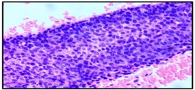
Figure 1: Photomicrograph showing cervical intraepithelial neoplasia 3 (CIN3)
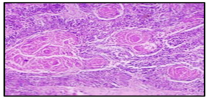
Figure 2: Photomicrograph showing well differentiated squamous cell carcinoma (H&E 100X)
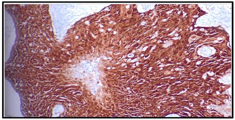
Figure 3: Photomicrograph of p16 showing strong positivity in poorly differentiated squamous cell carcinoma cervix (IHC 400X)
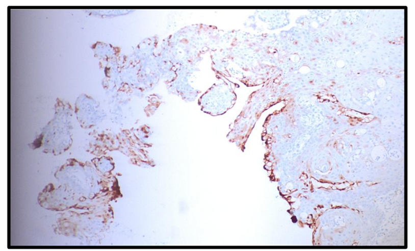
Figure 4: Photomicrograph of p16 showing weak positivity in cervical intraepithelial neoplasia 2 (CIN 2) (IHC 100X)
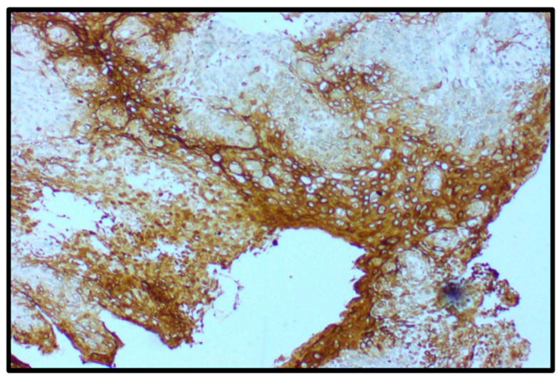
Figure 5: Photomicrograph of elafin showing strong nuclear and cytoplasmic positivity in moderately differentiated squamous cell carcinoma cervix (IHC 100X)
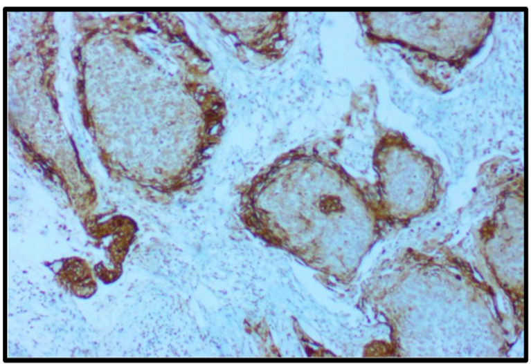
Figure 6: Photomicrograph showing strong membranous positivity for elafin expression in cervical intraepithelial neoplasia 2 (CIN 2) (IHC, 100X).
RESULTS:
The majority of patients belonged to the 46–55 years age group (45.5%), followed by 36–45 years (36.4%). Most specimens were obtained by biopsy (90.9%), while hysterectomy specimens accounted for 9.1%. Clinically, cervical squamous intraepithelial lesions were diagnosed in 63.6% of cases, whereas squamous cell carcinoma (SCC) of the cervix constituted 36.4%. Among SCC cases (n = 20), moderately differentiated SCC was the predominant histological grade (70.0%), with well- and poorly differentiated SCC each accounting for 15.0%. Among squamous intraepithelial lesions (n = 35), CIN 1 was the most frequent grade (54.3%), followed by CIN 3 (34.3%) and CIN 2 (11.4%) as shown in table 1.
Table 1: Demographic characteristics, procedure details, clinical diagnosis, and histopathological grading of the study population (N = 55)
|
Parameter |
Category |
Frequency (n) |
Percentage (%) |
|
Age group (years) (N=55) |
22–35 |
8 |
14.5 |
|
36–45 |
20 |
36.4 |
|
|
46–55 |
25 |
45.5 |
|
|
56–70 |
2 |
3.6 |
|
|
Procedure (N=55) |
Biopsy |
50 |
90.9 |
|
Hysterectomy |
5 |
9.1 |
|
|
Clinical diagnosis (N=55) |
Cervical squamous intraepithelial lesions |
35 |
63.6 |
|
Squamous cell carcinoma cervix |
20 |
36.4 |
|
|
Histological grade of SCC (n=20) |
Well differentiated SCC |
3 |
15.0 |
|
Moderately differentiated SCC |
14 |
70.0 |
|
|
Poorly differentiated SCC |
3 |
15.0 |
|
|
Grade of squamous intraepithelial lesions (n=35) |
CIN 1 |
19 |
54.3 |
|
CIN 2 |
4 |
11.4 |
|
|
CIN 3 |
12 |
34.3 |
In evaluation of P16 immunohistochemistry using Allred scoring system, an intensity score of 2 was the most frequent finding (45.5%), followed by an intensity score of 3 (32.7%). For the proportion score, a score of 2 was the most common (32.7%), whereas scores of 4, 1, and 5 were observed in 21.8%, 14.5%, and 14.5% of cases, respectively. Regarding the final Allred score, score 4 was the most prevalent (23.6%), followed by score 3 (21.8%), score 7 (16.4%), and score 8 (14.5%). A final Allred score of 0 was observed in 5.5% of cases, while scores 2, 5, and 6 were each identified in a smaller proportion of patients. This distribution indicates that most cases demonstrated intermediate to high p16 expression based on the Allred scoring system (table 2).
Table 2: Distribution of Cases according to p16 IHC Final score (Allred Score)
|
Parameter |
Category |
Frequency (n) |
Percentage (%) |
|
Intensity score |
0 |
3 |
5.5 |
|
1 |
9 |
16.4 |
|
|
2 |
25 |
45.5 |
|
|
3 |
18 |
32.7 |
|
|
Proportion score |
0 |
3 |
5.5 |
|
1 |
8 |
14.5 |
|
|
2 |
18 |
32.7 |
|
|
3 |
6 |
10.9 |
|
|
4 |
12 |
21.8 |
|
|
5 |
8 |
14.5 |
|
|
Final Allred score |
0 |
3 |
5.5 |
|
2 |
2 |
3.6 |
|
|
3 |
12 |
21.8 |
|
|
4 |
13 |
23.6 |
|
|
5 |
4 |
7.3 |
|
|
6 |
4 |
7.3 |
|
|
7 |
9 |
16.4 |
|
|
8 |
8 |
14.5 |
As per Elafin immunohistochemistry expression at the plasma cell membrane, negative or weak staining (Negative/+1) was observed in 54.5% of cases, while moderate to strong expression (+2/+3) was noted in 45.5%. Nuclear expression was nearly equally distributed, with 50.9% of cases demonstrating moderate to strong staining (+2/+3) and 49.1% showing negative or weak expression. Similarly, cytoplasmic expression revealed a slight predominance of moderate to strong staining (+2/+3) in 52.7% of cases compared with 47.3% exhibiting negative or weak staining (Negative/+1) as shown in graph 1.
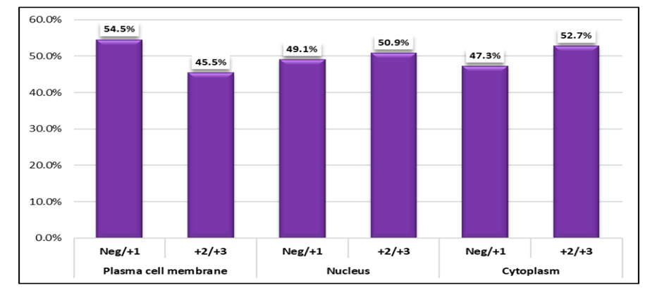
Graph 1: Distribution of Elafin immunohistochemistry (IHC) expression according to subcellular localization in the study population (N = 55)
Cervical squamous intraepithelial lesions predominantly demonstrated intensity score 2 (62.9%), proportion score 2 (51.4%), and final Allred score 4 (37.1%), whereas squamous cell carcinoma cases predominantly exhibited intensity score 3 (85.0%), proportion scores 4 (60.0%) and 5 (40.0%), and final Allred scores 7 (45.0%) and 8 (40.0%). The distributions of intensity score (χ² = 39.51), proportion score (χ² = 55.00), and final Allred score (χ² = 51.76) differed significantly between the two diagnostic groups (p < 0.001 for all), indicating significantly higher p16 expression in squamous cell carcinoma compared with cervical squamous intraepithelial lesions (table 3).
Table 3: Association of p16 immunohistochemistry (IHC) Allred scores with clinical diagnosis
|
Parameter |
Score |
Cervical squamous intraepithelial lesions (n = 35) |
Squamous cell carcinoma cervix (n = 20) |
p value |
||
|
|
|
No. |
% |
No. |
% |
|
|
Intensity score |
0 |
3 |
8.6 |
0 |
0.0 |
<0.001 |
|
1 |
9 |
25.7 |
0 |
0.0 |
||
|
2 |
22 |
62.9 |
3 |
15.0 |
||
|
3 |
1 |
2.9 |
17 |
85.0 |
|
|
|
Proportion score |
0 |
3 |
8.6 |
0 |
0.0 |
<0.001 |
|
1 |
8 |
22.9 |
0 |
0.0 |
||
|
2 |
18 |
51.4 |
0 |
0.0 |
||
|
3 |
6 |
17.1 |
0 |
0.0 |
||
|
4 |
0 |
0.0 |
12 |
60.0 |
||
|
5 |
0 |
0.0 |
8 |
40.0 |
||
|
Final Allred score |
0 |
3 |
8.6 |
0 |
0.0 |
<0.001 |
|
2 |
2 |
5.7 |
0 |
0.0 |
||
|
3 |
12 |
34.3 |
0 |
0.0 |
||
|
4 |
13 |
37.1 |
0 |
0.0 |
||
|
5 |
4 |
11.4 |
0 |
0.0 |
||
|
6 |
1 |
2.9 |
3 |
15.0 |
||
|
7 |
0 |
0.0 |
9 |
45.0 |
||
|
8 |
0 |
0.0 |
8 |
40.0 |
||
Intensity score 3 was observed in all well-differentiated (100%) and poorly differentiated (100%) SCC cases and in the majority of moderately differentiated SCC cases (78.6%). Similarly, proportion scores 4 and 5 predominated across all histological grades. Final Allred scores 7 and 8 were the most frequently observed scores irrespective of tumor grade. However, no statistically significant association was found between histological grade and p16 intensity score (χ² = 1.51, p = 0.469), proportion score (χ² = 1.05, p = 0.591), or final Allred score (χ² = 2.34, p = 0.674), indicating that p16 expression did not vary significantly with the degree of tumor differentiation among SCC cases (table 4).
Table 4: Association of p16 IHC Final score with Histological Grade of SCC
|
Parameter |
Score |
Well differentiated SCC (n = 3) |
Moderately differentiated SCC (n = 14) |
Poorly differentiated SCC (n = 3) |
p-value |
|||
|
|
|
No. |
% |
No. |
% |
No. |
% |
|
|
Intensity score |
0 |
0 |
0.0 |
0 |
0.0 |
0 |
0.0 |
0.469 |
|
1 |
0 |
0.0 |
0 |
0.0 |
0 |
0.0 |
||
|
2 |
0 |
0.0 |
3 |
21.4 |
0 |
0.0 |
||
|
3 |
3 |
100.0 |
11 |
78.6 |
3 |
100.0 |
||
|
Proportion score |
0 |
0 |
0.0 |
0 |
0.0 |
0 |
0.0 |
0.591 |
|
1 |
0 |
0.0 |
0 |
0.0 |
0 |
0.0 |
||
|
2 |
0 |
0.0 |
0 |
0.0 |
0 |
0.0 |
||
|
3 |
0 |
0.0 |
0 |
0.0 |
0 |
0.0 |
||
|
4 |
2 |
66.7 |
9 |
64.3 |
1 |
33.3 |
||
|
5 |
1 |
33.3 |
5 |
35.7 |
2 |
66.7 |
||
|
Final Allred score |
0 |
0 |
0.0 |
0 |
0.0 |
0 |
0.0 |
0.674 |
|
2 |
0 |
0.0 |
0 |
0.0 |
0 |
0.0 |
||
|
3 |
0 |
0.0 |
0 |
0.0 |
0 |
0.0 |
||
|
4 |
0 |
0.0 |
0 |
0.0 |
0 |
0.0 |
||
|
5 |
0 |
0.0 |
0 |
0.0 |
0 |
0.0 |
||
|
6 |
0 |
0.0 |
3 |
21.4 |
0 |
0.0 |
||
|
7 |
2 |
66.7 |
6 |
42.9 |
1 |
33.3 |
||
|
8 |
1 |
33.3 |
5 |
35.7 |
2 |
66.7 |
||
The majority of CIN 1 cases demonstrated an intensity score of 2 (52.6%), whereas 75.0% of both CIN 2 and CIN 3 cases also exhibited an intensity score of 2. A significant association was observed between proportion score and CIN grade (χ² = 14.23, p = 0.027), with higher-grade lesions showing a greater proportion of score 3 expression. Although final Allred score 3 predominated in CIN 1 (47.4%), final score 4 was the most frequent in both CIN 2 (50.0%) and CIN 3 (58.3%). However, no significant association was found between CIN grade and either intensity score (χ² = 5.77, p = 0.449) or final Allred score (χ² = 15.70, p = 0.109) as shown in table 5.
Table 5: Association of p16 IHC Final score with CIN
|
Parameter |
Score |
CIN 1 (n=19) |
CIN 2 (n=4) |
CIN 3 (n=12) |
p-value |
|||
|
|
|
No. |
% |
No. |
% |
No. |
% |
|
|
Intensity score |
0 |
3 |
15.8 |
0 |
0.0 |
0 |
0.0 |
0.449 |
|
1 |
6 |
31.6 |
1 |
25.0 |
2 |
16.7 |
||
|
2 |
10 |
52.6 |
3 |
75.0 |
9 |
75.0 |
||
|
3 |
0 |
0.0 |
0 |
0.0 |
1 |
8.3 |
||
|
Proportion score |
0 |
3 |
15.8 |
0 |
0.0 |
0 |
0.0 |
0.027 |
|
1 |
7 |
36.8 |
1 |
25.0 |
0 |
0.0 |
||
|
2 |
8 |
42.1 |
3 |
75.0 |
7 |
58.3 |
||
|
3 |
1 |
5.3 |
0 |
0.0 |
5 |
41.7 |
||
|
4 |
0 |
0.0 |
0 |
0.0 |
0 |
0.0 |
||
|
5 |
0 |
0.0 |
0 |
0.0 |
0 |
0.0 |
||
|
Final Allred score |
0 |
3 |
15.8 |
0 |
0.0 |
0 |
0.0 |
0.109 |
|
2 |
2 |
10.5 |
0 |
0.0 |
0 |
0.0 |
||
|
3 |
9 |
47.4 |
2 |
50.0 |
1 |
8.3 |
||
|
4 |
4 |
21.1 |
2 |
50.0 |
7 |
58.3 |
||
|
5 |
1 |
5.3 |
0 |
0.0 |
3 |
25.0 |
||
|
6 |
0 |
0.0 |
0 |
0.0 |
1 |
8.3 |
||
|
7 |
0 |
0.0 |
0 |
0.0 |
0 |
0.0 |
||
|
8 |
0 |
0.0 |
0 |
0.0 |
0 |
0.0 |
||
Plasma membrane expression differed significantly between the groups (χ²=11.76, p=0.001), with moderate to strong staining (+2/+3) observed more frequently in cervical squamous intraepithelial lesions (62.9%) than in squamous cell carcinoma (15.0%). In contrast, nuclear (χ² = 10.64, p = 0.001) and cytoplasmic (χ² = 13.13, p < 0.001) Elafin expression were significantly higher in squamous cell carcinoma, where +2/+3 staining was observed in 80.0% and 85.0% of cases, respectively, compared with 34.3% of cervical squamous intraepithelial lesions for both compartments (table 6).
Table 6: Association of Elafin IHC Final score with Clinical Diagnosis
|
Subcellular localization |
Expression score |
Cervical squamous intraepithelial lesions (n = 35) |
Squamous cell carcinoma cervix (n = 20) |
p value |
||
|
|
|
No. |
% |
No. |
% |
|
|
Plasma cell membrane |
Negative/+1 |
13 |
37.1 |
17 |
85.0 |
0.001 |
|
+2/+3 |
22 |
62.9 |
3 |
15.0 |
||
|
Nucleus |
Negative/+1 |
23 |
65.7 |
4 |
20.0 |
0.001 |
|
+2/+3 |
12 |
34.3 |
16 |
80.0 |
||
|
Cytoplasm |
Negative/+1 |
23 |
65.7 |
3 |
15.0 |
<0.001 |
|
+2/+3 |
12 |
34.3 |
17 |
85.0 |
||
Negative or weak plasma membrane expression (Negative/+1) predominated across all histological grades, being observed in 66.7% of well-differentiated SCC, 85.7% of moderately differentiated SCC, and 100.0% of poorly differentiated SCC cases. In contrast, moderate to strong nuclear (+2/+3) and cytoplasmic (+2/+3) expression was observed more frequently, particularly in moderately differentiated (85.7%) and poorly differentiated (100.0%) SCC. However, no statistically significant association was identified between histological grade and plasma membrane expression (χ² = 1.33, p = 0.515), nuclear expression (χ² = 5.12, p = 0.077), or cytoplasmic expression (χ² = 1.33, p = 0.515), suggesting that Elafin expression was not significantly associated with the degree of tumor differentiation among SCC cases (table 7).
Table 7: Association of Elafin IHC Final score with Histological Grade of SCC
|
Parameter |
Score |
Well differentiated SCC (n = 3) |
Moderately differentiated SCC (n = 14) |
Poorly differentiated SCC (n = 3) |
p-value |
|||
|
|
|
No. |
% |
No. |
% |
No. |
% |
|
|
Plasma cell membrane |
Negative/+1 |
2 |
66.7 |
12 |
85.7 |
3 |
100.0 |
0.515 |
|
|
+2/+3 |
1 |
33.3 |
2 |
14.3 |
0 |
0.0 |
|
|
Nucleus |
Negative/+1 |
2 |
66.7 |
2 |
14.3 |
0 |
0.0 |
0.077 |
|
|
+2/+3 |
1 |
33.3 |
12 |
85.7 |
3 |
100.0 |
|
|
Cytoplasm |
Negative/+1 |
1 |
33.3 |
2 |
14.3 |
0 |
0.0 |
0.515 |
|
|
+2/+3 |
2 |
66.7 |
12 |
85.7 |
3 |
100.0 |
|
The present study evaluated Elafin IHC expression across different grades of squamous intraepithelial lesions (SIL)—CIN 1, CIN 2, and CIN 3—revealing highly significant differences in subcellular localization patterns. For plasma membrane expression, moderate to strong Elafin staining (+2/+3) was observed in 94.7% of CIN 1 and 100% of CIN 2 cases, whereas 100% of CIN 3 cases showed only weak or negative expression (Neg/+1), indicating a significant inverse trend with increasing lesion severity (χ² = 30.94, p < 0.001). In contrast, nuclear Elafin expression showed a strong positive correlation with severity: all CIN 1 and CIN 2 cases (100%) had Neg/+1 staining, while 100% of CIN 3 cases demonstrated +2/+3 staining (χ² = 35.00, p < 0.001). A similar pattern was seen in cytoplasmic expression, with 100% of CIN 3 cases showing strong staining (+2/+3) and all CIN 1 and CIN 2 cases showing weak or no expression (χ² = 35.00, p < 0.001) as shown in graph 2.
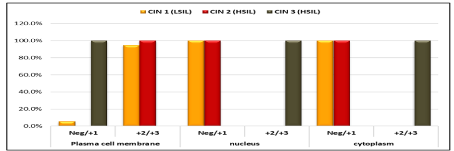
Graph 2: Association of Elafin IHC Final score with SIL Grades\
A statistically significant negative correlation was observed between Elafin expression in the plasma cell membrane and p16 scores (rho = –0.618, p < 0.001), suggesting that higher membrane-associated Elafin expression is associated with lower p16 expression. In contrast, significant positive correlations were noted between Elafin expression in the nucleus (rho = 0.619, p < 0.001) and cytoplasm (rho = 0.655, p < 0.001) with p16 scores, indicating that increased nuclear and cytoplasmic Elafin expression is associated with higher p16 expression levels (table 8).
Table 8: Correlation (Spearman) of Elafin IHC Final score with p-16 final score
|
Elafin IHC expression |
Final score p16 |
|
|
Spearman's correlation coefficient (ρ) |
p-value |
|
|
Plasma cell membrane |
-0.618 |
<0.001 |
|
Nucleus |
0.619 |
<0.001 |
|
Cytoplasm |
0.655 |
<0.001 |
DISCUSSION:
In the present study, the majority of cases belonged to the 46–55-year age group (45.5%), with a mean age of 47.05±11.67 years. Similar age distributions have been reported by Moschen et al16, Karube et al17, Tamrakar and Shrestha18, Kujur et al19, and Savani et al20, who demonstrated that cervical neoplasia predominantly affects middle-aged women. Although cervical lesions are increasingly detected in younger women, invasive disease remains most common in the fourth and fifth decades of life.
Biopsy was the most common diagnostic procedure in our study (90.9%), whereas hysterectomy accounted for only 9.1% of cases. This observation is consistent with previous reports showing that cervical biopsy remains the primary diagnostic modality, while hysterectomy is reserved for selected patients18,19. Cervical squamous intraepithelial lesions (SIL) constituted 63.6% of cases, while squamous cell carcinoma (SCC) accounted for 36.4%. The predominance of premalignant lesions in our cohort contrasts with studies by Umate et al21 and Dahiya et al22, which reported a higher prevalence of invasive SCC, possibly reflecting differences in screening practices and timing of diagnosis.
Among SCC cases, moderately differentiated SCC was the predominant histological subtype (70.0%), followed by well-differentiated and poorly differentiated SCC (15.0% each). Similar findings were reported by Gaikwad et al23 and Patil et al24, who also observed a predominance of moderately differentiated tumors. Among SIL cases, CIN 1 was the most frequent lesion (54.3%), followed by CIN 3 (34.3%) and CIN 2 (11.4%). This pattern is consistent with observations by Loopik et al25, who identified CIN 1 as the most common lesion and emphasized its high regression potential. Nevertheless, CIN 2 and CIN 3 remain clinically significant because of their greater risk of progression to invasive carcinoma26.
Evaluation of p16 expression using the Allred scoring system demonstrated that most cases showed intermediate-to-high expression. Moderate intensity staining (score 2) and an Allred score of 4 were the most frequent findings. Previous studies by Singh et al27 and Saigal et al28 reported strong and diffuse p16 positivity in high-grade lesions and SCC, supporting the role of p16 as a surrogate marker of HPV-associated oncogenic transformation.
A significant association was observed between p16 expression and clinical diagnosis. SCC cases predominantly exhibited strong staining intensity, high proportion scores, and Allred scores of 7–8, whereas SIL cases demonstrated lower scores. These findings are consistent with those of Singh et al27, Kishore and Patil29 and Saigal et al28, all of whom demonstrated progressive increases in p16 expression from low-grade lesions to invasive carcinoma. The results further support the utility of p16 immunohistochemistry in distinguishing invasive SCC from precursor lesions.
No significant association was identified between p16 expression and histological grade of SCC. Similar observations have been reported by Kalyani et al29 and Kishore and Patil30, indicating that although p16 is highly sensitive for identifying HPV-related SCC, its expression does not vary significantly according to tumor differentiation. Therefore, p16 appears to be more valuable as a diagnostic rather than a grading marker.
When p16 expression was evaluated across CIN grades, a significant association was observed only for the proportion score, with higher-grade lesions demonstrating a greater proportion of p16-positive cells. Ferdous et al31 similarly reported increased p16 positivity in high-grade squamous intraepithelial lesions, supporting the role of p16 in differentiating CIN 2/3 from CIN 1 and aiding diagnostic accuracy.
Elafin immunohistochemistry revealed distinct compartment-specific expression patterns. Membranous expression was relatively lower, whereas nuclear and cytoplasmic expression were observed in approximately half of the study population. Compared with the findings of Longatto-Filho et al32, our cohort demonstrated relatively higher nuclear and cytoplasmic Elafin expression, suggesting possible differences in tumor biology, disease stage, or population characteristics.
A significant association was observed between Elafin expression and clinical diagnosis. SCC cases showed significantly reduced membranous expression but increased nuclear and cytoplasmic expression compared with SIL cases. These findings indicate a progressive shift in Elafin localization during malignant transformation. Although Longatto-Filho et al32 reported overall downregulation of Elafin in cervical cancer, our compartment-specific analysis suggests that intracellular redistribution of Elafin may be associated with disease progression. Therefore, Elafin may provide complementary diagnostic information when evaluated alongside established markers such as p16 and SCC antigen.
Evaluation of Elafin expression across SCC grades demonstrated no statistically significant association with tumor differentiation, although a trend toward increased nuclear and cytoplasmic staining and reduced membranous expression was observed in poorly differentiated tumors. Similar findings were reported by Longatto-Filho et al32 and Westin et al33, suggesting that Elafin localization may reflect tumor progression rather than histological grade.
Among SIL cases, Elafin expression showed a striking association with lesion severity. Membranous staining predominated in CIN 1 and CIN 2 lesions, whereas CIN 3 lesions demonstrated complete loss of membranous expression and strong nuclear and cytoplasmic positivity. Similar observations by Longatto-Filho et al32 support the hypothesis that redistribution of Elafin from the cell membrane to intracellular compartments may represent an important event in cervical neoplastic progression.
Overall, the present study confirms the diagnostic value of p16 in cervical neoplasia and demonstrates that Elafin exhibits distinct subcellular localization patterns associated with progression from CIN to invasive SCC. The combined assessment of p16 and Elafin may therefore improve characterization of cervical premalignant and malignant lesions and warrants further evaluation in larger studies.
CONCLUSION:
The findings of this study highlight p16 as a reliable immunohistochemical marker in distinguishing precancerous lesions from invasive carcinoma. Elafin expression showed an inverse correlation with p16 at the plasma membrane, while nuclear and cytoplasmic expression of Elafin correlated positively with p16. Combined evaluation of p16 and Elafin may improve diagnostic accuracy and prognostic assessment in cervical lesions. Since, Elafin also showed varying expression patterns, its prognostic significance requires further investigation. So, large-scale studies are warranted to validate these biomarkers and explore their potential synergistic role in predicting disease progression.
REFERENCES:
1. Muraleedharan VR. Patterns in the prevalence and wealth-based inequality of cervical cancer screening in India. BMC Womens Health. 2023;23(1).
2. Naiknaware SV. Cancer Cervix: An Experience at Tertiary Healthcare Centre in India. Deleted J. 2024;4(4):150–4.
3. Ramamoorthy T, Kulothungan V, Sathishkumar K, et al. Burden of cervical cancer in India: estimates of years of life lost, years lived with disability and disability adjusted life years at national and subnational levels using the National Cancer Registry Programme data. Reprod Health. 2024;21:111.
4. Phukan P, Dutta A, Deuri A, Choudhury G, Borpujari P, Bhattacharyya N, et al. Prevalence of 14 high-risk human papillomavirus subtypes among volunteers of cervical cancer screening by Papanicolaou smear cytology from a tertiary care institute of Assam, India. Ann Oncol Res Ther. 2024.
5. Farzanehpour M, Muhammadnejad A, Akhavan S, Razavi AN, Jalilvand S, Salimi V, et al. P16INK4A immunohistochemistry as a gold standard for cervical cancer and precursor lesions screening. Iran J Public Health. 2020;49(2):312.
6. Mishra S, Mitra S, Kumari A, Tiwari HC. Study of expression of P-16 and Galectin-3 in cervical intraepithelial neoplasia (CIN) and invasive squamous cell carcinoma of uterine cervix. Int J Pharm Clin Res. 2023;15(5):2158–67.
7. Sandhu PS, Nibhoria S, Sandhu VK, Kaur H. Immunohistochemical expression of p16INK4a in premalignant lesions and malignant tumours of cervix. Int J Reprod Contracept Obstet Gynecol. 2023;12(6):1813–9.
8. Pari P, Bhat RV. A study on expression of p16INK4a by immunohistochemistry in cervical dysplasia and Invasive carcinoma of cervix. Arch Cytol Histopathol Res. 2020;4(4):310–4.
9. He Y, Shi J, Zhao H, Wang Y, Zhang C, Han S, et al. p16INK4A flow cytometry of exfoliated cervical cells: Its role in quantitative pathology and clinical diagnosis of squamous intraepithelial lesions. Clin Transl Med. 2023;13(3).
10. Ismail R, Gaballah A, Salama A, Shakweer MM, Meckawy GR, Faisal MM. Prognostic Impact of Human Papilloma Virus Infection on Cervical Cancer Patients Reflected by p16ink4a Expression: Single Institution Experience. Asian Pac J Cancer Biol. 2023;8(2):119–25.
11. Longatto-Filho A, Fregnani JH, Mafra da Costa A, de Araujo-Souza PS, ScapulatempoNeto C, Herbster S, et al. Evaluation of elafin immunohistochemical expression as marker of cervical cancer severity. Acta Cytol. 2021;65(2):165–74.
12. Yoshida N, Egami H, Yamashita J, Takai EI, Tamori Y, Fujino N, et al. Immunohistochemical expression of SKALP/elafin in squamous cell carcinoma of human lung. Oncol Rep. 2002;9(3):495–501.
13. Verrier T, Solhonne B, Sallenave JM, Garcia-Verdugo I. The WAP protein Trappin2/Elafin: a handyman in the regulation of inflammatory and immune responses. Int J Biochem Cell Biol. 2012;44(8):1377–80.
14. Chen L, Qing J, Xiao Y, Huang X, Chi Y, Chen Z. TIM-1 promotes proliferation and metastasis, and inhibits apoptosis, in cervical cancer through the PI3K/AKT/p53 pathway. BMC Cancer. 2022;22(1).
15. Zhang G, Cao S, Wang P, Zhu X, Yuan Z. Human papillomavirus status and P16INK4a expression in cervical precancerous lesions. BMC Cancer. 2020;20:1-8.
16. Moschen H, Méndez M, Soderini A, Aragona A, Bustos J, Améstica G, Retamozo J. 195 Can age be considered as prognostic factor of survival in patients with uterine cervical cancer? Int J Gynecol Cancer. 2019;29:A85.
17. Karube A, Saito F, Nagao D, Otomo M, Tamura D, Kimura N. Influence of the HPV16/18 infection on the age distribution of cervical cancer patients. J Jpn Assoc Rural Med. 2014;63:87–92.
18. Tamrakar SR, Shrestha SP. Study of cervical biopsy over a decade at a tertiary level hospital, Nepal. Nepal Med Coll J. 2019;21(1):50–4.
19. Kujur P, Jha RK, Singh S. Histopathological study of cervical biopsies in a tertiary care hospital of Chhattisgarh. Trop J Path Micro. 2021;7(3):147–51.
20. Zhao D, Li B, Wang Y, Liu S, Zhang Y, Yuan G. Clinical outcomes in early cervical cancer patients treated with nerve plane–sparing laparoscopic radical hysterectomy. J Minim Invasive Gynecol. 2020;27(3):687–96.
21. Umate P, Thengal DS, Kurdukar D. Study of clinical profile of cervical cancer patients: a hospital based study. Int J Reprod Contracept Obstet Gynecol. 2017;6(11):4873–8.
22. Dahiya N, Bachani D, Acharya AS, Sharma DN, Gupta S, Haresh KP. Sociodemographic, reproductive and clinical profile of women diagnosed with advanced cervical cancer in a tertiary care institute of Delhi. J Obstet Gynaecol India. 2016;66(Suppl 1):518–25.
23. Gaikwad SL, Valand AG, Agarwal NU. Clinico-histopathological analysis of lesions of uterine cervix in Ambejogai city of Maharashtra: A 2 year study at tertiary level hospital. J Diagn Pathol Oncol. 2016;1(2):32–5.
24. Patil N, Deshmukh V, Rathid A, Jyoti D, Chavan S. Clinicopathological correlation of cervical carcinoma: a tertiary hospital-based study. Int J Sci Stud. 2019;6(10):1–4.
25. Loopik DL, Bentley HA, Eijgenraam MN, Inthout J, Bekkers RLM, Bentley JR. The natural history of cervical intraepithelial neoplasia grades 1, 2, and 3: a systematic review and meta-analysis. J Low Genit Tract Dis. 2021;25(3):221–31.
26. Bergqvist L, Virtanen A, Kalliala I, Bützow R, Jakobsson M, Heinonen A, et al. Predictors for regression and progression of actively surveilled cervical intraepithelial neoplasia grade 2: a prospective cohort study. Acta Obstet Gynecol Scand. 2025.
27. Singh P, Kaushik S, Thakur B, Acharya S, Bhardwaj A, Bahal N. Evaluation of variable p16 immunostaining patterns, Ki-67 indices and HPV status in cervical SILs and squamous cell carcinomas: an institutional experience. Indian J Pathol Microbiol. 2023;66(1):63–9.
28. Saigal A, Gulati A, Kaushik R, Dattal D. Role of p16 expression in preneoplastic and neoplastic lesions of cervix. Int J Res Med Sci. 2024.
29. Kalyani R, Raghuveer C, Sheela S. Expression of P16 biomarker in squamous cell carcinoma of uterine cervix and its association with clinico-pathological parameters: a cross-sectional study. Biomed Res Ther. 2020;7:3954–61.
30. Kishore V, Patil A. Expression of p16INK4A protein in cervical intraepithelial neoplasia and invasive carcinoma of uterine cervix. J Clin Diagn Res. 2017;11(9):EC17–20.
31. Ferdous NE, Sharmin F, Haque DKM, Akhter L, Ahmed R, Shimu SJ, Yousuf S, Nessa A. Value of p16 expression in the detection of high-grade squamous intraepithelial lesion of cervix. Sch J App Med Sci. 2023;11(1):242–9.
32. Longatto-Filho A, Fregnani JH, Mafrada Costa A, et al. Supplementary material for: evaluation of Elafin immunohistochemical expression as marker of cervical cancer severity [dataset]. Karger Publishers. 2020; Available from: https://doi.org/10.6084/m9.figshare.13325465.v1.
33. Westin U, Nyström M, Ljungcrantz I, Eriksson B, Ohlsson K. The presence of elafin, SLPI, IL1-RA and STNFalpha RI in head and neck squamous cell carcinomas and their relation to the degree of tumour differentiation. Mediators Inflamm. 2002;11(1):7–12.