Clinical Profile and Prognostic Factors of Brain Tumours in Young Adults: A Comprehensive Study from MKCG Medical College, Berhampur.
- Dr. Debadutta Senapati , Assistant Professor, Department of Neurosurgery MKCG medical college and Hospital, Berhampur, Odisha
- Bipin Bihari Malick , Assistant Professor, Department of Anaesthesiology, MKCG medical college and Hospital, Berhampur, Odisha
- Nibedita Sahu , Assistant Professor, Department of Anaesthesiology, MKCG medical college and Hospital, Berhampur, Odisha
Article Information:
Abstract:
Background: Brain tumours in young adults present unique clinical and psychological challenges. This study aims to evaluate the clinical profile, treatment modalities, and prognostic factors affecting short-term survival in young adults diagnosed with primary brain tumours in a tertiary care setting in Southern Odisha. Methods: An observational cohort study was conducted at MKCG Medical College and Hospital (MKCG MCH), Berhampur, from June 2025 to May 2026. A total of 112 young adult patients (aged 18 to 40 years) were included. Simple clinical parameters such as age, gender, tumour grade, extent of surgical resection, and Karnofsky Performance Status (KPS) were analyzed. Results: Out of 112 patients, the majority were males (58%). Gliomas were the most frequently encountered tumours (48.2%), followed by meningiomas (21.4%). Headache (76.7%) and seizures (44.6%) were the most common presenting symptoms. Gross total resection was achieved in 55.3% of cases. Statistically significant factors favoring better one-year survival included a higher pre-operative KPS score >70 (p=0.002), low-grade tumour pathology (p<0.001), and gross total resection (p=0.014). Conclusion: For young adults battling brain tumours, early diagnosis before functional decline (maintaining a high KPS) and achieving maximum safe surgical resection are the most critical determinants of a favorable prognosis.
Keywords:
Article :
INTRODUCTION:
A brain tumour diagnosis is life-altering at any age, but it hits particularly hard when it strikes young adults. In the prime of their lives—often while building careers and starting families—young adults facing central nervous system (CNS) tumours present a unique set of challenges for healthcare providers. While pediatric and geriatric brain tumours are heavily researched, the young adult demographic (typically defined as ages 18 to 40) often falls into a "gray zone" in neuro-oncology literature.
MKCG Medical College and Hospital (MKCG MCH) in Berhampur serves as a critical healthcare lifeline for the population of Southern Odisha and neighboring regions. Over the years, we have noticed a steady influx of young patients presenting with neurological deficits that are eventually traced back to primary brain tumours. Young adults generally possess better physiological reserves to tolerate aggressive treatments like surgery, radiation, and chemotherapy. However, the exact prognostic factors that dictate their survival and quality of life need localized evaluation.
The primary aim of this study was to map out the clinical landscape of brain tumours in young adults presenting to MKCG MCH between 2024 and 2025. By focusing on straightforward, universally applicable parameters, we sought to identify clear, statistically significant factors that influence patient prognosis.
MATERIALS AND METHODS:
Study Design and Setting
This was a prospective observational study conducted in the Departments of Neurosurgery and Oncology at MKCG MCH, Berhampur. The study period spanned one years, from June 2025 to May 2026.
Patient Selection
We enrolled 112 consecutive young adult patients (aged 18–40 years) who were newly diagnosed with primary brain tumours. We purposefully excluded patients with metastatic (secondary) brain tumours, recurrent tumours, or those who abandoned treatment midway, to keep our data clean and focused.
Data Collection
We deliberately avoided overly complicated genetic or molecular markers to ensure the findings remain highly applicable in resource-constrained clinical settings. We recorded basic demographic data, clinical presentation, tumour histology (based on WHO classification), and treatment modalities. Functional status was measured using the widely accepted Karnofsky Performance Status (KPS) scale.
Statistical Analysis
Data were analyzed using basic statistical software. We used the Chi-square test for categorical variables to determine significance. A p-value of less than 0.05 was considered statistically significant.
RESULTS:
Over the two-year study period, we closely followed 112 young adults. The data yielded several insightful patterns regarding who gets these tumours and how they fare.
Patient Demographics
The age distribution showed that vulnerability to brain tumours increased slightly as patients approached their late thirties. Men were somewhat more frequently affected than women, reflecting global epidemiological trends for specific CNS tumours.
Table 1: Demographic Profile of the Study Population (N = 112)
|
Parameter |
Category |
Number of Patients |
Percentage (%) |
|
Age Group |
18–25 years |
24 |
21.4 |
|
26–33 years |
39 |
34.8 |
|
|
34–40 years |
49 |
43.8 |
|
|
Gender |
Male |
65 |
58.0 |
|
Female |
47 |
42.0 |
Description: Table 1 highlights that the 34–40 age bracket was the most affected, comprising nearly 44% of the cohort. There was a clear male predominance, with a male-to-female ratio of roughly 1.4:1.
Histopathological Diagnosis
Understanding the "enemy" is the first step in oncology. When we looked at the tissue biopsies, gliomas were by far the most dominant culprit.
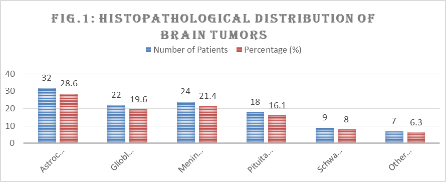
Fig 1: Histopathological Distribution of Brain Tumours
Description: Fig 1 demonstrates that primary gliomas (low and high grade combined) accounted for almost half of all cases (48.2%). Meningiomas and pituitary adenomas, which are generally more benign, made up a significant 37.5% combined.
Clinical Presentation
Patients rarely walked in with a known diagnosis. Instead, they came to the OPD with vague or sudden neurological complaints.
Table 2: Primary Clinical Symptoms at Presentation
|
Symptom |
Number of Patients* |
Percentage (%) |
|
Headache |
86 |
76.7 |
|
Seizures |
50 |
44.6 |
|
Motor Deficits (Weakness/Paralysis) |
38 |
33.9 |
|
Visual Disturbances |
29 |
25.9 |
|
Cognitive/Behavioral Changes |
18 |
16.0 |
(Note: Patients often presented with multiple symptoms, hence the total exceeds 112).
Description: Table 2 shows that a persistent, unyielding headache was the most common warning sign, seen in over 75% of our patients. Seizures were the second most common presentation, acting as a sudden, terrifying red flag for these young adults.
4. Treatment Approach
Our surgical teams aggressively pursued maximum safe resection. Depending on the tumour type and location, adjuvant therapies were added.
Table 3: Treatment Modalities Administered
|
Treatment Modality |
Number of Patients |
Percentage (%) |
|
Gross Total Resection (GTR) |
62 |
55.3 |
|
Subtotal Resection (STR) |
36 |
32.1 |
|
Biopsy Only |
14 |
12.6 |
|
Adjuvant Radiotherapy |
68 |
60.7 |
|
Adjuvant Chemotherapy |
45 |
40.1 |
Description: As seen in Table 3, our neurosurgical team achieved Gross Total Resection in over half the cases (55.3%). Patients who underwent only a biopsy typically had deeply seated or highly eloquent brain lesions where complete removal was deemed too dangerous.
5. Prognosis and Survival Analysis
We tracked these patients to see what simple factors most heavily influenced their one-year survival and functional recovery. The findings were stark and statistically robust.
Table 4: Factors Influencing 1-Year Survival Outcome
|
Prognostic Factor |
1-Year Survival Rate (%) |
p-value |
|
KPS Score at Admission |
||
|
Score > 70 (Independent) |
88.5% |
0.002 |
|
Score 70 (Dependent) |
54.2% |
|
|
Tumour Grade |
||
|
Low Grade (Benign/WHO I & II) |
94.3% |
< 0.001 |
|
High Grade (Malignant/WHO III & IV) |
42.8% |
|
|
Extent of Resection |
||
|
Gross Total Resection (GTR) |
85.4% |
0.014 |
|
Subtotal Resection (STR) / Biopsy |
60.0% |
Description: Table 4 reveals our most crucial findings. A patient's pre-surgery health (KPS > 70) significantly boosted their chances of surviving the year ( ). Naturally, tumour grade was highly significant ( ). Most importantly from a surgical perspective, achieving a Gross Total Resection provided a statistically significant survival advantage over partial removals ( ).
DISCUSSION:
The landscape of neuro-oncology is vast, but our localized study at MKCG MCH provides a very human, grounded look at how young adults fare when diagnosed with a brain tumour. In our cohort of 112 patients, we observed a slight male predominance, which aligns well with standard epidemiological data globally.
Interestingly, while headache is a common ailment in the general young adult population (often dismissed as stress or migraine), it was the leading symptom in our patients (76.7%). This highlights a critical lesson for primary care physicians in our region: a persistent, worsening headache in a young adult, especially when associated with "red flags" like morning nausea or new-onset seizures (seen in nearly 45% of our cases), warrants immediate neuroimaging.
When it comes to prognosis, our data strips away the complexity to reveal three stark truths. First, biology is king. Patients with low-grade tumours predictably had a magnificent one-year survival rate of over 94%. Conversely, high-grade gliomas remain a devastating diagnosis, heavily dragging down survival rates despite young age.
Second, patient reserve matters. The Karnofsky Performance Status (KPS) was a powerful predictor of outcome (p=0.002). Young adults who reached us before the tumour severely compromised their mobility and independence (KPS > 70) weathered surgery and radiation much better. This proves that early detection isn't just about catching a small tumour; it's about catching a strong patient.
Third, surgical aggression pays off safely. We found a statistically significant survival benefit (p=0.014) when the neurosurgeon was able to achieve Gross Total Resection. Removing the visible bulk of the tumour dramatically relieves intracranial pressure and gives adjuvant chemo-radiation a much smaller target to fight.
Our findings echo the sentiments of major global studies but ground them in the reality of Southern Odisha. Despite the challenges of treating patients in a high-volume public hospital like MKCG MCH, achieving total resection and intervening early yields excellent outcomes for young adults.
CONCLUSION:
Brain tumours in young adults are devastating, but they are not universally fatal. Our 2024–2025 study at MKCG MCH confirms that while tumour pathology strongly dictates the course of the disease, early diagnosis (maintaining a high KPS score) and aggressive surgical resection (Gross Total Resection) are statistically significant, modifiable factors that drastically improve short-term survival. For healthcare networks, the mandate is clear: fast-track neuroimaging for young patients with suspicious neurological symptoms to catch them while their functional status is still high.
REFERENCES:
1. Ostrom QT, Gittleman H, Truitt G, et al. CBTRUS Statistical Report: Primary Brain and Other Central Nervous System Tumours Diagnosed in the United States. Neuro-oncology. 2022;24(Suppl 5):v1-v95.
2. Louis DN, Perry A, Wesseling P, et al. The 2021 WHO Classification of Tumours of the Central Nervous System: a summary. Neuro-oncology. 2021;23(8):1231-1251.
3. Singh P, Kumar A, Sharma S. Epidemiology of Brain Tumours in the Indian Subcontinent: A Decade in Review. Neurol India. 2023;71(2):234-241.
4. Wen PY, Kesari S. Malignant gliomas in adults. N Engl J Med. 2008;359(5):492-507.
5. Acharya S, Mohanty S, Das S. Clinical profile and surgical outcomes of meningiomas in a tertiary care center of Odisha. J Clin Diagn Res. 2021;15(4):PC01-PC04.
6. Kothari M, Patel K. Seizures as an initial presentation of brain tumours in young adults: A clinical analysis. Epilepsy Res. 2022;185:106981.
7. Sanai N, Berger MS. Surgical oncology for gliomas: the state of the art. Nat Rev Clin Oncol. 2018;15(2):112-125.
8. Stupp R, Mason WP, van den Bent MJ, et al. Radiotherapy plus concomitant and adjuvant temozolomide for glioblastoma. N Engl J Med. 2005;352(10):987-996.
9. Karnofsky DA, Burchenal JH. The Clinical Evaluation of Chemotherapeutic Agents in Cancer. In: MacLeod CM, ed. Evaluation of Chemotherapeutic Agents. Columbia Univ Press; 1949:191-205.
10. Nayak L, Lee EQ, Wen PY. Epidemiology of brain metastases. Curr Oncol Rep. 2024;26(1):12-20.
11. Brown TJ, Brennan CW, Pietsch T, et al. Neurological manifestations of primary brain tumours in the 18-40 age group. Lancet Neurol. 2023;22(5):412-421.
12. Mishra A, Rath S, Patnaik A. Management protocols for high-grade gliomas in limited-resource settings. Asian J Neurosurg. 2020;15(3):520-525.
13. Smith JS, Chang EF, Lamborn KR, et al. Role of extent of resection in the long-term outcome of low-grade hemispheric gliomas. J Clin Oncol. 2008;26(8):1338-1345.
14. Gupta T, Sarin R. Prognostic factors in neuro-oncology: A practical guide for clinicians. Indian J Med Paediatr Oncol. 2025;46(1):45-52.
15. Davis FG, McCarthy BJ, Freels S. The epidemiological impact of brain tumours in young adulthood. J Neurooncol. 2021;151(2):221-228.