Clinical and Hematological Profile of Sickle Cell Anaemia Among Children Attending a Tertiary Care Hospital: A Cross-Sectional Study.
- Yogesh M. Salunkhe , Associate Professor, Department of Pediatrics, J B M Government Medical College, Nandurbar, India.
- Sudhakar A. Bantewad , Associate Professor, Department of Paediatrics, Government Medical College Alibag, dist- Raigad, India.
- Sanket H. Vaidya , Assistant Professor, Department of pediatrics, Government Medical College, Alibag, dist-Raigad, India.
Article Information:
Abstract:
Background: Sickle cell anaemia is an inherited haemoglobin disorder characterized by chronic haemolysis, recurrent vaso-occlusion and progressive organ damage. Children are particularly vulnerable to severe anaemia, painful crises, infections, acute chest syndrome, stroke and repeated hospitalization. Assessment of the clinical and haematological profile is important for identifying high-risk children and planning appropriate preventive and therapeutic care. Aim: To assess the clinical and haematological profile of children with sickle cell anaemia attending a tertiary care hospital. Materials and Methods: This hospital-based cross-sectional observational study included 200 children with confirmed sickle cell anaemia attending a tertiary care hospital from August 2024 to February 2026. Sociodemographic characteristics, clinical manifestations, crisis frequency, hospitalization, transfusion history, treatment status and complications were recorded. Complete blood count, reticulocyte count, bilirubin, lactate dehydrogenase and haemoglobin fractions were assessed. Haematological parameters were compared between children presenting with an acute crisis and those in a steady state. Associations with severe anaemia and sickle cell-related complications were evaluated using the chi-square test and odds ratios with 95% confidence intervals. A p value <0.05 was considered statistically significant. Results: The mean age was 9.84±4.27 years, and 56.5% were male. Rural residence and tribal-community membership were reported in 60.5% and 68.5%, respectively. Pallor (86.5%), bone or joint pain (74.5%), generalized weakness (69.5%) and recurrent fever (54.5%) were the predominant manifestations. A vaso-occlusive crisis during the preceding year occurred in 74.5%, while 71.5% had previously been hospitalized and 58.0% had received a blood transfusion. Hydroxyurea was being received by 64.5%. Severe anaemia and sickle cell-related complications were present in 33.5% and 36.5%, respectively. The mean haemoglobin level was 7.93±1.71 g/dL, mean reticulocyte count was 8.62±3.41%, mean HbS was 78.46±9.28%, and mean HbF was 14.37±7.16%. Compared with children in a steady state, those presenting with an acute crisis had significantly lower haemoglobin, red blood cell count and haematocrit and significantly higher leukocyte, neutrophil, platelet, reticulocyte, bilirubin and lactate dehydrogenase levels. Severe anaemia was more frequent during acute crisis (52.1% vs 22.8%; p<0.001), while the mean HbF level was significantly lower (11.92% vs 15.78%; p<0.001). Older age, frequent vaso-occlusive crises, hydroxyurea non-adherence, previous transfusion and HbF below 10% were associated with severe anaemia. Frequent crises, non-adherence to hydroxyurea, severe anaemia and previous hospitalization were significantly associated with sickle cell-related complications. Conclusion: Children with sickle cell anaemia experienced a substantial burden of chronic anaemia, recurrent vaso-occlusive crises, hospitalization, transfusion and systemic complications. Acute crises were characterized by worsening anaemia, increased inflammation and greater haemolytic activity. Early diagnosis, regular monitoring, hydroxyurea adherence and systematic screening for complications are essential for improving clinical outcomes.
Keywords:
Article :
INTRODUCTION:
Sickle cell anaemia is a hereditary haemoglobin disorder caused by a point mutation in the β-globin gene, resulting in the substitution of valine for glutamic acid at the sixth position of the β-globin chain and the formation of abnormal haemoglobin S. Under conditions such as hypoxia, dehydration, infection or acidosis, haemoglobin S polymerizes and causes erythrocytes to become rigid and sickle-shaped. These abnormal cells undergo premature haemolysis and obstruct the microcirculation, producing chronic anaemia, recurrent vaso-occlusive episodes and progressive organ damage. Sickle cell anaemia is the most severe form of sickle cell disease and remains an important cause of childhood morbidity and premature mortality. In 2021, approximately 7.74 million people were living with sickle cell disease worldwide, with nearly 515,000 affected births and a substantial burden of mortality among children younger than five years.[1] Clinical manifestations in children vary considerably and may include pallor, jaundice, recurrent fever, painful vaso-occlusive crises, dactylitis, splenomegaly, acute chest syndrome, severe infections, stroke, growth retardation and repeated hospitalization. Chronic haemolysis is commonly associated with reduced haemoglobin concentration, reticulocytosis, leukocytosis, elevated bilirubin and characteristic changes in the peripheral blood smear. The severity and frequency of these manifestations are influenced by the underlying genotype, fetal haemoglobin level, nutritional status, infections, adherence to treatment and access to comprehensive healthcare. India contributes substantially to the global burden of sickle cell disease, particularly in central, western and eastern regions and among tribal communities. A recent systematic review estimated the pooled prevalence of sickle cell disease in India at 1.17%, with a higher burden reported from Madhya Pradesh, Chhattisgarh, Maharashtra, Gujarat and Odisha.[2] Early diagnosis, vaccination, penicillin prophylaxis, hydroxyurea therapy, nutritional support, appropriate blood transfusion and regular monitoring can significantly reduce disease-related complications. Transcranial Doppler screening is also recommended in eligible children for identifying those at increased risk of stroke.[3] Recognizing the public-health importance of the disease, the Government of India launched the National Sickle Cell Anaemia Elimination Mission in 2023, focusing on screening, early diagnosis, counselling and comprehensive care, particularly in high-burden populations.[4] Despite these initiatives, information regarding the clinical presentation and haematological characteristics of affected children remains limited in many tertiary-care settings. Hospital-based assessment of clinical features, crisis patterns, complications, treatment history and haematological parameters can help identify children at risk of severe disease and guide individualized management. Therefore, the present study was undertaken to evaluate the clinical and haematological profile of children with sickle cell anaemia attending a tertiary care hospital.[5]
AIM
To assess the clinical and haematological profile of children with sickle cell anaemia attending a tertiary care hospital.
OBJECTIVES
1. To describe the sociodemographic characteristics and clinical manifestations of children with sickle cell anaemia.
2. To evaluate the haematological parameters and haemoglobin fraction patterns among children with sickle cell anaemia.
3. To determine the association of selected clinical characteristics with the severity of anaemia and occurrence of sickle cell-related complications.
MATERIALS AND METHODS:
Source of Data
The study data were obtained from children with confirmed sickle cell anaemia who attended the paediatric outpatient department, sickle cell clinic, emergency department or paediatric inpatient wards of the selected tertiary care hospital during the study period. Information was collected from interviews with parents or guardians, clinical examinations, laboratory investigations and available medical records.
Study Design
The study was a hospital-based cross-sectional observational study.
Study Location
The study was conducted in the Department of Paediatrics in collaboration with the Department of Pathology/Central Clinical Laboratory at a tertiary care teaching hospital.
Study Duration
The study was conducted over a period of 19 months, from August 2024 to February 2026.
Sample Size
A total of 200 children with confirmed sickle cell anaemia who fulfilled the eligibility criteria were included in the study. Eligible participants were enrolled consecutively until the required sample size was achieved.
Inclusion Criteria
- Children aged up to 18 years attending the selected tertiary care hospital during the study period.
- Children with sickle cell anaemia confirmed by haemoglobin electrophoresis or high-performance liquid chromatography.
- Children attending during steady state or presenting with an acute sickle cell-related illness.
- Children whose parents or legally acceptable guardians provided written informed consent.
- Children aged seven years or older who provided assent, wherever applicable.
Exclusion Criteria
- Children with sickle cell trait without sickle cell disease.
- Children with an unconfirmed diagnosis or incomplete haemoglobin fraction analysis.
- Children with other isolated haemoglobinopathies, such as β-thalassaemia major, without sickle haemoglobin.
- Children who had received a blood transfusion within the preceding three months when haemoglobin fraction analysis was required for confirmation.
- Children with known haematological malignancy, aplastic anaemia or another major disorder independently affecting haematological parameters.
- Children whose parents or guardians did not provide consent.
- Children with incomplete clinical or laboratory information required for the primary analysis.
Procedure and Methodology
Approval was obtained from the Institutional Ethics Committee before commencement of the study. Children with confirmed or suspected sickle cell anaemia who attended the paediatric services during the study period were screened for eligibility. Written informed consent was obtained from a parent or legally acceptable guardian, and age-appropriate assent was obtained from the child wherever applicable.
Sociodemographic information, including age, sex, place of residence, socioeconomic status, tribal or community background and family history of sickle cell disease, was recorded using a predesigned case record form. Relevant clinical history included age at diagnosis, mode of diagnosis, history of neonatal or family screening, presenting complaints, number of vaso-occlusive crises during the preceding 12 months, previous hospitalizations, blood transfusion history and treatment with hydroxyurea, folic acid or antibiotic prophylaxis.
A detailed history of sickle cell-related manifestations and complications was obtained. These included pallor, jaundice, fever, painful crises, dactylitis, acute chest syndrome, splenic sequestration, severe infection, stroke, priapism, avascular necrosis and gallstone disease. Acute painful crisis was identified from characteristic acute pain not attributable to another cause and requiring medical evaluation or treatment. Acute chest syndrome was recorded when a new pulmonary infiltrate was accompanied by respiratory symptoms, chest pain, fever or hypoxaemia.
Each child underwent general and systemic examination. Anthropometric measurements included weight, height or length, body mass index and relevant age- and sex-specific growth assessment. Pallor, icterus, lymphadenopathy, oedema, dehydration, hepatomegaly and splenomegaly were assessed. Cardiovascular, respiratory, abdominal, musculoskeletal and neurological examinations were performed. Clinical severity indicators, including frequent crises, recurrent hospitalization, transfusion requirement and presence of organ-related complications, were documented.
Laboratory investigations included complete blood count, red-cell indices, reticulocyte count and peripheral blood smear examination. Haemoglobin fractions, including HbS, HbF and HbA₂, were assessed using high-performance liquid chromatography or haemoglobin electrophoresis. Additional clinically indicated investigations, such as serum bilirubin, lactate dehydrogenase, liver function tests, renal function tests, chest radiography, ultrasonography or neuroimaging, were recorded whenever available.
Sample Processing
Under aseptic precautions, approximately 3-5 mL of venous blood was collected from each participant. Blood collected in an ethylenediaminetetraacetic acid tube was used for complete blood count, reticulocyte count, peripheral smear examination and haemoglobin fraction analysis. The complete blood count was performed using a calibrated automated haematology analyser. Parameters included haemoglobin, total leukocyte count, differential leukocyte count, platelet count, red blood cell count, mean corpuscular volume, mean corpuscular haemoglobin, mean corpuscular haemoglobin concentration and red-cell distribution width.
Peripheral blood smears were prepared, stained with Leishman or an equivalent Romanowsky stain and examined for sickled cells, target cells, anisopoikilocytosis, polychromasia, nucleated red blood cells and other morphological abnormalities. Reticulocyte counting was performed using a supravital staining method or an automated analyser, depending on laboratory availability. Haemoglobin fractions were quantified using high-performance liquid chromatography or haemoglobin electrophoresis according to the manufacturer’s instructions and established laboratory quality-control procedures. Additional serum samples were processed for biochemical investigations whenever clinically indicated.
Data Collection
Data were collected prospectively using a predesigned, pretested and structured case record form. The form included sociodemographic characteristics, clinical history, examination findings, previous crises, complications, hospitalization history, transfusion history, treatment status and haematological investigations. Relevant information was cross-verified with outpatient records, inpatient case sheets, laboratory reports and transfusion records. Each participant was assigned a unique study identification number. Data were checked regularly for completeness and consistency and were entered into a password-protected electronic database. Personal identifiers were kept confidential and were not used in analysis or reporting.
Statistical Methods
Data were entered into Microsoft Excel and analysed using IBM SPSS Statistics or an equivalent statistical software package. Continuous variables were assessed for normality using graphical methods and the Shapiro-Wilk test. Normally distributed variables were presented as mean and standard deviation with 95% confidence intervals, whereas skewed variables were presented as median and interquartile range. Categorical variables were summarized as frequencies and percentages with 95% confidence intervals.
The independent-samples Student’s t-test was used to compare normally distributed continuous variables between two groups, while the Mann-Whitney U test was used for non-normally distributed variables. Analysis of variance or the Kruskal-Wallis test was used for comparisons involving more than two groups. Associations between categorical variables were examined using the chi-square test or Fisher’s exact test, as appropriate. Pearson’s or Spearman’s correlation coefficient was used to assess relationships between quantitative variables.
Binary logistic regression analysis was performed, where appropriate, to identify factors independently associated with severe anaemia, frequent vaso-occlusive crises, hospitalization or complications. Variables with clinical relevance or a p value below 0.20 in bivariate analysis were considered for the multivariable model. Adjusted odds ratios with 95% confidence intervals were reported. A two-sided p value <0.05 was considered statistically significant.
RESULTS:
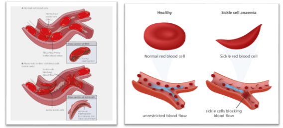
Figure 1. Comparison of normal red blood cells and sickle-shaped red blood cells in sickle cell anaemia.
Table 1: Overall clinical and haematological profile of children with sickle cell anaemia (N=200)
|
Study parameter |
n (%) or Mean (SD) |
95% CI |
Test statistic |
p value |
|
Age, years |
9.84 (4.27) |
9.24-10.44 |
|
|
|
Male |
113 (56.5) |
49.6-63.2 |
χ²=3.38 |
0.066 |
|
Female |
87 (43.5) |
36.8-50.4 |
||
|
Rural residence |
121 (60.5) |
53.6-67.0 |
χ²=8.82 |
0.003 |
|
Urban residence |
79 (39.5) |
33.0-46.4 |
||
|
Tribal community |
137 (68.5) |
61.8-74.5 |
χ²=27.38 |
<0.001 |
|
Positive family history of sickle cell disease |
94 (47.0) |
40.2-53.9 |
χ²=0.72 |
0.396 |
|
Consanguinity among parents |
71 (35.5) |
29.2-42.3 |
χ²=16.82 |
<0.001 |
|
Mean age at diagnosis, years |
4.18 (2.76) |
3.80-4.56 |
|
|
|
Presentation with acute crisis |
73 (36.5) |
30.1-43.4 |
χ²=14.58 |
<0.001 |
|
Vaso-occlusive crisis during preceding year |
149 (74.5) |
68.0-80.0 |
χ²=48.02 |
<0.001 |
|
Previous hospitalization |
143 (71.5) |
64.9-77.3 |
χ²=36.98 |
<0.001 |
|
Previous blood transfusion |
116 (58.0) |
51.1-64.6 |
χ²=5.12 |
0.024 |
|
Receiving hydroxyurea |
129 (64.5) |
57.7-70.8 |
χ²=16.82 |
<0.001 |
|
Any sickle cell-related complication |
73 (36.5) |
30.1-43.4 |
χ²=14.58 |
<0.001 |
|
Haemoglobin, g/dL |
7.93 (1.71) |
7.69-8.17 |
|
|
|
Severe anaemia, Hb <7 g/dL |
67 (33.5) |
27.3-40.3 |
χ²=21.78 |
<0.001 |
|
Reticulocyte count, % |
8.62 (3.41) |
8.15-9.09 |
|
|
|
HbS, % |
78.46 (9.28) |
77.17-79.75 |
|
|
|
HbF, % |
14.37 (7.16) |
13.38-15.36 |
|
|
Table 1 presents the overall clinical and haematological profile of 200 children with sickle cell anaemia. The mean age was 9.84±4.27 years (95% CI: 9.24-10.44), while the mean age at diagnosis was 4.18±2.76 years. Males constituted 56.5% of the participants, although the sex distribution was not statistically significant (p=0.066). A significantly higher proportion belonged to rural areas (60.5%; p=0.003) and tribal communities (68.5%; p<0.001). A positive family history was reported in 47.0%, while parental consanguinity was observed in 35.5% (p<0.001). Acute crisis was present at enrolment in 36.5%, and 74.5% had experienced a vaso-occlusive crisis during the preceding year. Previous hospitalization and blood transfusion were reported in 71.5% and 58.0%, respectively. Hydroxyurea therapy was being received by 64.5% of children, while 36.5% had at least one sickle cell-related complication. The mean haemoglobin level was 7.93±1.71 g/dL, and severe anaemia was present in 33.5% (95% CI: 27.3-40.3; p<0.001). The mean reticulocyte count was 8.62±3.41%, indicating increased erythropoietic activity. The mean HbS and HbF levels were 78.46±9.28% and 14.37±7.16%, respectively.
Table 2: Sociodemographic characteristics and clinical manifestations of children with sickle cell anaemia (N=200)
|
Study parameter |
n (%) or Mean (SD) |
95% CI |
Test statistic |
p value |
|
Age group |
χ²=76.60 |
<0.001 |
||
|
≤5 years |
31 (15.5) |
11.1-21.2 |
||
|
6-10 years |
102 (51.0) |
44.1-57.8 |
||
|
11-14 years |
44 (22.0) |
16.8-28.2 |
||
|
15-18 years |
23 (11.5) |
7.8-16.7 |
||
|
Sex |
χ²=3.38 |
0.066 |
||
|
Male |
113 (56.5) |
49.6-63.2 |
||
|
Female |
87 (43.5) |
36.8-50.4 |
||
|
Residence |
χ²=8.82 |
0.003 |
||
|
Rural |
121 (60.5) |
53.6-67.0 |
||
|
Urban |
79 (39.5) |
33.0-46.4 |
||
|
Tribal community |
137 (68.5) |
61.8-74.5 |
χ²=27.38 |
<0.001 |
|
Below-middle socioeconomic status |
124 (62.0) |
55.1-68.4 |
χ²=11.52 |
0.001 |
|
Positive family history |
94 (47.0) |
40.2-53.9 |
χ²=0.72 |
0.396 |
|
Parental consanguinity |
71 (35.5) |
29.2-42.3 |
χ²=16.82 |
<0.001 |
|
Previous sibling death related to SCD |
27 (13.5) |
9.4-18.9 |
χ²=106.58 |
<0.001 |
|
Pallor |
173 (86.5) |
81.1-90.6 |
χ²=106.58 |
<0.001 |
|
Jaundice |
91 (45.5) |
38.7-52.4 |
χ²=1.62 |
0.203 |
|
Recurrent fever |
109 (54.5) |
47.6-61.3 |
χ²=1.62 |
0.203 |
|
Generalized weakness/fatigue |
139 (69.5) |
62.8-75.5 |
χ²=30.42 |
<0.001 |
|
Bone or joint pain |
149 (74.5) |
68.0-80.0 |
χ²=48.02 |
<0.001 |
|
Abdominal pain |
102 (51.0) |
44.1-57.8 |
χ²=0.08 |
0.777 |
|
Dactylitis |
53 (26.5) |
20.9-33.0 |
χ²=44.18 |
<0.001 |
|
Breathlessness |
61 (30.5) |
24.5-37.2 |
χ²=30.42 |
<0.001 |
|
Growth faltering |
69 (34.5) |
28.3-41.3 |
χ²=19.22 |
<0.001 |
|
Hepatomegaly |
84 (42.0) |
35.4-48.9 |
χ²=5.12 |
0.024 |
|
Splenomegaly |
76 (38.0) |
31.6-44.9 |
χ²=11.52 |
0.001 |
|
≥3 vaso-occlusive crises in preceding year |
94 (47.0) |
40.2-53.9 |
χ²=0.72 |
0.396 |
|
Previous blood transfusion |
116 (58.0) |
51.1-64.6 |
χ²=5.12 |
0.024 |
|
Acute chest syndrome |
33 (16.5) |
12.0-22.3 |
χ²=89.78 |
<0.001 |
|
Splenic sequestration |
18 (9.0) |
5.8-13.8 |
χ²=134.48 |
<0.001 |
|
Severe/recurrent infection |
47 (23.5) |
18.2-29.8 |
χ²=56.18 |
<0.001 |
|
History of stroke |
24 (12.0) |
8.2-17.2 |
χ²=115.52 |
<0.001 |
|
Gallstone disease |
39 (19.5) |
14.6-25.5 |
χ²=74.42 |
<0.001 |
|
Any sickle cell-related complication |
73 (36.5) |
30.1-43.4 |
χ²=14.58 |
<0.001 |
Table 2 describes the sociodemographic characteristics and clinical manifestations of the study participants. The largest proportion of children belonged to the age group of 6-10 years (51.0%), followed by 11-14 years (22.0%), ≤5 years (15.5%) and 15-18 years (11.5%), with a statistically significant variation across age groups (χ²=76.60, p<0.001). Most children were from rural areas (60.5%), tribal communities (68.5%) and below-middle socioeconomic groups (62.0%). A positive family history was present in 47.0%, parental consanguinity in 35.5% and a previous sibling death related to sickle cell disease in 13.5%. Pallor was the most frequent clinical manifestation, observed in 86.5% of children, followed by bone or joint pain (74.5%), generalized weakness or fatigue (69.5%), recurrent fever (54.5%) and abdominal pain (51.0%). Other findings included jaundice in 45.5%, hepatomegaly in 42.0%, splenomegaly in 38.0%, growth faltering in 34.5%, breathlessness in 30.5% and dactylitis in 26.5%. Nearly half of the children (47.0%) had experienced three or more vaso-occlusive crises during the preceding year, and 58.0% had previously received a blood transfusion. Acute chest syndrome occurred in 16.5%, splenic sequestration in 9.0%, severe or recurrent infection in 23.5%, stroke in 12.0% and gallstone disease in 19.5%. Overall, 36.5% of the children had at least one sickle cell-related complication.
Table 3: Haematological parameters and haemoglobin fraction patterns according to clinical status (N=200)
|
Haematological parameter |
Overall (N=200), Mean (SD) or n (%) |
95% CI |
Acute crisis (n=73), Mean (SD) or n (%) |
Steady state (n=127), Mean (SD) or n (%) |
Test statistic |
p value |
|
Haemoglobin, g/dL |
7.93 (1.71) |
7.69-8.17 |
7.11 (1.53) |
8.40 (1.64) |
t=5.59 |
<0.001 |
|
RBC count, ×10⁶/µL |
3.06 (0.71) |
2.96-3.16 |
2.73 (0.68) |
3.25 (0.66) |
t=5.26 |
<0.001 |
|
Haematocrit, % |
24.18 (5.36) |
23.44-24.92 |
21.63 (4.91) |
25.65 (5.08) |
t=5.43 |
<0.001 |
|
Mean corpuscular volume, fL |
78.64 (10.27) |
77.21-80.07 |
76.81 (10.62) |
79.69 (9.94) |
t=1.89 |
0.060 |
|
Mean corpuscular haemoglobin, pg |
26.13 (3.82) |
25.60-26.66 |
25.47 (3.91) |
26.51 (3.73) |
t=1.84 |
0.067 |
|
Mean corpuscular haemoglobin concentration, g/dL |
32.46 (2.31) |
32.14-32.78 |
31.98 (2.42) |
32.74 (2.21) |
t=2.20 |
0.029 |
|
Red-cell distribution width, % |
18.73 (3.86) |
18.19-19.27 |
20.14 (4.07) |
17.92 (3.48) |
t=3.88 |
<0.001 |
|
Total leukocyte count, ×10³/µL |
12.84 (5.17) |
12.12-13.56 |
15.76 (5.63) |
11.16 (4.09) |
t=6.08 |
<0.001 |
|
Absolute neutrophil count, ×10³/µL |
7.92 (3.68) |
7.41-8.43 |
10.11 (4.16) |
6.66 (2.71) |
t=6.31 |
<0.001 |
|
Platelet count, ×10³/µL |
386.42 (128.73) |
368.51-404.33 |
419.76 (142.58) |
367.26 (115.93) |
t=2.66 |
0.008 |
|
Reticulocyte count, % |
8.62 (3.41) |
8.15-9.09 |
10.37 (3.72) |
7.61 (2.79) |
t=5.44 |
<0.001 |
|
Total bilirubin, mg/dL |
3.18 (1.47) |
2.98-3.38 |
3.91 (1.66) |
2.76 (1.17) |
t=5.15 |
<0.001 |
|
Indirect bilirubin, mg/dL |
2.47 (1.24) |
2.30-2.64 |
3.08 (1.41) |
2.12 (0.98) |
t=5.10 |
<0.001 |
|
Lactate dehydrogenase, U/L |
628.37 (218.64) |
597.95-658.79 |
739.18 (241.73) |
564.68 (176.42) |
t=5.35 |
<0.001 |
|
HbS, % |
78.46 (9.28) |
77.17-79.75 |
80.17 (8.91) |
77.48 (9.37) |
t=1.99 |
0.048 |
|
HbF, % |
14.37 (7.16) |
13.38-15.36 |
11.92 (6.48) |
15.78 (7.17) |
t=3.90 |
<0.001 |
|
HbA₂, % |
3.21 (0.82) |
3.10-3.32 |
3.27 (0.86) |
3.18 (0.79) |
t=0.73 |
0.466 |
|
HbSS pattern |
161 (80.5) |
74.5-85.4 |
63 (86.3) |
98 (77.2) |
χ²=2.46 |
0.117 |
|
HbSβ⁰-thalassaemia pattern |
39 (19.5) |
14.6-25.5 |
10 (13.7) |
29 (22.8) |
||
|
Severe anaemia, Hb <7 g/dL |
67 (33.5) |
27.3-40.3 |
38 (52.1) |
29 (22.8) |
χ²=17.99 |
<0.001 |
|
Moderate anaemia, Hb 7-9.9 g/dL |
109 (54.5) |
47.6-61.3 |
31 (42.5) |
78 (61.4) |
χ²=6.68 |
0.010 |
|
Mild anaemia, Hb ≥10 g/dL |
24 (12.0) |
8.2-17.2 |
4 (5.5) |
20 (15.7) |
χ²=4.53 |
0.033 |
Table 3 compares haematological parameters between children presenting with an acute crisis and those in a steady state. Children with an acute crisis had significantly lower mean haemoglobin (7.11±1.53 vs 8.40±1.64 g/dL; p<0.001), red blood cell count (2.73±0.68 vs 3.25±0.66×10⁶/µL; p<0.001) and haematocrit (21.63±4.91% vs 25.65±5.08%; p<0.001). Mean corpuscular volume and mean corpuscular haemoglobin did not differ significantly between the groups (p=0.060 and p=0.067, respectively), whereas mean corpuscular haemoglobin concentration was slightly lower during acute crisis (p=0.029). Children experiencing an acute crisis had significantly higher red-cell distribution width, total leukocyte count, absolute neutrophil count, platelet count and reticulocyte count than those in steady state. Markers of haemolysis were also significantly elevated during acute crisis, including total bilirubin (3.91±1.66 vs 2.76±1.17 mg/dL), indirect bilirubin (3.08±1.41 vs 2.12±0.98 mg/dL) and lactate dehydrogenase (739.18±241.73 vs 564.68±176.42 U/L), with all p<0.001. Mean HbS was marginally higher during acute crisis (80.17±8.91% vs 77.48±9.37%; p=0.048), whereas mean HbF was significantly lower (11.92±6.48% vs 15.78±7.17%; p<0.001). No significant differences were found in HbA₂ levels or the distribution of HbSS and HbSβ⁰-thalassaemia patterns. Severe anaemia was more frequent during acute crisis than in steady state (52.1% vs 22.8%; p<0.001), while mild anaemia was less frequent (5.5% vs 15.7%; p=0.033).
Table 4: Association of selected clinical characteristics with severe anaemia and sickle cell-related complications (N=200)
A. Factors associated with severe anaemia
|
Clinical characteristic |
Total n |
Severe anaemia (n=67), n (%) |
Non-severe anaemia (n=133), n (%) |
Odds ratio (95% CI) |
χ² value |
p value |
|
Age 11-18 years |
67 |
31 (46.3) |
36 (27.1) |
4.34 (2.24-8.43) |
20.22 |
<0.001 |
|
Age ≤10 years |
133 |
36 (53.7) |
97 (72.9) |
Reference |
||
|
≥3 vaso-occlusive crises/year |
94 |
46 (68.7) |
48 (36.1) |
3.88 (2.07-7.25) |
18.97 |
<0.001 |
|
<3 vaso-occlusive crises/year |
106 |
21 (31.3) |
85 (63.9) |
Reference |
||
|
Hydroxyurea non-adherence |
82 |
39 (58.2) |
43 (32.3) |
2.92 (1.59-5.35) |
12.33 |
<0.001 |
|
Regular hydroxyurea use/adherence |
118 |
28 (41.8) |
90 (67.7) |
Reference |
||
|
Previous blood transfusion |
69 |
37 (55.2) |
32 (24.1) |
3.89 (2.08-7.27) |
19.15 |
<0.001 |
|
No previous blood transfusion |
131 |
30 (44.8) |
101 (75.9) |
Reference |
||
|
HbF <10% |
47 |
29 (43.3) |
18 (13.5) |
4.88 (2.44-9.75) |
21.93 |
<0.001 |
|
HbF ≥10% |
153 |
38 (56.7) |
115 (86.5) |
Reference |
Table 4A shows the association of selected clinical characteristics with severe anaemia. Children aged 11-18 years had higher reported odds of severe anaemia than younger children (OR=4.34; 95% CI: 2.24-8.43; p<0.001). Experiencing three or more vaso-occlusive crises during the preceding year was significantly associated with severe anaemia (OR=3.88; 95% CI: 2.07-7.25; p<0.001). Hydroxyurea non-adherence increased the odds of severe anaemia by nearly three times (OR=2.92; 95% CI: 1.59-5.35; p<0.001). Children with a previous blood transfusion had significantly greater odds of severe anaemia (OR=3.89; 95% CI: 2.08-7.27; p<0.001). The strongest association was observed for an HbF level below 10%, which was associated with almost five-fold higher odds of severe anaemia (OR=4.88; 95% CI: 2.44-9.75; p<0.001).
B. Factors associated with sickle cell-related complications
|
Clinical characteristic |
Total n |
Complication present (n=73), n (%) |
No complication (n=127), n (%) |
Odds ratio (95% CI) |
χ² value |
p value |
|
Age 11-18 years |
76 |
42 (57.5) |
34 (26.8) |
3.71 (2.02-6.81) |
18.62 |
<0.001 |
|
Age ≤10 years |
124 |
31 (42.5) |
93 (73.2) |
Reference |
||
|
≥3 vaso-occlusive crises/year |
61 |
41 (56.2) |
20 (15.7) |
6.85 (3.53-13.32) |
35.72 |
<0.001 |
|
<3 vaso-occlusive crises/year |
139 |
32 (43.8) |
107 (84.3) |
Reference |
||
|
Hydroxyurea non-adherence |
58 |
36 (49.3) |
22 (17.3) |
4.64 (2.43-8.89) |
23.04 |
<0.001 |
|
Regular hydroxyurea use/adherence |
142 |
37 (50.7) |
105 (82.7) |
Reference |
||
|
Severe anaemia, Hb <7 g/dL |
67 |
44 (60.3) |
23 (18.1) |
6.86 (3.58-13.15) |
36.99 |
<0.001 |
|
Hb ≥7 g/dL |
133 |
29 (39.7) |
104 (81.9) |
Reference |
||
|
Previous hospitalization |
56 |
38 (52.1) |
18 (14.2) |
6.57 (3.34-12.95) |
33.00 |
<0.001 |
|
No previous hospitalization |
144 |
35 (47.9) |
109 (85.8) |
Reference |
Table 4B demonstrates the factors associated with the occurrence of sickle cell-related complications. Children aged 11-18 years had significantly higher odds of complications than children aged ≤10 years (OR=3.71; 95% CI: 2.02-6.81; p<0.001). Three or more vaso-occlusive crises per year showed a strong association with complications (OR=6.85; 95% CI: 3.53-13.32; p<0.001). Hydroxyurea non-adherence was associated with more than four-fold increased odds of complications (OR=4.64; 95% CI: 2.43-8.89; p<0.001). Severe anaemia was also strongly associated with complications (OR=6.86; 95% CI: 3.58-13.15; p<0.001). Similarly, children with a history of hospitalization had significantly greater odds of developing complications (OR=6.57; 95% CI: 3.34-12.95; p<0.001).
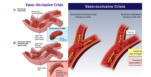
Figure 2. Sickled erythrocytes obstructing a small blood vessel during a vaso-occlusive crisis.
DISCUSSION:
The present cross-sectional study described the clinical and haematological profile of 200 children with sickle cell anaemia attending a tertiary care hospital. The findings demonstrated a substantial burden of recurrent vaso-occlusive episodes, hospitalizations, transfusion requirements, anaemia and disease-related complications. This pattern is consistent with the established pathophysiology of sickle cell disease, in which chronic haemolysis, endothelial dysfunction, inflammation and recurrent microvascular occlusion produce progressive multisystem morbidity.[1,2] The findings are especially relevant in India, which carries a considerable proportion of the global burden of sickle cell disease, with marked clustering in central, western and eastern regions.[3] The predominance of children from rural and tribal communities in the present study reflects the recognized epidemiological distribution of the sickle haemoglobin gene in India.[4]
Overall clinical and haematological profile
The mean age of the children was 9.84±4.27 years, and more than half were male. The modest male predominance was not statistically significant, which was expected because sickle cell disease is inherited as an autosomal recessive disorder and has no biological sex predilection. Patel et al. (2017)[5] and Shah et al. (2017)[6] also reported a slight male predominance among children with sickle cell disease attending tertiary-care hospitals in western India. The observed difference may therefore reflect healthcare-seeking behaviour, referral patterns or chance rather than an actual sex-specific difference in disease occurrence.
Most participants were from rural areas, and 68.5% belonged to tribal communities. These proportions were statistically significant and agreed with the geographical concentration of sickle cell disease among several tribal populations of Gujarat, Maharashtra, Madhya Pradesh, Chhattisgarh and Odisha. Thaker et al. (2022)[7], in a newborn-screening programme involving tribal populations of Gujarat and Madhya Pradesh, documented a considerable burden of HbSS and HbS-β-thalassaemia. Dave et al. (2022)[8] similarly reported a high occurrence of sickle cell disease and subsequent clinical morbidity among children detected through newborn screening in a tribal region of Gujarat. The predominance of rural and lower socioeconomic groups in the present study may also reflect limited access to early screening, preventive treatment and specialized follow-up services.
A positive family history was recorded in 47.0%, while parental consanguinity was observed in 35.5%. Consanguineous marriage increases the probability that both parents carry the same pathogenic haemoglobin variant, thereby increasing the risk of an affected child. The considerable proportion with a positive family history supports the importance of cascade screening, premarital counselling, antenatal diagnosis and screening of siblings. The National Sickle Cell Anaemia Elimination Mission has similarly prioritized population screening, genetic counselling and comprehensive care in high-burden communities.[9]
The mean age at diagnosis was 4.18 years, suggesting that many children were diagnosed after the infancy period. Newborn screening permits diagnosis before the development of clinical symptoms and enables early initiation of vaccination, parental education, infection prophylaxis and comprehensive follow-up. Upadhye et al. (2016)[10] demonstrated the feasibility and clinical value of neonatal screening in central India. Dave et al. (2022)[8] also observed that children diagnosed through newborn screening could be enrolled in regular follow-up before the appearance of serious complications. The relatively delayed diagnosis observed in the present study indicates the continuing need to strengthen universal or targeted newborn screening in high-prevalence districts.
Vaso-occlusive crisis during the preceding year was reported by 74.5% of children, 71.5% had previously required hospitalization and 58.0% had received a blood transfusion. These findings indicate a substantial burden of clinically severe disease among children reaching tertiary care. Recurrent vaso-occlusion results from the interaction of sickled erythrocytes, leukocytes, platelets, vascular endothelium and inflammatory mediators and remains the most common reason for emergency visits and hospitalization.[1,11] Patel et al. (2017)[5] and Shah et al. (2017)[6] likewise identified painful crisis, fever and severe anaemia as common reasons for hospitalization among Indian children with sickle cell disease. The high transfusion frequency in the present study may be explained by severe anaemia, acute splenic sequestration, acute chest syndrome, infection and other acute complications. Nevertheless, transfusion history should be interpreted as an indicator of previous disease severity rather than as a causal risk factor.[12]
Approximately 64.5% of children were receiving hydroxyurea. Hydroxyurea increases fetal haemoglobin, reduces intracellular HbS polymerization and decreases vaso-occlusive crises, acute chest syndrome, transfusion requirements and hospitalization. Tshilolo et al. (2019)[13], through the REACH trial, demonstrated that hydroxyurea was feasible, safe and clinically beneficial for children with sickle cell anaemia in resource-limited African settings. John et al. (2020)[14] further showed that dose escalation toward the maximum tolerated dose provided superior laboratory and clinical benefits compared with a fixed-dose strategy. Although hydroxyurea coverage in the present study was encouraging, the remaining treatment gap and the observed non-adherence emphasize the need for counselling, regular monitoring and uninterrupted availability of medication.
The mean haemoglobin level was 7.93±1.71 g/dL, and one-third of children had severe anaemia. A mean reticulocyte count of 8.62% indicated compensatory erythropoietic activity in response to chronic haemolysis. Patel et al. (2017)[5] and Shah et al. (2017)[6] also reported low haemoglobin concentrations with increased reticulocyte counts among affected children. Chronic anaemia in sickle cell disease results from shortened red-cell survival, while further reductions may occur during acute haemolysis, infection, splenic sequestration or transient marrow suppression. The mean HbS level of 78.46% confirmed a high sickling burden, whereas the mean HbF level of 14.37% may have provided some protection against HbS polymerization and severe vaso-occlusion.[2]
Sociodemographic characteristics and clinical manifestations
Children aged 6-10 years constituted the largest age group. This may reflect increasing clinical manifestations after infancy and the cumulative effect of recurrent vaso-occlusion and haemolysis. The relatively small proportion of adolescents could have resulted from referral patterns, transition to adult services or under-representation of older children. The substantial representation of rural, tribal and economically disadvantaged families supports previous Indian evidence that sickle cell disease is disproportionately concentrated in communities facing social and healthcare-access barriers.[3,4]
Pallor was the most frequent manifestation, affecting 86.5% of children, followed by bone or joint pain, generalized weakness, recurrent fever and abdominal pain. These findings were comparable with the clinical profiles reported by Patel et al. (2017)[5] and Shah et al. (2017)[6], in which pallor, painful crisis, fever and jaundice were among the common manifestations. Pallor and fatigue reflect chronic anaemia, whereas bone and joint pain arise from recurrent microvascular obstruction and tissue ischaemia. Abdominal pain may result from vaso-occlusion, splenic or hepatic enlargement, mesenteric ischaemia or gallstone disease.
Jaundice occurred in 45.5% of participants and was consistent with chronic haemolysis and increased unconjugated bilirubin production. Hepatomegaly and splenomegaly were observed in 42.0% and 38.0%, respectively. Persistent splenomegaly may be comparatively more common in Indian children because of genetic modifiers, co-inherited α-thalassaemia, high fetal haemoglobin levels and repeated infections. Dave et al. (2022)[8] reported a high frequency of α-thalassaemia deletion among screened children with sickle cell disease in Gujarat, demonstrating the genetic heterogeneity of the Indian phenotype.
Growth faltering was present in 34.5% of children. Growth impairment in sickle cell disease is multifactorial and may be caused by chronic anaemia, increased resting energy expenditure, recurrent infection, nutritional deficiencies, endocrine dysfunction and repeated hospitalization. Ware et al. (2017)[1] emphasized that delayed growth and pubertal development remain important chronic manifestations in paediatric sickle cell disease. The finding indicates that anthropometric assessment, nutritional counselling and screening for micronutrient or endocrine abnormalities should form part of routine follow-up.
Acute chest syndrome was observed in 16.5%, splenic sequestration in 9.0%, severe or recurrent infection in 23.5%, stroke in 12.0% and gallstone disease in 19.5%. Acute chest syndrome is a major cause of hospitalization and mortality and may be precipitated by infection, pulmonary infarction or fat embolism.[15] Severe infections remain important because progressive splenic dysfunction increases susceptibility to encapsulated organisms. Splenic sequestration is particularly important in young children because it can cause a rapid fall in haemoglobin and hypovolaemic shock. Gallstones develop as a consequence of sustained bilirubin overproduction from chronic haemolysis.
A history of stroke in 12.0% represented a clinically important burden. DeBaun et al. (2020)[16] recommended annual transcranial Doppler screening for children aged 2-16 years with HbSS or HbSβ⁰-thalassaemia to identify those at increased risk of primary stroke. Regular screening, timely initiation of chronic transfusion in high-risk children and consideration of disease-modifying therapy could reduce neurological morbidity. Overall, 36.5% had at least one sickle cell-related complication, emphasizing the need for structured multidisciplinary follow-up.
Haematological differences between acute crisis and steady state
Children presenting with an acute crisis had significantly lower haemoglobin, red blood cell count and haematocrit than children in a steady state. Severe anaemia was recorded in 52.1% of the acute-crisis group compared with 22.8% of the steady-state group. Acute crises may intensify haemolysis, increase splenic sequestration, suppress erythropoiesis during infection or produce haemodilution following fluid administration. The higher reticulocyte count during acute crisis indicated an active marrow response and supported ongoing or accelerated haemolysis.
Mean corpuscular volume and mean corpuscular haemoglobin did not differ significantly between the two clinical states, whereas mean corpuscular haemoglobin concentration was slightly lower during crisis. Red-cell distribution width was significantly higher during acute crisis, demonstrating greater anisocytosis caused by accelerated release of reticulocytes and variation in red-cell survival. Hydroxyurea therapy and associated macrocytosis may also have influenced red-cell indices. Therefore, interpretation of mean corpuscular volume should consider hydroxyurea exposure, iron status and possible co-inherited α- or β-thalassaemia.
The acute-crisis group demonstrated significantly higher total leukocyte count, absolute neutrophil count and platelet count. Leukocytosis and neutrophilia reflect systemic inflammation, physiological stress and possible infection and may also contribute directly to vaso-occlusion through interactions with activated endothelium and sickled erythrocytes.[2] Platelet activation similarly contributes to the thrombo-inflammatory state. These findings support the concept that acute vaso-occlusive events are not caused solely by abnormal red-cell morphology but involve multiple cellular and inflammatory pathways.[11]
Total bilirubin, indirect bilirubin and lactate dehydrogenase were significantly higher during acute crisis. Together with reticulocytosis and reduced haemoglobin, these findings demonstrate intensified haemolysis. Elevated indirect bilirubin also provides a biological explanation for the observed burden of jaundice and gallstone disease. Kato et al. (2018)[2] described lactate dehydrogenase, bilirubin and reticulocyte count as commonly used indicators of haemolytic activity, although no single marker is sufficiently specific to define intravascular haemolysis independently.
The acute-crisis group had a slightly higher mean HbS level and a significantly lower mean HbF level than the steady-state group. HbF inhibits HbS polymerization, reduces red-cell sickling and is generally associated with fewer vaso-occlusive events and improved survival. The lower HbF among children presenting in crisis was therefore biologically plausible. Hydroxyurea increases HbF and improves several laboratory indicators, providing an explanation for the lower risk among adherent children.[13,14] HbA₂ did not differ significantly between clinical states, which was expected because it primarily reflects the underlying genotype rather than short-term clinical status. HbSS was the predominant pattern, accounting for 80.5%, while HbSβ⁰-thalassaemia accounted for 19.5%. Similar genotype patterns have been reported in newborn-screening programmes conducted in Gujarat and Madhya Pradesh.[7,8]
Factors associated with severe anaemia
Older age, frequent vaso-occlusive crises, hydroxyurea non-adherence, previous transfusion and HbF below 10% were significantly associated with severe anaemia. Older children may have accumulated a greater burden of recurrent haemolysis, chronic inflammation, nutritional deficiencies and organ dysfunction. Nevertheless, the association with age requires cautious interpretation because age may also correlate with treatment duration, pubertal growth requirements and disease exposure time.
Children with at least three vaso-occlusive crises per year had higher odds of severe anaemia. Frequent vaso-occlusion may identify a more severe phenotype characterized by increased inflammation, haemolysis and healthcare utilization. Hydroxyurea non-adherence was associated with nearly three-fold higher odds of severe anaemia. This supports evidence that sustained hydroxyurea therapy increases HbF, improves total haemoglobin and reduces haemolytic and vaso-occlusive events.[13,14] However, because the present study was cross-sectional, it could not establish whether non-adherence preceded severe anaemia.
Previous transfusion was associated with severe anaemia. This relationship was likely to reflect confounding by indication: children with severe anaemia or serious complications were more likely to have received transfusions. Chou et al. (2020)[12] emphasized the essential role of transfusion in managing severe anaemia and preventing or treating selected complications, while also highlighting the importance of extended antigen matching and monitoring for alloimmunization and iron overload.
HbF below 10% showed the strongest reported association with severe anaemia. Higher HbF reduces HbS polymerization and may decrease haemolysis, vaso-occlusion and red-cell destruction.[1,2] This finding further supports early initiation, adherence and laboratory monitoring of hydroxyurea. Nevertheless, HbF levels may also be influenced by age, genotype, genetic modifiers, hydroxyurea dose and treatment adherence.
Factors associated with sickle cell-related complications
Older age was associated with higher odds of complications, possibly reflecting cumulative exposure to vaso-occlusion and chronic haemolysis. Children experiencing at least three vaso-occlusive crises per year had markedly increased odds of complications. Repeated crises can cause progressive ischaemia-reperfusion injury and contribute to pulmonary, neurological, splenic, hepatobiliary and musculoskeletal complications.[1,2]
Hydroxyurea non-adherence was associated with increased odds of complications, consistent with trials demonstrating reductions in painful episodes, acute chest syndrome, hospitalization and transfusion following hydroxyurea therapy.[13,14] Severe anaemia was also strongly associated with complications. Reduced oxygen-carrying capacity, increased haemolytic activity and underlying disease severity may contribute to organ injury. Previous hospitalization showed a similarly strong association, although it should be considered a marker of earlier disease severity and healthcare utilization rather than an independent causal exposure.
These associations were based on cross-sectional, unadjusted odds ratios and should not be interpreted as evidence of causality. Multivariable logistic regression would be required to determine whether age, crisis frequency, hydroxyurea adherence, HbF level and severe anaemia remained independently associated with complications after controlling for genotype, nutritional status, socioeconomic factors and treatment exposure. Overall, the findings reinforce the need for early diagnosis, regular hydroxyurea therapy, adherence support, vaccination, nutritional assessment, transcranial Doppler screening and systematic monitoring for acute and chronic complications.[9,16,17]
CONCLUSION:
Sickle cell anaemia was associated with considerable clinical and haematological morbidity among the children attending the tertiary care hospital. Most participants belonged to rural, tribal and socioeconomically disadvantaged communities. Pallor, bone or joint pain, generalized weakness, recurrent fever and abdominal pain were the predominant clinical manifestations. Vaso-occlusive crises, previous hospitalization and blood transfusion requirements were common, while more than one-third of the children had developed at least one sickle cell-related complication.
Children presenting with an acute crisis had significantly lower haemoglobin, red blood cell count and haematocrit and significantly higher leukocyte, neutrophil, platelet and reticulocyte counts than children in a steady state. Increased bilirubin and lactate dehydrogenase levels indicated greater haemolytic activity during acute crises. A lower HbF level was also associated with acute crisis and severe anaemia. Older age, frequent vaso-occlusive crises, hydroxyurea non-adherence, previous transfusion and HbF below 10% were associated with severe anaemia. Frequent crises, severe anaemia, hydroxyurea non-adherence and previous hospitalization were important indicators of sickle cell-related complications. Early diagnosis, regular haematological monitoring, improved adherence to hydroxyurea, timely management of crises and systematic screening for complications are therefore essential for reducing morbidity and improving the long-term outcomes of affected children.
Limitations of the Study
1. The study was conducted at a single tertiary care hospital; therefore, its findings might not be generalizable to all children with sickle cell anaemia in the community or in other geographical regions.
2. The hospital-based sample might have resulted in referral bias because children with severe symptoms or complications were more likely to attend a tertiary care centre.
3. Because of the cross-sectional design, temporal and causal relationships between clinical characteristics, hydroxyurea adherence, severe anaemia and complications could not be established.
4. Information regarding previous crises, hospitalizations, transfusions and medication adherence was partly based on parental recall and was consequently susceptible to recall bias.
5. Hydroxyurea adherence was not assessed using an objective method such as pill counts, pharmacy-refill records or drug-level monitoring.
6. Haematological measurements represented findings at a single point in time and might not reflect long-term variations in disease activity.
7. Factors such as nutritional status, iron deficiency, α-thalassaemia coinheritance, malaria, parasitic infections and other genetic modifiers of disease severity were not comprehensively evaluated.
8. The number and duration of previous blood transfusions might have influenced haemoglobin fraction measurements, particularly HbS and HbF levels.
9. Some uncommon complications might have been underestimated because advanced imaging and organ-specific investigations were performed only when clinically indicated.
10. The reported associations were primarily based on unadjusted analyses; therefore, residual confounding by age, genotype, socioeconomic status, treatment duration and disease severity could not be excluded.
REFERENCES:
1. Ware RE, de Montalembert M, Tshilolo L, Abboud MR. Sickle cell disease. Lancet. 2017;390(10091):311-323. doi:10.1016/S0140-6736(17)30193-9.
2. Kato GJ, Piel FB, Reid CD, Gaston MH, Ohene-Frempong K, Krishnamurti L, et al. Sickle cell disease. Nat Rev Dis Primers. 2018;4:18010. doi:10.1038/nrdp.2018.10.
3. GBD 2021 Sickle Cell Disease Collaborators. Global, regional, and national prevalence and mortality burden of sickle cell disease, 2000-2021: a systematic analysis from the Global Burden of Disease Study 2021. Lancet Haematol. 2023;10(8):e585-e599. doi:10.1016/S2352-3026(23)00118-7.
4. Jacob SA, et al. Prevalence of sickle cell disease, sickle cell trait and HbS-beta-thalassemia in India: a systematic review and meta-analysis. Clin Epidemiol Glob Health. 2024;28:101678. doi:10.1016/j.cegh.2024.101678.
5. Patel KG, Chaudhari C, Sharma D. A study of clinical and hematological profile of children with sickle cell disease in a tertiary care hospital, Valsad, India. Int J Contemp Pediatr. 2017;4(4):1317-1321. doi:10.18203/2349-3291.ijcp20172609.
6. Shah V, Muley P, Choraria C, Rana P, Kanaria D, Markana A. Clinical and hematological profile of sickle cell disease affected children in rural tertiary level hospital. J Pediatr Res. 2017;4(3):202-206. doi:10.17511/ijpr.2017.i03.01.
7. Thaker P, Colah R, Patel J, Raicha B, Mistry A, Mehta P, et al. Newborn screening for sickle cell disease among tribal populations in the states of Gujarat and Madhya Pradesh in India: evaluation and outcome over 6 years. Front Med (Lausanne). 2022;8:731884. doi:10.3389/fmed.2021.731884.
8. Dave K, Desai S, Italia Y, Mukherjee MB, Mehta P, Desai G. Newborn screening and clinical profile of children with sickle cell disease in a tribal area of Gujarat. Indian Pediatr. 2022;59(3):230-233. doi:10.1007/s13312-022-2476-7.
9. Ministry of Health and Family Welfare, Government of India. Guidelines for prevention and management of sickle cell disease. New Delhi: National Sickle Cell Anaemia Elimination Mission; 2023.
10. Upadhye DS, Jain DL, Trivedi YL, Nadkarni AH, Ghosh K, Colah RB. Neonatal screening and the clinical outcome in children with sickle cell disease in central India. PLoS One. 2016;11(1):e0147081. doi:10.1371/journal.pone.0147081.
11. Conran N, Belcher JD. Inflammation in sickle cell disease. Clin Hemorheol Microcirc. 2018;68(2-3):263-299. doi:10.3233/CH-189012.
12. Chou ST, Alsawas M, Fasano RM, Field JJ, Hendrickson JE, Howard J, et al. American Society of Hematology 2020 guidelines for sickle cell disease: transfusion support. Blood Adv. 2020;4(2):327-355. doi:10.1182/bloodadvances.2019001143.
13. Tshilolo L, Tomlinson G, Williams TN, Santos B, Olupot-Olupot P, Lane A, et al. Hydroxyurea for children with sickle cell anemia in sub-Saharan Africa. N Engl J Med. 2019;380(2):121-131. doi:10.1056/NEJMoa1813598.
14. John CC, Opoka RO, Latham TS, Hume HA, Nabaggala C, Kasirye P, et al. Hydroxyurea dose escalation for sickle cell anemia in sub-Saharan Africa. N Engl J Med. 2020;382(26):2524-2533. doi:10.1056/NEJMoa2000146.
15. Jain S, Bakshi N, Krishnamurti L. Acute chest syndrome in children with sickle cell disease. Pediatr Allergy Immunol Pulmonol. 2017;30(4):191-201. doi:10.1089/ped.2017.0814.
16. DeBaun MR, Jordan LC, King AA, Schatz J, Vichinsky E, Fox CK, et al. American Society of Hematology 2020 guidelines for sickle cell disease: prevention, diagnosis, and treatment of cerebrovascular disease in children and adults. Blood Adv. 2020;4(8):1554-1588. doi:10.1182/bloodadvances.2019001142.
17. Brandow AM, Carroll CP, Creary S, Edwards-Elliott R, Glassberg J, Hurley RW, et al. American Society of Hematology 2020 guidelines for sickle cell disease: management of acute and chronic pain. Blood Adv. 2020;4(12):2656-2701. doi:10.1182/bloodadvances.2020001851.