Evaluation of Serum Magnesium in Patients with Acute Exacerbation of Chronic Obstructive Pulmonary Disease.
- Narra Swetha , Senior Resident, Department of Respiratory Medicine, Bhaskara Medical College, Hyderabad, Telangana, India
- K. Ramesh Kumar , Professor and Head, Department of Respiratory Medicine, Bhaskara Medical College, Hyderabad, Telangana, India
- Akhil Kumar , Senior Resident, Department of Respiratory Medicine, Bhaskara Medical College, Hyderabad, Telangana, India
Article Information:
Abstract:
Background: Magnesium is an essential intracellular cation that regulates bronchial smooth-muscle tone, neuromuscular transmission, and inflammatory signalling. Hypomagnesemia is increasingly recognised as a potentially modifiable contributor to poor outcomes in chronic obstructive pulmonary disease (COPD). This study evaluated serum magnesium concentrations in patients hospitalised with an acute exacerbation of COPD (AECOPD) and examined the association between magnesium deficiency and clinical outcomes. Methods: In this prospective, single-centre observational study, 50 patients admitted with AECOPD (GOLD criteria) were enrolled. Admission serum magnesium, length of hospital stay, annual exacerbation frequency, dyspnea severity (modified Medical Research Council [mMRC] scale), comorbidities, nutritional status, and spirometry (FEV₁/FVC) were recorded. Patients were stratified as hypomagnesemic (<1.7 mg/dL) or normomagnesemic (1.7–2.2 mg/dL). Comparisons used Student's t-test and ANOVA, with p<0.05 considered significant. Results: Hypomagnesemia was present in 30 of 50 patients (60%; mean 1.61 mg/dL). Compared with normomagnesemic patients, hypomagnesemic patients had significantly longer hospital stays (10 ± 2.1 vs 7 ± 1.5 days; p<0.001), more frequent exacerbations (3.5 ± 1.2 vs 2.0 ± 0.8 per year; p<0.005), and a markedly higher burden of severe dyspnea (mMRC grade 3–4: 56% vs 0%; p<0.001). Airflow limitation was greater in the hypomagnesemic group (FEV₁/FVC 0.514 ± 0.03 vs 0.55 ± 0.04; p<0.001). Diabetes (60% vs 23%; p=0.002), hypertension (53% vs 30%), and malnutrition (67% vs 33%) were also more prevalent among hypomagnesemic patients. Conclusion: Hypomagnesemia is common in AECOPD and is associated with prolonged hospitalisation, more frequent exacerbations, greater symptom burden, and poorer lung function. Serum magnesium is an inexpensive, correctable biomarker; routine measurement at admission and targeted supplementation warrant evaluation as strategies to improve outcomes in AECOPD.
Keywords:
Article :
INTRODUCTION:
Chronic obstructive pulmonary disease (COPD) is a leading cause of morbidity and mortality worldwide and is projected to remain among the top causes of death for the foreseeable future. It is characterised by persistent respiratory symptoms and progressive airflow limitation arising from chronic airway inflammation and parenchymal destruction, most commonly driven by tobacco smoke and biomass-fuel exposure 1. The clinical course of COPD is punctuated by acute exacerbations (AECOPD)—episodes of acute worsening of respiratory symptoms that require additional therapy. Exacerbations accelerate lung-function decline, impair quality of life, account for the majority of COPD-related health-care costs, and are a major driver of hospitalisation and mortality 1.
Beyond the well-established roles of infection and airway inflammation, there is growing interest in modifiable metabolic and electrolyte disturbances that may influence exacerbation severity and recovery. Magnesium, the second most abundant intracellular cation, is a cofactor for more than 300 enzymatic reactions and is essential for adenosine-triphosphate metabolism, neuromuscular conduction, and the regulation of calcium flux across cell membranes 3. In the airway, magnesium promotes bronchial smooth-muscle relaxation by antagonising calcium entry, stabilising mast cells, and attenuating the release of pro-inflammatory mediators 2. These properties underpin the long-standing therapeutic use of intravenous magnesium sulfate in severe asthma and its emerging evaluation in COPD exacerbations 2,3.
Hypomagnesemia is common in hospitalised patients and may be especially frequent in COPD owing to poor dietary intake, systemic inflammation, chronic use of loop and thiazide diuretics, β₂-agonist therapy (which drives magnesium into cells), corticosteroids, and coexisting diabetes mellitus 3,11. Despite this biological plausibility, serum magnesium is rarely measured routinely during AECOPD, and its prognostic significance remains incompletely defined, with some studies reporting strong associations with exacerbation frequency and readmission while others find no relationship with severity 4–9. Against this background, the present study was undertaken to determine the prevalence of hypomagnesemia in patients hospitalised with AECOPD and to evaluate its association with length of hospital stay, exacerbation frequency, dyspnea severity, pulmonary function, comorbidities, and nutritional status.
MATERIALS AND METHODS:
Study design and setting
This was a prospective, observational study conducted in the Department of Respiratory Medicine, Bhaskara Medical College, Hyderabad, Telangana, India, over an 18-month period from July 2022 to January 2024. Consecutive patients admitted with a diagnosis of AECOPD were screened for eligibility.
Participants
Fifty patients aged 45–80 years with an established diagnosis of COPD, confirmed according to Global Initiative for Chronic Obstructive Lung Disease (GOLD) spirometric criteria (post-bronchodilator FEV₁/FVC < 0.70), and presenting with an acute exacerbation were enrolled 1. An exacerbation was defined as an acute worsening of respiratory symptoms—increased dyspnea, cough, or sputum volume/purulence—beyond normal day-to-day variation and requiring a change in regular medication.
Exclusion criteria were applied to minimise confounding of serum magnesium: chronic kidney disease or renal failure, hepatic failure, congestive heart failure, known primary magnesium-wasting disorders, patients receiving magnesium supplementation at admission, and pregnancy. Patients with these conditions were excluded because impaired renal handling, fluid overload, and hepatic dysfunction independently alter magnesium homeostasis.
Measurements and definitions
A venous blood sample was drawn at admission, before administration of intravenous fluids or magnesium-containing therapy, and serum magnesium was measured by the calmagite colorimetric method. Hypomagnesemia was defined as a serum magnesium concentration below 1.7 mg/dL, and the normal reference range was taken as 1.7–2.2 mg/dL. Patients were stratified into a hypomagnesemic group and a normomagnesemic group for all comparisons.
The following variables were recorded for each patient: age, sex, length of hospital stay (days), number of exacerbations in the preceding 12 months, dyspnea severity graded on the modified Medical Research Council (mMRC) scale, presence of comorbidities (diabetes mellitus and hypertension), nutritional status (assessed clinically and by body-mass index, with malnutrition defined as BMI < 18.5 kg/m²), and spirometric airflow limitation expressed as the post-bronchodilator FEV₁/FVC ratio.
Statistical analysis
Data were analysed using IBM SPSS Statistics version 26. Continuous variables are presented as mean ± standard deviation and categorical variables as counts and percentages. Between-group differences were tested using Student's independent-samples t-test for continuous variables and the chi-square test for categorical variables; ANOVA was used for comparisons across more than two subgroups. A two-tailed p-value < 0.05 was considered statistically significant.
Ethical considerations
The study was conducted in accordance with the principles of the Declaration of Helsinki. Approval was obtained from the Institutional Ethics Committee, and written informed consent was obtained from all participants prior to enrolment.
RESULTS:
Baseline demographic and clinical characteristics
Fifty patients with AECOPD were enrolled. The cohort was predominantly male (70%), consistent with the smoking-related epidemiology of COPD in this region, and most patients were in the sixth and seventh decades of life, with the 61–65-year (24%) and 56–60-year (20%) age bands most frequently represented. Baseline characteristics are summarised in Table 1 and the demographic profile is shown in Figure 6.
Table 1. Baseline demographic and clinical characteristics of the study cohort (n = 50).
|
Characteristic |
Category |
Value |
|
Total patients |
— |
50 |
|
Sex |
Male |
35 (70%) |
|
|
Female |
15 (30%) |
|
Predominant age bands |
61–65 years |
24% |
|
|
56–60 years |
20% |
|
Age range |
— |
45–80 years |
|
Magnesium status |
Hypomagnesemia (<1.7 mg/dL) |
30 (60%) |
|
|
Normal (1.7–2.2 mg/dL) |
20 (40%) |
|
Mean serum Mg (hypomagnesemic) |
|
1.61 mg/dL |
Values are n (%) unless otherwise stated.
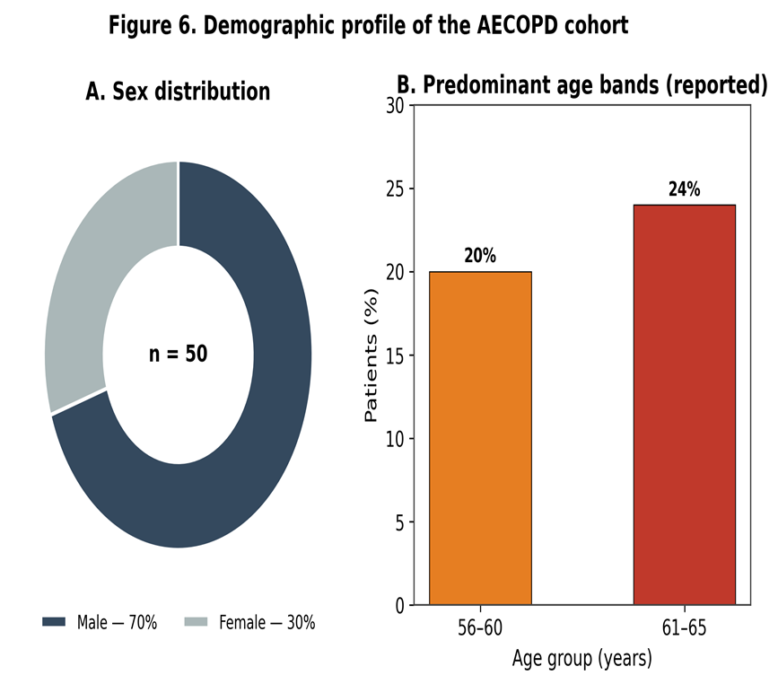
Figure 6. Demographic profile of the AECOPD cohort: (A) sex distribution and (B) predominant reported age bands.
3.2 Prevalence of hypomagnesemia
Hypomagnesemia was highly prevalent, affecting 30 of 50 patients (60%), with a mean serum magnesium concentration of 1.61 mg/dL in the deficient group (Figure 1). The remaining 20 patients (40%) had magnesium concentrations within the normal reference range.
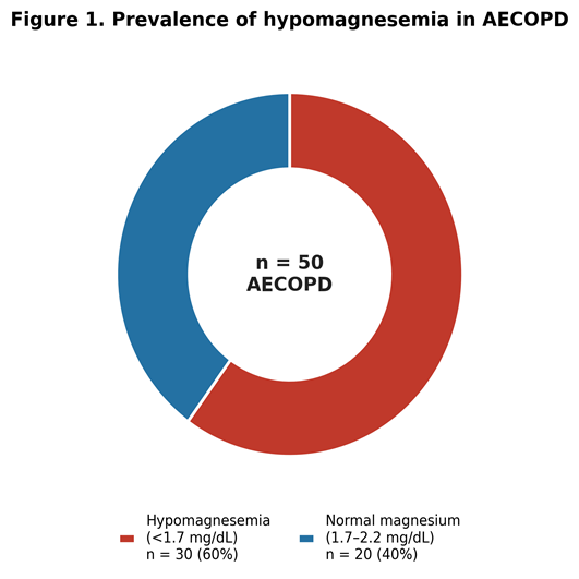
Figure 1. Prevalence of hypomagnesemia among patients hospitalised with AECOPD (n = 50).
3.3 Hospitalisation and exacerbation burden
Magnesium status was strongly associated with the burden of hospitalisation. Hypomagnesemic patients had a significantly longer mean hospital stay than normomagnesemic patients (10 ± 2.1 vs 7 ± 1.5 days; p<0.001) and a higher annual exacerbation frequency (3.5 ± 1.2 vs 2.0 ± 0.8 exacerbations per year; p<0.005) (Figure 2, Table 2). These findings indicate that magnesium deficiency identifies a subgroup of patients with a more relapsing and resource-intensive disease course.
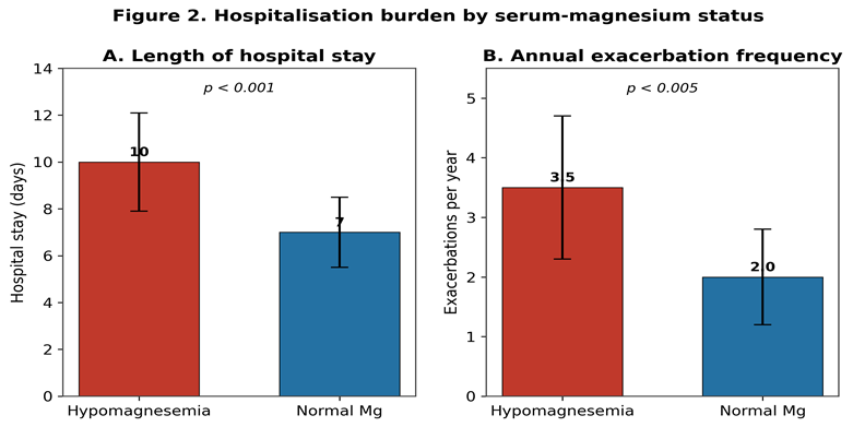
Figure 2. Length of hospital stay (A) and annual exacerbation frequency (B) by serum-magnesium status. Bars show mean ± SD.
3.4 Dyspnea severity
Symptom burden differed markedly between groups. Severe dyspnea (mMRC grade 3–4) was reported by 56% of hypomagnesemic patients but by none of the normomagnesemic patients (0%; p<0.001) (Figure 3), underscoring the association between magnesium deficiency and greater functional limitation during exacerbation.
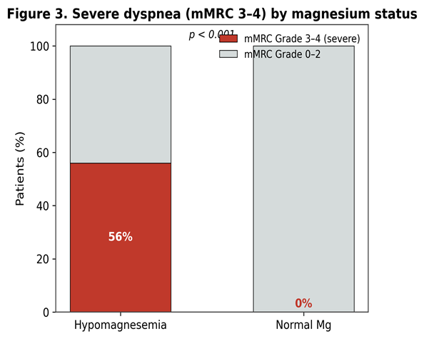
Figure 3. Proportion of patients with severe dyspnea (mMRC grade 3–4) by magnesium status.
3.5 Comorbidities and nutritional status
Metabolic comorbidities and malnutrition clustered in the hypomagnesemic group. Diabetes mellitus was present in 60% of hypomagnesemic patients versus 23% of normomagnesemic patients (p=0.002), and hypertension in 53% versus 30%. Malnutrition was also more common among hypomagnesemic patients (67% vs 33%) (Figure 4). This pattern is consistent with the bidirectional relationship between magnesium deficiency and insulin resistance and with reduced dietary intake in advanced COPD 11.
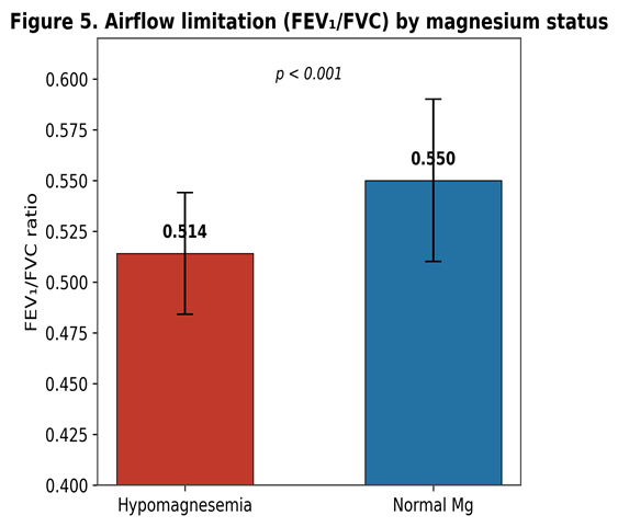
Figure 4. Prevalence of diabetes, hypertension, and malnutrition by magnesium status. p = 0.002 for diabetes.
3.6 Pulmonary function
Airflow limitation was more severe in magnesium-deficient patients. The mean post-bronchodilator FEV₁/FVC ratio was significantly lower in the hypomagnesemic group than in the normomagnesemic group (0.514 ± 0.03 vs 0.55 ± 0.04; p<0.001) (Figure 5), supporting a link between low magnesium and reduced airway patency.
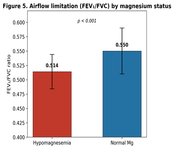
Figure 5. Post-bronchodilator FEV₁/FVC ratio by magnesium status. Bars show mean ± SD.
3.7 Summary of group comparisons
Table 2. Comparison of clinical outcomes between hypomagnesemic and normomagnesemic patients.
|
Parameter |
Hypomagnesemia (n = 30) |
Normal Mg (n = 20) |
p-value |
|
Hospital stay (days) |
10 ± 2.1 |
7 ± 1.5 |
<0.001 |
|
Exacerbations / year |
3.5 ± 1.2 |
2.0 ± 0.8 |
<0.005 |
|
Severe dyspnea (mMRC 3–4) |
56% |
0% |
<0.001 |
|
FEV₁/FVC ratio |
0.514 ± 0.03 |
0.55 ± 0.04 |
<0.001 |
|
Diabetes mellitus |
60% |
23% |
0.002 |
|
Hypertension |
53% |
30% |
— |
|
Malnutrition |
67% |
33% |
— |
Continuous data are mean ± SD; categorical data are percentages. Dashes indicate p-values not reported for those parameters.
Table 3. Present findings in the context of published studies on magnesium and COPD exacerbations.
|
Study (year) |
Design / n |
Hypomagnesemia |
Principal finding |
|
Aziz et al. (2005) [4] |
Retrospective |
Lower in AECOPD |
Serum Mg lower in exacerbation vs stable COPD (0.77 vs 0.91 mmol/L) |
|
Bhatt et al. (2008) [5] |
Prospective |
— |
Low Mg independently predicts frequent readmission |
|
Gumus et al. (2014) [6] |
Prospective |
— |
Low Mg associated with higher exacerbation frequency |
|
Sonagara (2022) [7] |
Cross-sectional, 100 |
57% |
Hypomagnesemia linked to longer hospital stay |
|
Najafabadi et al. (2026) [10] |
Cross-sectional |
7.4% |
No association of total serum Mg with severity |
|
Present study (2024) |
Prospective, 50 |
60% |
Low Mg linked to longer stay, more exacerbations, worse dyspnea & FEV₁/FVC |
DISCUSSION:
In this prospective study of 50 patients hospitalised with AECOPD, hypomagnesemia was present in 60% of patients and was consistently associated with a worse clinical profile: longer hospital stays, more frequent exacerbations, more severe dyspnea, greater airflow limitation, and a higher burden of diabetes, hypertension, and malnutrition. Taken together, these findings support serum magnesium as a simple, inexpensive, and potentially correctable marker of disease severity in AECOPD.
Biological plausibility and mechanisms
Several mechanisms link magnesium deficiency to worse respiratory outcomes. Magnesium acts as a physiological calcium antagonist: by limiting calcium influx into airway smooth-muscle cells and reducing acetylcholine release at cholinergic nerve terminals, it promotes bronchodilation and dampens bronchial hyper-responsiveness 2. Low magnesium therefore favours bronchoconstriction, which may manifest as the reduced FEV₁/FVC and greater dyspnea observed in our deficient patients. Magnesium also stabilises mast cells, modulates neutrophil oxidative burst, and attenuates the release of pro-inflammatory cytokines; hypomagnesemia is associated with a low-grade systemic inflammatory state that could amplify the airway inflammation central to COPD exacerbations 3. These pleiotropic actions provide a coherent biological basis for the associations reported here.
The high prevalence of hypomagnesemia in our cohort is itself explicable by the therapeutic and metabolic context of COPD. Long-term use of β₂-agonists shifts magnesium intracellularly, loop and thiazide diuretics increase renal magnesium loss, corticosteroids promote urinary excretion, and reduced dietary intake in breathless, cachectic patients limits replacement 3. The clustering of diabetes in the hypomagnesemic group is consistent with the well-described bidirectional relationship in which insulin resistance promotes renal magnesium wasting, and magnesium deficiency in turn worsens insulin signalling—a self-reinforcing cycle relevant to the metabolic comorbidity of COPD 11.
Comparison with the literature
Our prevalence estimate of 60% is broadly concordant with previous reports. Aziz et al. found significantly lower serum magnesium during exacerbations than in stable COPD (0.77 vs 0.91 mmol/L) 4, and Sonagara reported hypomagnesemia in 57% of AECOPD patients, with a longer hospital stay in the deficient group 7—closely mirroring both our prevalence and our length-of-stay findings. Bhatt et al. identified serum magnesium as an independent predictor of frequent readmissions due to AECOPD 5, and Gumus et al. demonstrated an association between low magnesium and higher exacerbation frequency 6, paralleling the elevated annual exacerbation rate in our hypomagnesemic patients. At the therapeutic end, a Cochrane review concluded that adding intravenous magnesium sulfate to standard care for COPD exacerbations may reduce hospital admissions and shorten length of stay, although the evidence base remains limited 8, and a clinical trial of oral magnesium supplementation in stable COPD explored its role in symptom control and muscle function 9. Population data further show that higher dietary magnesium intake is associated with better FEV₁ and FVC and lower odds of COPD 12, reinforcing the biological relevance of magnesium status to lung function.
Not all evidence is concordant, and this tension should be acknowledged. A recent cross-sectional study by Najafabadi et al. found a low prevalence of hypomagnesemia (7.4%) and no association between total serum magnesium and exacerbation severity, and argued that ionised or intracellular magnesium may be a more informative measure than total serum magnesium 10. Differences in populations, assay methods, magnesium thresholds, and the timing of sampling relative to treatment likely contribute to this heterogeneity. Our use of admission samples drawn before intravenous therapy, and exclusion of renal and hepatic failure, strengthens the internal validity of the associations we report, but the discordant literature underscores that total serum magnesium is an imperfect surrogate for whole-body magnesium status.
Clinical implications
From a practical standpoint, serum magnesium is inexpensive, widely available, and rapidly measurable. Our findings support incorporating admission magnesium measurement into the routine assessment of AECOPD, particularly in patients with diabetes, chronic diuretic use, malnutrition, or frequent exacerbations, who are at greatest risk of deficiency. Identification of hypomagnesemia offers a correctable target: repletion is low-cost and generally well tolerated, and randomised evidence already supports intravenous magnesium sulfate as an adjunct that may reduce admissions and shorten stay in exacerbations 8. Whether correcting hypomagnesemia prospectively improves outcomes in unselected AECOPD patients is an important question for interventional trials.
Strengths and limitations
Strengths of this study include its prospective design, the measurement of magnesium on admission before confounding therapy, and the exclusion of major conditions that independently disturb magnesium homeostasis. Several limitations should be acknowledged. First, this was a single-centre study with a modest sample of 50 patients, which limits statistical power for secondary comparisons (for example, hypertension and malnutrition, where p-values were not reported) and generalisability. Second, the observational design cannot establish causality; low magnesium may be a marker of overall disease severity rather than a direct cause of poor outcomes. Third, only total serum magnesium was measured, which correlates imperfectly with intracellular and ionised magnesium and may misclassify true whole-body status 10. Finally, no interventional arm was included, so the effect of magnesium repletion on outcomes could not be assessed. Larger, multicentre, prospective studies—ideally incorporating ionised magnesium and a supplementation intervention—are needed to confirm these associations and test causality.
CONCLUSION:
Hypomagnesemia is common in patients hospitalised with acute exacerbations of COPD and is associated with prolonged hospitalisation, more frequent exacerbations, greater symptom burden, and poorer lung function, together with a higher prevalence of diabetes, hypertension, and malnutrition. Serum magnesium is thus a simple, low-cost, and potentially modifiable biomarker of severity in AECOPD. Routine measurement at admission and correction of deficiency merit prospective evaluation as strategies to reduce exacerbation frequency, shorten hospital stay, and improve lung function in this population.
REFERENCES:
1. Global Initiative for Chronic Obstructive Lung Disease (GOLD). Global Strategy for the Diagnosis, Management, and Prevention of Chronic Obstructive Pulmonary Disease: 2024 Report. Available from: https://goldcopd.org
2. Gourgoulianis KI, Chatziparasidis G, Chatziefthimiou A, Molyvdas PA. Magnesium as a relaxing factor of airway smooth muscles. J Aerosol Med. 2001;14(3):301–307.
3. de Baaij JHF, Hoenderop JGJ, Bindels RJM. Magnesium in man: implications for health and disease. Physiol Rev. 2015;95(1):1–46.
4. Aziz HS, Blamoun AI, Shubair MK, Ismail MM, DeBari VA, Khan MA. Serum magnesium levels and acute exacerbation of chronic obstructive pulmonary disease: a retrospective study. Ann Clin Lab Sci. 2005;35(4):423–427.
5. Bhatt SP, Khandelwal P, Nanda S, Stoltzfus JC, Fioravanti GT. Serum magnesium is an independent predictor of frequent readmissions due to acute exacerbation of chronic obstructive pulmonary disease. Respir Med. 2008;102(7):999–1003.
6. Gumus A, Haziroglu M, Gunes Y. Association of serum magnesium levels with frequency of acute exacerbations in chronic obstructive pulmonary disease: a prospective study. Pulm Med. 2014;2014:329476.
7. Sonagara MK. Correlation between serum magnesium level and acute exacerbation in patients with chronic obstructive pulmonary disease (COPD). Cureus. 2022;14(6):e26229.
8. Ni H, Aye SZ, Naing C. Magnesium sulfate for acute exacerbations of chronic obstructive pulmonary disease. Cochrane Database Syst Rev. 2022;(5):CD013506.
9. Zanforlini BM, Ceolin C, Trevisan C, et al. Clinical trial on the effects of oral magnesium supplementation in stable-phase COPD patients. Aging Clin Exp Res. 2022;34(1):167–174.
10. Siroos Najafabadi G, Kalantari F, Kalantari E. Association between serum magnesium levels and severity of acute exacerbations in COPD: a cross-sectional study. Egypt J Bronchol. 2026;20:30.
11. Gommers LMM, Hoenderop JGJ, Bindels RJM, de Baaij JHF. Hypomagnesemia in type 2 diabetes: a vicious circle? Diabetes. 2016;65(1):3–13.
12. Lin ZF, Lin HW, Liao WZ, Huang ZM, Liao XY, Wang YY, Guo XG. The association between dietary magnesium intake with chronic obstructive pulmonary disease and lung function in US population: a cross-sectional study. Biol Trace Elem Res. 2024;202(7):3062–3072.