A Clinical Study of Neurological Manifestations in HIV Individuals.
- Uday Kumar Karnati , MD General Medicine, Nizams Institute of Medical Sciences
- Surya Thogalla , MD General Medicine, Senior Resident, Nizam’s Institute of Medical Sciences
- Sharanya Vydugula , MD General Medicine, Assistant professor, Nizam’s Institute of Medical Sciences Hyderabad, Telangana
- Subbalaxmi V S Malladi , MD General Medicine, Professor of General Medicine, Nizam’s Institute of Medical Sciences
- Krishna Chaithanya Reddy Bollipalli , MD General Medicine, Nizams Institute of Medical Sciences.
Article Information:
Abstract:
Background: Neurological complications contribute substantially to morbidity and mortality in people living with HIV, particularly in settings with a high burden of opportunistic infections. Methods: This prospective cross-sectional study included 50 HIV-positive patients admitted to the Internal Medicine and Neurology wards of a tertiary-care centre in Hyderabad. Clinical and neurological evaluation, MMSE assessment, CD4 T-lymphocyte counts, and relevant CSF and neuroimaging investigations were performed. Results: Neurological manifestations were present in 17 (34.0%) patients. Altered sensorium was the most common presentation (58.8%), followed by headache (35.3%). Neurotuberculosis accounted for 64.7% of neurological diagnoses. Mean CD4 count was lower in patients with neurological manifestations than in those without, although the difference was not significant (179.4±176 vs 275±261 cells/µL; p=0.138). Mean MMSE score was significantly lower in the neurological group (23.5±2.5 vs 27.06±1.14; p<0.001). Among patients with neurological involvement, lower CD4 counts were significantly associated with poorer clinical outcome (p=0.0015). Conclusion: Neurological manifestations were common in hospitalized HIV-positive patients, with neurotuberculosis predominating. Lower cognitive scores and declining CD4 counts were associated with adverse outcomes.
Keywords:
Article :
INTRODUCTION:
Human immunodeficiency virus (HIV) infection can involve all parts of the nervous system and cause a wide range of central and peripheral nervous system diseases. Neurological disease can occur as a direct consequence of HIV in the nervous system, through immune-mediated processes, opportunistic infections and malignancies related to immunosuppression, as a result of toxicity of treatment, or as a consequence of vascular disease [1,2]. While the advent of combination antiretroviral therapy (ART) has dramatically changed the natural history of HIV infection and the prevalence of some opportunistic neurological disorders, neurological complications remain a significant cause of morbidity, disability, diminished quality of life and mortality in people living with HIV [3,4].
The type of neurological involvement is heavily dependent on the level of immunosuppression. While some HIV-associated neurological syndromes can be seen over a broader spectrum of immune function, declining CD4 T-lymphocyte counts increase the risk of opportunistic infections and other secondary CNS disorders [3,4]. The clinical picture is therefore variable and can involve meningitis or encephalitis, focal intracranial lesions, seizures, altered sensorium, cerebrovascular syndromes, myelopathy, peripheral neuropathies and cognitive impairment. Furthermore, HIV-associated neurocognitive disorders are clinically relevant even in the ART era and can vary from asymptomatic neurocognitive impairment to more severe functional disturbance [5]. Therefore, neurological evaluation in HIV should be combined with immune status, CSF analysis, neuroimaging and other specific tests.
Opportunistic infections are a significant cause of neurological disease in people with advanced HIV infection. Cerebral toxoplasmosis is one of the most common causes of focal brain lesions in immunocompromised patients, and is often associated with headache, altered mental status, seizures, or focal neurologic deficits; neuroimaging typically shows multiple enhancing lesions [6]. Cryptococcosis, cytomegalovirus infection, progressive multifocal leukoencephalopathy, herpesvirus-associated disease, and tuberculosis are other important CNS infections and disorders [1,2]. They can have similar clinical and radiological features, and can be difficult to diagnose in a timely manner.
In areas where tuberculosis and HIV are present, tuberculosis becomes a special concern. Disseminated and extrapulmonary tuberculosis, especially central nervous system involvement, is significantly more common in HIV-infected individuals due to their immune deficiency [7]. Tuberculous meningitis is the most serious neurological manifestation of TB and is a significant diagnostic and therapeutic challenge in HIV infected people. Profound immunosuppression may affect its presentation and outcome, and HIV-associated tuberculous meningitis remains a significant cause of mortality despite antimicrobial and antiretroviral therapy [7,8]. The burden of TB in people living with HIV is also high in India, highlighting the ongoing clinical importance of HIV–tuberculosis coinfection in this context [9].
The neurological presentation of HIV is dependent on the host immune status, the presence of opportunistic infections, treatment exposure and geographical epidemiology, and therefore, it is important to characterize the local clinical spectrum. Furthermore, there are correlations between neurological involvement, CD4 T-cell count, cognitive performance and short-term clinical outcome which could be useful in identifying patients at higher risk of adverse outcome. Therefore, the present study was conducted at a tertiary care centre in Hyderabad to find out the frequency and spectrum of neurological complications in HIV infected patients, to describe the clinical and etiological profile of these patients and to assess the association of these complications with the CD4 T-lymphocyte count, cognitive status and in-hospital outcome.
MATERIALS AND METHODS:
Study design and setting
This prospective cross-sectional study was conducted at Nizam’s Institute of Medical Sciences (NIMS), Hyderabad, among HIV-positive patients admitted to the Internal Medicine and Neurology wards between October 2020 and September 2021. A total of 50 HIV-positive patients were included.
Eligibility criteria
Inclusion criterion: All HIV-positive patients admitted to the medical wards were eligible for inclusion.
Exclusion criterion: Patients with an immunocompromised state attributable to any cause other than HIV infection were excluded.
Clinical evaluation
After informed consent was obtained from the patient or an appropriate relative, each participant underwent detailed history taking and clinical examination, including neurological assessment. Cognitive function was assessed using the Mini-Mental State Examination (MMSE). Relevant demographic characteristics, presenting neurological symptoms, previous illnesses, clinical findings, etiological diagnosis, and in-hospital outcome were recorded.
Laboratory and neurological investigations
HIV serology was performed using the enzyme-linked fluorescent assay (ELFA) method on the bioMérieux VIDAS platform. CD4 T-lymphocyte counts were measured by flow cytometry. Routine investigations performed in the study population included complete haemogram, blood glucose, urea, creatinine, serum electrolytes, liver-function tests including bilirubin, AST, ALT, serum alkaline phosphatase and albumin, and chest radiography. Laboratory testing was performed in a single laboratory.
Patients presenting with neurological manifestations underwent further investigations according to the clinical presentation. These included cerebrospinal fluid examination, CT brain, MRI brain, nerve-conduction studies, creatine phosphokinase estimation, electroencephalography, and VDRL testing, as clinically indicated. Funduscopic examination was performed in cooperative patients.
Outcome measures
The principal outcome was the presence and spectrum of neurological manifestations among HIV-positive patients. Neurological diagnoses and clinical presentations were characterized, and CD4 counts were compared between patients with and without neurological manifestations. Among patients with neurological involvement, the relationship of CD4 count with clinical outcome and tuberculous meningitis was also assessed. MMSE scores were compared according to neurological status.
Statistical analysis
Categorical variables were summarized as n (%), while continuous variables were expressed as mean ± standard deviation (SD). Associations of neurological manifestations with sex and age group were assessed using the Pearson chi-square test. Continuous variables between two groups were compared using the independent-samples/Welch t test, as appropriate, while CD4 counts across the three clinical-outcome groups were compared using one-way analysis of variance (ANOVA). All tests were two-sided, and p<0.05 was considered statistically significant.
Ethical considerations
Informed consent was obtained from the patient or an appropriate relative before enrollment. The dissertation documents institutional ethics committee approval for the study.
RESULTS:
Study population and neurological involvement
A total of 50 HIV-positive patients were included in the study, of whom 17 (34.0%) had neurological manifestations and 33 (66.0%) had no neurological involvement. Men constituted 35 (70.0%) of the study population and women 15 (30.0%). Neurological manifestations were present in 10/35 (28.6%) men and 7/15 (46.7%) women, with no significant association between sex and neurological involvement (χ²=1.53, p=0.216).
Most patients were aged 31–45 years, 24 (48.0%), followed by >45 years, 20 (40.0%), and <30 years, 6 (12.0%). The prevalence of neurological manifestations did not differ significantly across age groups (χ²=0.94, p=0.624) (Table 1).
Table 1. Demographic characteristics according to neurological involvement
|
Characteristic |
Total, n (%) |
Neurological manifestations, n (%) |
No neurological manifestations, n (%) |
Test statistic |
p-value |
|
Overall |
50 (100) |
17 (34.0) |
33 (66.0) |
— |
— |
|
Sex |
χ²=1.53, df=1 |
0.216 |
|||
|
Male |
35 (70.0) |
10 (28.6) |
25 (71.4) |
||
|
Female |
15 (30.0) |
7 (46.7) |
8 (53.3) |
||
|
Age group |
χ²=0.94, df=2 |
0.624 |
|||
|
<30 years |
6 (12.0) |
3 (50.0) |
3 (50.0) |
||
|
31–45 years |
24 (48.0) |
7 (29.2) |
17 (70.8) |
||
|
>45 years |
20 (40.0) |
7 (35.0) |
13 (65.0) |
Percentages for neurological status are calculated within each sex or age category.
All 50 (100%) patients reported heterosexual exposure as the presumed route of HIV transmission. Among the 17 patients with neurological manifestations, previous pulmonary tuberculosis was documented in 3 (17.6%), while 1 (5.9%) had a history of non-Hodgkin lymphoma following chemotherapy and radiotherapy.
Clinical presentation and etiological spectrum
Altered sensorium was the most frequent neurological presentation, occurring in 10 (58.8%) of the 17 patients with neurological manifestations, followed by headache in 6 (35.3%). Hemiplegia and seizures were each present in 3 (17.6%), cerebellar symptoms in 2 (11.8%), and paresthesias in 1 (5.9%). Because individual patients could have more than one presenting symptom, these categories were not mutually exclusive.
Neurotuberculosis predominated among the etiological diagnoses. Tuberculous meningitis alone was diagnosed in 6 (35.3%), tuberculoma alone in 4 (23.5%), and combined tuberculous meningitis with tuberculoma in 1 (5.9%). Overall, 11 (64.7%) patients with neurological involvement had neurotuberculosis. CNS toxoplasmosis was identified in 2 (11.8%), while HSV encephalitis, CMV cerebellitis, progressive multifocal leukoencephalopathy, and lumbosacral plexopathy were each identified in 1 (5.9%) patient (Table 2; Figure 1).
Table 2. Clinical presentation and etiological diagnoses among patients with neurological manifestations
|
Variable |
n (%) |
|
Clinical presentation* |
|
|
Altered sensorium |
10 (58.8) |
|
Headache |
6 (35.3) |
|
Hemiplegia |
3 (17.6) |
|
Seizures |
3 (17.6) |
|
Cerebellar symptoms |
2 (11.8) |
|
Paresthesias |
1 (5.9) |
|
Etiological diagnosis |
|
|
Tuberculous meningitis |
6 (35.3) |
|
Tuberculoma |
4 (23.5) |
|
Tuberculous meningitis + tuberculoma |
1 (5.9) |
|
CNS toxoplasmosis |
2 (11.8) |
|
HSV encephalitis |
1 (5.9) |
|
CMV cerebellitis |
1 (5.9) |
|
Progressive multifocal leukoencephalopathy |
1 (5.9) |
|
Lumbosacral plexopathy |
1 (5.9) |
*Clinical presentations were multiple-response variables and therefore do not sum to 100%.
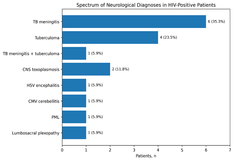
Figure 1. Spectrum of neurological diagnoses among HIV-positive patients with neurological manifestations.
Neurological investigations
CSF examination was performed in 10 patients. Elevated protein with lymphocytic predominance was observed in 6 (60.0%), elevated protein with polymorph predominance in 2 (20.0%), and normal CSF findings in 2 (20.0%). Toxoplasma antibodies were positive in two patients.
CT brain was performed in 13 patients, of whom 3 (23.1%) had multiple calcified granulomas, 1 (7.7%) had bilateral frontal, basifrontal, and temporal hypoattenuating lesions, and 9 (69.2%) had a normal CT scan. MRI brain was also performed in 13 patients; the major abnormalities included meningeal enhancement suggestive of tuberculous meningitis in five patients, multiple ring-enhancing lesions in four, toxoplasmosis-related brainstem/parieto-occipital/cerebellar hyperintensities in one, white-matter lesions suggestive of progressive multifocal leukoencephalopathy in one, and small-vessel ischemic changes in one.
Table 3. Major cerebrospinal fluid and neuroimaging findings
|
Investigation/finding |
n (%) |
|
CSF examination (n=10) |
|
|
Elevated protein with lymphocytic predominance |
6 (60.0) |
|
Elevated protein with polymorph predominance |
2 (20.0) |
|
Normal CSF |
2 (20.0) |
|
Positive toxoplasma antibodies |
2† |
|
CT brain (n=13) |
|
|
Multiple calcified granulomas |
3 (23.1) |
|
Bilateral frontal/basifrontal/temporal hypoattenuating lesions |
1 (7.7) |
|
Normal CT brain |
9 (69.2) |
|
MRI brain (n=13) |
|
|
Meningeal enhancement suggestive of TB meningitis |
5 |
|
Multiple ring-enhancing lesions |
4 |
|
Toxoplasmosis-related hyperintensities |
1 |
|
White-matter hyperintensities suggestive of PML |
1 |
|
Small-vessel ischemic changes |
1 |
†Toxoplasma antibody positivity represents an additional CSF test result rather than a mutually exclusive CSF category.
CD4 count and cognitive status
CD4 counts were available in 16 of 17 patients with neurological manifestations and all 33 patients without neurological involvement. Mean CD4 count was lower among patients with neurological manifestations than among those without neurological manifestations (179.4±176 vs 275±261 cells/µL), although the difference was not statistically significant (Welch t=−1.51, p=0.138).
Among patients with tuberculous meningitis for whom CD4 count was available, the mean CD4 count was 212±205 cells/µL, compared with 275±261 cells/µL among patients without neurological manifestations; this difference was also not significant (Welch t=−0.66, p=0.526). Mean MMSE score was significantly lower in patients with neurological manifestations than in those without neurological involvement (23.5±2.5 vs 27.06±1.14; Welch t=−5.58, p<0.001) (Table 4).
Table 4. CD4 count and MMSE according to neurological status
|
Measure |
Neurological manifestations |
No neurological manifestations |
Test statistic |
p-value |
|
CD4 count, cells/µL |
179.4±176 (n=16) |
275±261 (n=33) |
Welch t=−1.51 |
0.138 |
|
MMSE score |
23.5±2.5 (n=17) |
27.06±1.14 (n=33) |
Welch t=−5.58 |
<0.001 |
|
CD4 count: TB meningitis vs no neurological manifestations |
212±205 (n=6) |
275±261 (n=33) |
Welch t=−0.66 |
0.526 |
Values are mean±SD unless otherwise indicated.
Clinical outcome and relationship with CD4 count
Among the 17 patients with neurological manifestations, 9 (52.9%) died, 6 (35.3%) showed no clinical improvement, and 2 (11.8%) improved.
CD4 count differed significantly according to clinical outcome. Among the 16 patients for whom CD4 data were available, mean CD4 count was lowest among those who died (65.4±70.1 cells/µL; n=8), intermediate among those with no improvement (233.5±145.0 cells/µL; n=6), and highest among those who improved (474±178 cells/µL; n=2). The overall difference was statistically significant (one-way ANOVA, F (2,13) =11.20, p=0.0015) (Table 5; Figure 2).
Table 5. Clinical outcome and CD4 count among patients with neurological manifestations
|
Clinical outcome |
Patients, n (%) |
CD4 data available, n |
Mean CD4±SD, cells/µL |
Test statistic |
p-value |
|
Death |
9 (52.9) |
8 |
65.4±70.1 |
||
|
No improvement |
6 (35.3) |
6 |
233.5±145.0 |
||
|
Improved |
2 (11.8) |
2 |
474±178 |
||
|
Overall comparison |
17 (100) |
16 |
— |
F(2,13)=11.20 |
0.0015 |
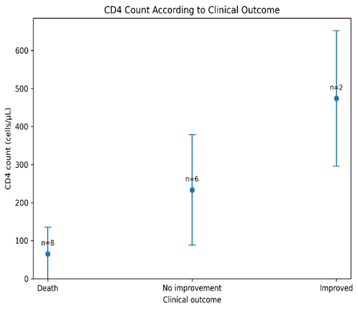
Figure 2. CD4 count according to clinical outcome among patients with neurological manifestations. Values represent mean ± SD.
Overall, neurological manifestations were present in approximately one-third of the study population, with neurotuberculosis accounting for nearly two-thirds of neurological diagnoses. Neurological involvement was associated with substantially lower MMSE scores and a high mortality rate, while progressively lower CD4 counts were strongly associated with poorer clinical outcome.
DISCUSSION:
In this study, 34.0% of the HIV-positive patients had neurological manifestations, with 64.7% of the neurological diagnoses being neurotuberculosis. Altered sensorium was the commonest presentation, followed by headache, and less common presentations were seizures and hemiplegia. The mean CD4 count was lower and MMSE scores were significantly lower in patients with neurological involvement compared to those without neurological manifestations. Importantly, there was a progressive decrease in CD4 count with poorer in-hospital outcome, with the lowest counts seen in those who died.
The incidence of neurological involvement in our study is similar to the earlier hospital based studies in India. Sonkar et al. reported neurological manifestations in 40.9% of HIV-positive patients, with headache, confusion or coma, and seizures among the common presenting features; meningitis was present in 67.4% of neurologically affected patients [10]. Headache was less common in our cohort, but the high percentage of patients with altered sensorium and the presence of seizures were similar, indicating that acute encephalopathic and meningoencephalitic presentations continue to be significant neurological presentations in hospitalized HIV patients.
Singh et al. also showed that there was a significant burden of neurological disease in the hospitalized HIV-positive population, and that CNS infections were a significant component of neurological morbidity. In their cohort, tuberculosis was the leading cause of CNS infections, and HIV-associated dementia was a significant neurological diagnosis [11]. The high prevalence of tuberculosis in CNS infections is similar to our observation that nearly two-thirds of the neurological diagnosis was for neurotuberculosis. Overt HIV-associated dementia, however, was less prominent in our patients, which may be due to differences in case selection, stage of disease, ART exposure, and diagnostic approach.
The predominance of neurotuberculosis in our study is also similar to that of Sharma et al. who found tuberculous meningitis in 43.9% of HIV-positive patients with CNS involvement. They reported a mean CD4 of about 110 cells/µL in their patients with tuberculous meningitis, with headache and seizures being some of the most common neurological symptoms [12]. The mean CD4 count in the patients with tuberculous meningitis in our study was higher, but there was a wide range. This indicates that CNS tuberculosis in HIV may not be limited to those with severe CD4 depletion and should be a key differential diagnosis in a broader spectrum of immune status in endemic areas.
However, the range of HIV associated neurological disease is wider than opportunistic CNS infection alone. In the study of non-opportunistic neurological manifestations, Deshpande and Patnaik found that 50.7% of affected patients had brain involvement, 35.8% had peripheral neuropathy, and 29.8% had stroke syndromes [13]. By contrast, our group was almost entirely comprised of infectious CNS disease, and only a few peripheral or noninfectious neurological diagnoses. This discrepancy may be partly due to the tertiary-care inpatient setting of the present study, which would tend to overrepresent patients with acute and severe opportunistic infections.
The same was observed by Kausadikar et al. who reported secondary CNS disease as the most common type of neurological involvement, with 30% of HIV-positive patients with CNS disease having tuberculous meningitis and a smaller proportion having cerebral toxoplasmosis [14]. The pattern of the study is similar to that of the higher proportion of neurotuberculosis, but overall, the results suggest that tuberculosis is a major cause of neurological morbidity among HIV patients in India. Variations in results between studies could be due to geographic variation in TB burden, referral patterns, ART status, diagnostic availability, and degree of immunosuppression at presentation.
Similar results have been reported from South India. Munamala and Pannem found that 33.6% of HIV infected patients with neurological symptoms had tuberculous meningitis and meningeal enhancement and ring-enhancing lesions were important neuroimaging features. They also noted that patients with severe immunosuppression, especially with low CD4 counts, had worse prognosis [15]. The MRI abnormalities that were most common in our neuroimaging findings were meningeal enhancement and multiple ring-enhancing lesions, which were similar to those seen in the other studies. Most importantly, our outcome analysis showed a clear gradient in CD4 count: patients who died had the lowest mean CD4 count, those who did not improve had intermediate values and those who improved had the highest values. This discovery indicates that immune status may be more relevant to prognosis of outcome than to the presence of neurological disease.
Another significant finding was cognitive performance. The mean MMSE scores were significantly poorer in patients with neurological manifestations than in patients without neurological involvement. Chan et al. reported that 22.7% of an HIV-positive cohort had HIV-associated neurocognitive disorder, and that lower baseline CD4 counts were more prevalent among those with HIV-associated neurocognitive disorder [16]. While these findings confirm the value of cognitive assessment in HIV, the results of the MMSE should not be used as a diagnostic criterion for HIV-associated neurocognitive disorder. Performance on the MMSE can be affected by acute CNS infection, alteration of sensorium, systemic illness and educational factors. However, the significant differences between the neurological and non-neurological groups indicate bedside cognitive screening could be helpful to supplement in hospitalized patients.
The association between CD4 count and neurological involvement in our study is complex and should be interpreted with caution. Although patients with neurological manifestations had lower mean CD4 counts than those without neurological involvement, the difference was not statistically significant. This may be due to the small sample size, the large range of CD4 levels and the heterogeneity of neurological diagnoses. The strong correlation between CD4 count and clinical outcome, however, was more evident, and suggested that severe immunosuppression is a biological marker of poor prognosis in patients with neurological complications.
The study is limited by its small, single-centre design, tertiary-care setting, and nonuniform use of neurological investigations. The diversity of the diagnoses also precludes disease comparisons, and MMSE may be affected by acute illness and educational level. However, the results indicate that there is a significant burden of neurological disease, especially neurotuberculosis, among HIV-positive patients admitted to hospital. The correlation of poor cognitive function and decreasing CD4 counts with poor clinical outcome emphasizes the need for early neurological evaluation and rapid assessment for CNS opportunistic infections.
CONCLUSION:
Neurological manifestations were common among hospitalized HIV-positive patients, with neurotuberculosis predominating. Lower MMSE scores and the strong association between lower CD4 counts and poor clinical outcome highlight the importance of early neurological evaluation and timely management of CNS complications.
REFERENCES:
1. Price RW. Neurological complications of HIV infection. Lancet. 1996;348(9025):445-452. https://doi.org/10.1016/S0140-6736(95)11035-6.
2. McArthur JC, Brew BJ, Nath A. Neurological complications of HIV infection. Lancet Neurol. 2005;4(9):543-555. https://doi.org/10.1016/S1474-4422(05)70165-4.
3. Kranick SM, Nath A. Neurologic complications of HIV-1 infection and its treatment in the era of antiretroviral therapy. Continuum (Minneap Minn). 2012;18(6 Infectious Disease):1319-1337. https://doi.org/10.1212/01.CON.0000423849.24900.ec.
4. Grill MF. Neurologic complications of human immunodeficiency virus. Continuum (Minneap Minn). 2021;27(4):963-991. https://doi.org/10.1212/CON.0000000000001035.
5. Antinori A, Arendt G, Becker JT, et al. Updated research nosology for HIV-associated neurocognitive disorders. Neurology. 2007;69(18):1789-1799. https://doi.org/10.1212/01.WNL.0000287431.88658.8b.
6. Dian S, Ganiem AR, Ekawardhani S. Cerebral toxoplasmosis in HIV-infected patients: a review. Pathog Glob Health. 2023;117(1):14-23. https://doi.org/10.1080/20477724.2022.2083977.
7. Marais S, Pepper DJ, Marais BJ, Török ME. HIV-associated tuberculous meningitis—diagnostic and therapeutic challenges. Tuberculosis (Edinb). 2010;90(6):367-374. https://doi.org/10.1016/j.tube.2010.08.006.
8. Purmohamad A, Azimi T, Nasiri MJ, Goudarzi M, Zangiabadian M, Sedighian H, Fooladi AAI. HIV-tuberculous meningitis co-infection: a systematic review and meta-analysis. Curr Pharm Biotechnol. 2021;22(7):960-968. https://doi.org/10.2174/1389201021666200730143906.
9. Rajasekaran S, Mahilmaran A, Annadurai S, Kumar S, Raja K. Manifestation of tuberculosis in patients with human immunodeficiency virus: a large Indian study. Ann Thorac Med. 2007;2(2):58-60. https://doi.org/10.4103/1817-1737.32231.
10. Sonkar SK, Gupta A, Atam V, Chaudhary SC, Tripathi AK, Sonkar GK. Clinical profile of neurological manifestation in human immunodeficiency virus-positive patients. N Am J Med Sci. 2012;4(11):596-599. https://doi.org/10.4103/1947-2714.103329.
11. Singh R, Kaur M, Arora D. Neurological complications in late-stage hospitalized patients with HIV disease. Ann Indian Acad Neurol. 2011;14(3):172-177. https://doi.org/10.4103/0972-2327.85878.
12. Sharma SR, Hussain M, Habung H. Neurological manifestations of HIV-AIDS at a tertiary care institute in North Eastern India. Neurol India. 2017;65(1):64-68. https://doi.org/10.4103/0028-3886.198203.
13. Deshpande AK, Patnaik MM. Nonopportunistic neurologic manifestations of the human immunodeficiency virus: an Indian study. J Int AIDS Soc. 2005;7(4):2. https://doi.org/10.1186/1758-2652-7-4-2.
14. Kausadikar SR, Chandak A, Jha P. Neurological manifestations in HIV positive patients attending tertiary care teaching hospital in Western India. Int J Adv Med. 2016;3(4):1055-1058. https://doi.org/10.18203/2349-3933.ijam20163748.
15. Munamala CSR, Pannem RB. A study on clinical profile of neurological manifestations among HIV infected patients attending a tertiary care hospital of south India. Int J Adv Med. 2018;5(6):1392-1396. https://doi.org/10.18203/2349-3933.ijam20184685.
16. Chan LG, Kandiah N, Chua A. HIV-associated neurocognitive disorders (HAND) in a South Asian population: contextual application of the 2007 criteria. BMJ Open. 2012;2(1):e000662. https://doi.org/10.1136/bmjopen-2011-000662.