Clinical and Radiological Profile of Recurrent Ischemic Stroke: A Retrospective Cross-Sectional Study at a Tertiary Care Centre in Bihar, India.
- Faiyaz Ahmad Ansari , Assistant Professor, Department of Neurology, Patna Medical College and Hospital, Patna (Bihar).
- Sanjay Kumar , Professor and Head, Department of Neurology, Patna Medical College and Hospital, Patna (Bihar).
- Devendra Kumar Sinha , Assistant Professor, Department of General Medicine, Patna Medical College and Hospital, Patna (Bihar).
Article Information:
Abstract:
Background: Recurrent ischemic stroke remains an important cause of additional neurological disability despite advances in acute stroke care and secondary prevention. Clinical risk factors and radiological patterns associated with recurrent events may vary across populations, and data from Bihar remain limited. This study evaluated the clinical and radiological profile of patients with recurrent ischemic stroke at a tertiary care centre in Bihar, India. Materials and Methods: This hospital-based retrospective cross-sectional study included all consecutive eligible adult patients with recurrent ischemic stroke managed at the Department of Neurology and General Medicine, Patna Medical College and Hospital, Patna, from August 2024 to July 2026. Demographic characteristics, clinical presentation, vascular risk factors, comorbidities, previous secondary preventive treatment, laboratory and cardiac findings, and neuroimaging and vascular characteristics were retrieved from medical records. Data were analyzed using descriptive statistics, with exploratory assessment of associations between major vascular risk factors and patterns of vascular involvement. Results: Of 138 records screened, 126 patients fulfilled the eligibility criteria. The mean age was 59.8 ± 12.6 years, and 60.3% were male. Hemiparesis/hemiplegia was the most frequent presentation (80.2%). Hypertension (61.9%) and diabetes mellitus (45.2%) were the predominant vascular risk factors, followed by smoking (30.2%) and dyslipidaemia (24.6%). Multiple infarcts were identified in 72.2% of patients, and the middle cerebral artery territory was most frequently involved (65.9%). Large-vessel disease predominated over small-vessel disease (62.7% vs. 37.3%). Among 112 patients with vascular imaging, 8.0% had ≥70% arterial stenosis. Dyslipidaemia (31.6% vs. 12.8%; p=0.017) and smoking (36.7% vs. 19.1%; p=0.038) were significantly more frequent among patients with large-vessel disease. Conclusion: Recurrent ischemic stroke in this tertiary-care cohort was characterized by a predominance of older male patients, a substantial burden of hypertension and diabetes mellitus, frequent multiple infarcts, and predominantly large-vessel involvement. Dyslipidaemia and smoking were associated with large-vessel disease, highlighting the importance of comprehensive vascular risk assessment and secondary preventive strategies in patients with previous ischemic stroke.
Keywords:
Article :
INTRODUCTION:
Stroke remains a major global health problem and a leading cause of mortality and long-term neurological disability. Survivors frequently experience persistent neurological deficits, functional dependence, reduced quality of life, and a continuing need for medical and rehabilitative care, resulting in a substantial socioeconomic burden [1]. Ischemic stroke accounts for the majority of stroke events and results from interruption of blood flow leading to cerebral, spinal, or retinal infarction [2]. Despite advances in acute stroke management and secondary prevention, recurrent ischemic stroke remains an important clinical challenge. A systematic review and meta-analysis by Mohan et al. demonstrated a substantial cumulative risk of stroke recurrence following an initial event [3]. Population-based data have similarly reported a one-year incidence of recurrent ischemic stroke of approximately 11% [4].
The risk is particularly high during the early period following the index event and persists over time [5]. Recurrent events may compound pre-existing neurological deficits, increase functional dependence, and adversely affect long-term outcomes. Recurrent ischemic stroke is influenced by both non-modifiable and potentially modifiable risk factors. Increasing age, sex, previous stroke characteristics, and underlying vascular pathology represent important non-modifiable determinants, whereas hypertension, diabetes mellitus, dyslipidaemia, smoking, cardiac disease, and inadequate control of vascular risk factors are potentially amenable to intervention [6,7]. Hypertension, poor glycaemic control, and dyslipidaemia are particularly relevant because they contribute to progressive vascular injury and atherosclerosis. Recognition and appropriate management of these factors following an initial stroke are therefore essential components of secondary prevention. The underlying mechanism and etiological subtype of ischemic stroke also influence recurrence. Large-artery atherosclerosis is particularly important because affected patients remain at risk of subsequent thromboembolic events. Lovett et al.
demonstrated a higher early recurrence risk among patients with large-artery atherosclerotic stroke than among those with other etiological subtypes, while small-vessel disease was associated with a comparatively lower risk [8]. Intracranial or extracranial arterial stenosis and potential cardiac sources of embolism may further contribute to recurrent cerebral ischemia. Radiological assessment is integral to the evaluation of recurrent ischemic stroke. Computed tomography (CT) and magnetic resonance imaging (MRI) can characterize the site, extent, number, and vascular distribution of cerebral infarctions, while vascular imaging can identify intracranial and extracranial arterial stenosis and other abnormalities relevant to recurrence. The distribution of infarcts across vascular territories and the presence of single or multiple lesions may also provide important clues regarding the underlying mechanism.
A hospital-based study from South India by Awati demonstrated a considerable burden of potentially modifiable risk factors among patients with recurrent ischemic stroke [9]. Hypertension, poor glycaemic control, dyslipidaemia, and ischemic heart disease were frequently observed, while large-vessel disease and anterior circulation involvement were prominent radiological findings. These observations highlight the importance of integrating clinical risk-factor assessment with neuroimaging and vascular evaluation in patients with recurrent stroke [9]. The burden of stroke in India remains substantial and has increased with demographic transition and the growing prevalence of vascular risk factors [10].
However, considerable geographical, socioeconomic, and healthcare-related heterogeneity exists across the country, and the clinical and radiological characteristics of recurrent ischemic stroke may vary between regions. Region-specific evidence is therefore important for understanding disease patterns and identifying potentially modifiable factors relevant to secondary prevention. Despite the clinical importance of recurrent stroke, published information on the combined clinical and radiological profile of recurrent ischemic stroke in Bihar remains limited. Characterization of demographic features, vascular risk factors, associated comorbidities, previous treatment, patterns of cerebral infarction, vascular territory involvement, and arterial stenosis may provide clinically relevant information for this population. Therefore, the present study was undertaken to evaluate the clinical and radiological profile of patients with recurrent ischemic stroke at a tertiary care centre in Bihar, India, with particular emphasis on demographic characteristics, vascular risk factors and comorbidities, treatment profile, and neuroimaging and vascular findings.
MATERIALS AND METHODS:
Study Design and Setting
This hospital-based retrospective cross-sectional study was conducted in the Department of Neurology and General Medicine, Patna Medical College and Hospital (PMCH), Patna, Bihar, India. Medical records of patients diagnosed with recurrent ischemic stroke during a period of two years were reviewed. All eligible patients fulfilling the predefined selection criteria during the study period were included.
Sample Size and Sampling Technique
A consecutive sampling technique was used. All patients diagnosed with recurrent ischemic stroke who fulfilled the predefined inclusion and exclusion criteria and were managed in the Department of Neurology and General Medicine, Patna Medical College and Hospital, Patna, during the study period from August 2026 to July 2026 were included in the study. As the study was retrospective and included all eligible cases during the defined study period, no fixed sample size was predetermined. The final sample size corresponded to the total number of eligible patients identified during the two-year study period.
Study Population
The study population comprised adult patients who had a documented history of a previous ischemic stroke and were subsequently admitted or evaluated at the Department of Neurology and General Medicine with another episode of ischemic stroke during the study period. For the purpose of the study, recurrent ischemic stroke was defined as a new episode of focal neurological dysfunction attributable to cerebral infarction occurring in a patient with a documented previous ischemic stroke, with the recurrent event supported by clinical findings and available neuroimaging records.
Inclusion Criteria
Patients were included if they were 18 years of age or older, had a documented history of a previous ischemic stroke, and subsequently presented with a recurrent ischemic stroke confirmed on the basis of clinical assessment and neuroimaging findings. Only records containing sufficient clinical and radiological information to establish the diagnosis of recurrent ischemic stroke were included in the final analysis.
Exclusion Criteria
Patients with a first-ever ischemic stroke, primary intracerebral haemorrhage or subarachnoid haemorrhage, transient ischemic attack without evidence of cerebral infarction, and focal neurological deficits attributable to causes other than ischemic stroke were excluded. Patients younger than 18 years and records with insufficient clinical or radiological information to confirm recurrent ischemic stroke were also excluded.
Case Identification and Data Collection
Eligible cases were identified retrospectively from the available inpatient and outpatient records of the Department of Neurology and General Medicine during the predefined two-year study period. Relevant information was retrieved from medical case records, discharge summaries, investigation reports, and available electronic or archived records. Data were entered into a structured data collection proforma designed for the study.
No additional investigations or interventions were performed specifically for the purpose of this study. Only information and investigations documented as part of routine clinical evaluation and management were included in the analysis.
Demographic and Clinical Variables
Demographic information including age and sex was recorded. Details regarding the previous ischemic stroke and the recurrent event were retrieved wherever available.
The presence of established vascular risk factors and associated comorbidities was documented from the medical records. These included hypertension, diabetes mellitus, dyslipidaemia, ischemic heart disease, chronic kidney disease, thyroid disorders, smoking, and alcohol consumption, wherever recorded.
Clinical features documented at the time of presentation with recurrent stroke were reviewed. These included focal neurological deficits such as hemiparesis or hemiplegia, cranial nerve involvement, sensory deficits, speech disturbances, altered sensorium, seizures, headache, vomiting, and visual disturbances, wherever applicable. Blood pressure and other relevant clinical parameters recorded at presentation were also documented.
Previous Treatment and Secondary Prevention
Available information regarding treatment following the previous ischemic stroke was retrieved from medical records, previous prescriptions, and discharge summaries. The use of single or dual antiplatelet therapy, statins, antihypertensive medications, oral hypoglycaemic agents, and insulin was recorded wherever documented. Information regarding medication adherence and control of vascular risk factors was included only when clearly documented in the available records.
Laboratory Evaluation
Results of laboratory investigations performed as part of routine clinical management were retrieved from the medical records. These included blood glucose parameters, renal function tests, serum electrolytes, and lipid profile, wherever available. Other relevant investigations documented in the records were also noted when clinically pertinent. Since the study was retrospective, laboratory parameters that were not available in the medical records were treated as missing data and were not inferred or substituted.
Cardiac Evaluation
Available findings from 12-lead electrocardiography (ECG) and two-dimensional echocardiography (2D-ECHO) performed during routine clinical evaluation were recorded. Relevant cardiac abnormalities, including ischemic heart disease and regional wall motion abnormalities, were documented wherever available. Other potential cardiac sources of cerebral embolism were recorded when documented in the clinical records.
Radiological Evaluation
Available neuroimaging records and reports were reviewed to characterize the recurrent ischemic stroke. Computed tomography (CT) and/or magnetic resonance imaging (MRI) of the brain, as available, were assessed for the site, distribution, number, and vascular territory of cerebral infarction. The vascular territory involved was categorized as middle cerebral artery (MCA), anterior cerebral artery (ACA), or posterior circulation territory, wherever identifiable. Infarcts were further characterized as single or multiple infarcts, and the presence of watershed infarction was recorded when documented. Available vascular imaging findings from magnetic resonance angiography (MRA), computed tomography angiography (CTA), and/or carotid and vertebral Doppler ultrasonography were retrieved. The presence, anatomical location, and degree of intracranial or extracranial arterial stenosis were recorded wherever these investigations had been performed. The pattern of vascular involvement, including large-vessel and small-vessel disease, was documented based on the available clinical and radiological records.
Study Variables
The principal variables evaluated included demographic characteristics, clinical presentation, vascular risk factors, associated comorbidities, previous secondary preventive treatment, laboratory parameters, cardiac findings, and radiological characteristics. Radiological variables included the location and vascular territory of infarction, single or multiple infarcts, large- or small-vessel involvement, watershed infarction, and the presence and degree of arterial stenosis.
Handling of Missing Data
As this was a retrospective record-based study, some clinical, laboratory, or radiological variables were not available for every patient. Variables that were not documented in the original medical records were considered missing and were not assumed to be absent. Analyses involving such variables were performed using the available data, and the corresponding denominator was reported wherever appropriate.
Statistical Analysis
The collected data were entered into Microsoft Excel and subsequently analyzed using IBM SPSS Statistics for Windows, version 25.0 (IBM Corp., Armonk, NY, USA). Continuous variables were assessed for distribution and summarized as mean ± standard deviation (SD) for normally distributed data or median with interquartile range (IQR) for non-normally distributed data. Categorical variables were expressed as frequencies and percentages. The clinical, demographic, and radiological characteristics of patients with recurrent ischemic stroke were primarily analyzed descriptively. Associations between categorical vascular risk factors and the pattern of vascular involvement were explored using Pearson's Chi-square test or Fisher's exact test, as appropriate. All tests were two-sided, and a p-value <0.05 was considered statistically significant.
RESULTS:
During the two-year study period, a total of 138 medical records of patients with a documented previous ischemic stroke and a subsequent cerebrovascular event were screened. Twelve records were excluded because of incomplete clinical or radiological documentation or failure to satisfy the predefined eligibility criteria. Thus, 126 patients with recurrent ischemic stroke were included in the final analysis.
Demographic Characteristics
The mean age of the study population was 59.8 ± 12.6 years, with the majority of patients belonging to the older age groups. Of the 126 patients, 29 (23.0%) were younger than 45 years, 39 (31.0%) were aged 45–60 years, and 58 (46.0%) were older than 60 years. There was a male predominance, with 76 (60.3%) males and 50 (39.7%) females (Table 1).
Table 1. Demographic characteristics of patients with recurrent ischemic stroke
|
Characteristic |
Number (n) |
Percentage (%) |
|
Age group (years) |
||
|
<45 |
29 |
23.0 |
|
45–60 |
39 |
31.0 |
|
>60 |
58 |
46.0 |
|
Sex |
||
|
Male |
76 |
60.3 |
|
Female |
50 |
39.7 |
|
Total |
126 |
100.0 |
|
Mean age (Years± SD) |
59.8 ± 12.6 years |
|
Clinical Profile at Presentation
Hemiparesis or hemiplegia was the most frequently documented neurological manifestation, occurring in 101 (80.2%) patients. Speech disturbances were present in 53 (42.1%) patients, while altered sensorium and cranial nerve involvement were documented in 27 (21.4%) and 25 (19.8%) patients, respectively. Sensory deficits were recorded in 21 (16.7%) patients. Less frequent manifestations included headache, vomiting, seizures, and visual disturbances (Table 2).
Table 2. Clinical presentation of patients with recurrent ischemic stroke
|
Clinical feature |
Number (n) |
Percentage (%) |
|
Hemiparesis/hemiplegia |
101 |
80.2 |
|
Speech disturbance |
53 |
42.1 |
|
Altered sensorium |
27 |
21.4 |
|
Cranial nerve involvement |
25 |
19.8 |
|
Sensory deficit |
21 |
16.7 |
|
Headache |
18 |
14.3 |
|
Vomiting |
14 |
11.1 |
|
Seizures |
11 |
8.7 |
|
Visual disturbance |
8 |
6.3 |
[More than one clinical manifestation could be present in an individual patient; therefore, percentages do not total 100%.]
Vascular Risk Factors and Associated Comorbidities
Hypertension was the most common vascular risk factor and was documented in 78 (61.9%) patients. Diabetes mellitus was present in 57 (45.2%), while dyslipidaemia was documented in 31 (24.6%) patients. Ischemic heart disease was present in 29 (23.0%) patients. A history of smoking was documented in 38 (30.2%), while alcohol consumption was recorded in 24 (19.0%) patients. Chronic kidney disease and thyroid disorders were comparatively less frequent (Table 3, Figure 1).
Table 3. Vascular risk factors and associated comorbidities
|
Risk factor/comorbidity |
Number (n) |
Percentage (%) |
|
Hypertension |
78 |
61.9 |
|
Diabetes mellitus |
57 |
45.2 |
|
Smoking |
38 |
30.2 |
|
Dyslipidaemia |
31 |
24.6 |
|
Ischemic heart disease |
29 |
23.0 |
|
Alcohol consumption |
24 |
19.0 |
|
Thyroid disorder |
9 |
7.1 |
|
Chronic kidney disease |
7 |
5.6 |
[Patients could have more than one vascular risk factor or associated comorbidity.
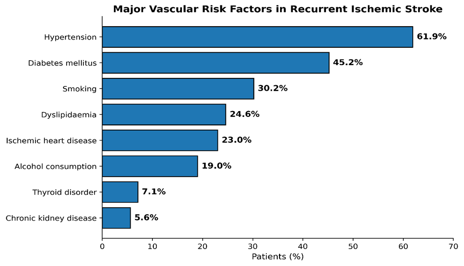
Figure 1. Distribution of major vascular risk factors and associated comorbidities among patients with recurrent ischemic stroke.
Secondary Preventive Treatment Following the Previous Stroke
Information regarding secondary preventive medication was available for 119 patients. Single antiplatelet therapy was documented in 88 (73.9%), while 25 (21.0%) were receiving dual antiplatelet therapy. Statin therapy was documented in 104 (87.4%) patients. Information regarding secondary preventive medication was available for 119 patients. Single antiplatelet therapy was documented in 88 (73.9%), dual antiplatelet therapy in 25 (21.0%), and statin therapy in 104 (87.4%). Antihypertensive therapy, oral hypoglycaemic agents, and insulin were documented in 68 (57.1%), 39 (32.8%), and 11 (9.2%) patients, respectively (Table 4).
Table 4. Secondary preventive treatment documented before recurrent ischemic stroke
|
Treatment |
Number (n) |
Percentage (%)* |
|
Single antiplatelet therapy |
88 |
73.9 |
|
Dual antiplatelet therapy |
25 |
21.0 |
|
Statin therapy |
104 |
87.4 |
|
Antihypertensive therapy |
68 |
57.1 |
|
Oral hypoglycaemic agents |
39 |
32.8 |
|
Insulin therapy |
11 |
9.2 |
[*Percentages are based on the 119 patients with available medication records. Treatment categories were not mutually exclusive.]
Laboratory and Cardiac Findings
Glycaemic parameters were available for 121 patients, of whom 55 (45.5%) showed abnormal glycaemic control (Table 5). Lipid profile was available for 112 patients, with dyslipidaemia documented in 31 (27.7%). Renal dysfunction was identified in 10 of 120 (8.3%) patients with available renal function tests. ECG findings were available for 123 patients, and atrial fibrillation was documented in 12 (9.8%). Two-dimensional echocardiography was available for 108 patients, of whom 28 (25.9%) had regional wall motion abnormalities.
Table 5. Selected laboratory and cardiac findings
|
Parameter |
Available records (n) |
Abnormal finding, n (%) |
|
Abnormal glycaemic parameters |
121 |
55 (45.5) |
|
Dyslipidaemia |
112 |
31 (27.7) |
|
Renal dysfunction |
120 |
10 (8.3) |
|
Atrial fibrillation on ECG |
123 |
12 (9.8) |
|
Regional wall motion abnormality on 2D-ECHO |
108 |
28 (25.9) |
[Percentages in this table were calculated using the number of patients with available results for each investigation as the denominator; therefore, they may differ from percentages based on the total study population.]
Radiological Profile
Neuroimaging demonstrated a predominance of multiple infarcts, which were identified in 91 (72.2%) patients, while 35 (27.8%) had a single infarct. The middle cerebral artery territory was most frequently involved, being documented in 83 (65.9%) patients. Anterior cerebral artery territory involvement was observed in 24 (19.0%), posterior circulation infarction in 32 (25.4%), and watershed infarction in 16 (12.7%) patients. More than one vascular territory could be involved in the same patient. Large-vessel disease was identified in 79 (62.7%) patients, whereas 47 (37.3%) showed a predominantly small-vessel pattern. Vascular imaging by MRA, CTA, and/or carotid-vertebral Doppler was available for 112 patients. Of these, 91 (81.3%) had no significant stenosis, 12 (10.7%) had 50–69% stenosis, and 9 (8.0%) had ≥70% stenosis (Table 6, Figure 2).
Table 6. Neuroimaging and vascular characteristics of recurrent ischemic stroke
|
Radiological characteristic |
Number (n) |
Percentage (%) |
|
Number of infarcts |
||
|
Single infarct |
35 |
27.8 |
|
Multiple infarcts |
91 |
72.2 |
|
Vascular territory involved |
||
|
MCA territory |
83 |
65.9 |
|
ACA territory |
24 |
19.0 |
|
Posterior circulation |
32 |
25.4 |
|
Watershed infarction |
16 |
12.7 |
|
Pattern of vascular involvement |
||
|
Large-vessel disease |
79 |
62.7 |
|
Small-vessel disease |
47 |
37.3 |
|
Arterial stenosis (n=112) |
||
|
No significant stenosis |
91 |
81.3 |
|
50–69% stenosis |
12 |
10.7 |
|
≥70% stenosis |
9 |
8.0 |
[Vascular territory categories were not mutually exclusive because some patients had infarcts involving more than one territory. Percentages for arterial stenosis were calculated using the 112 patients with available vascular imaging.
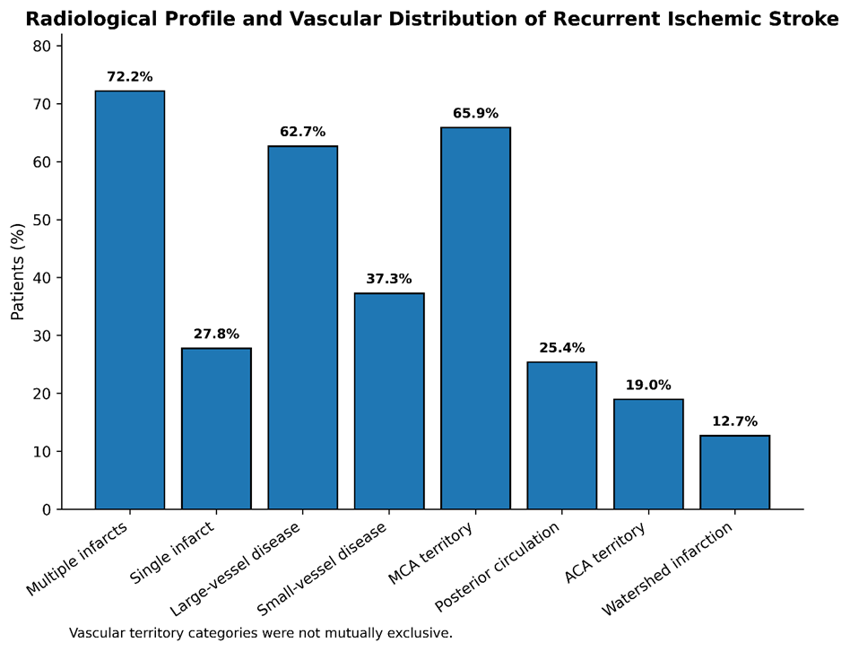
Figure 2. Radiological profile and vascular distribution of recurrent ischemic stroke, showing the distribution of single and multiple infarcts, large- and small-vessel disease, and involvement of major vascular territories.
Association Between Major Vascular Risk Factors and Pattern of Vascular Involvement.
Exploratory analysis showed that hypertension, diabetes mellitus, dyslipidaemia, ischemic heart disease, smoking, and atrial fibrillation were numerically more frequent among patients with large-vessel disease than among those with small-vessel disease. Dyslipidaemia (31.6% vs. 12.8%; p=0.017) and smoking (36.7% vs. 19.1%; p=0.038) were significantly associated with large-vessel disease. No statistically significant differences were observed for hypertension, diabetes mellitus, ischemic heart disease, or atrial fibrillation (Table 7, Figure 3).
Table 7. Association of major vascular risk factors with large- and small-vessel disease
|
Risk factor |
Large-vessel disease (n=79), n (%) |
Small-vessel disease (n=47), n (%) |
P-value |
|
Hypertension |
52 (65.8) |
26 (55.3) |
0.240 |
|
Diabetes mellitus |
39 (49.4) |
18 (38.3) |
0.227 |
|
Dyslipidaemia |
25 (31.6) |
6 (12.8) |
0.017* |
|
Ischemic heart disease |
20 (25.3) |
9 (19.1) |
0.426 |
|
Smoking |
29 (36.7) |
9 (19.1) |
0.038* |
|
Atrial fibrillation |
9 (11.4) |
3 (6.4) |
0.533** |
[*Values are expressed as n (%). P-values were calculated using Pearson’s Chi-square test, except for atrial fibrillation, for which Fisher’s exact test was used. p<0.05 was considered statistically significant. **Fisher’s exact test.]
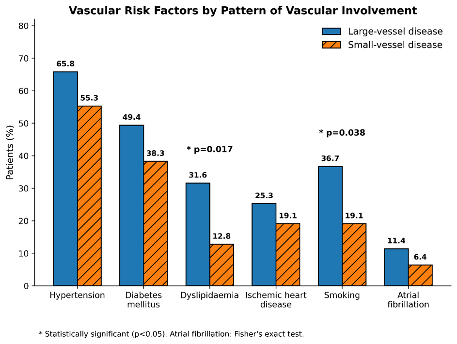
Figure 3. Comparison of major vascular risk factors between patients with large-vessel and small-vessel disease. Dyslipidaemia and smoking were significantly more frequent in patients with large-vessel disease (p<0.05).
Overall, the study population was characterized by a predominance of older male patients, a high burden of hypertension and diabetes mellitus, frequent multiple infarcts, and a predominance of large-vessel disease. Significant arterial stenosis was present in a smaller proportion of patients undergoing vascular imaging.
DISCUSSION:
The present study provides a combined clinical and radiological characterization of recurrent ischemic stroke in a tertiary-care population from Bihar. The principal observations were a predominance of older patients and males, a substantial burden of modifiable vascular risk factors, particularly hypertension and diabetes mellitus, frequent multiple infarcts, and predominance of large-vessel disease. The middle cerebral artery (MCA) territory was the most commonly involved vascular territory. Importantly, dyslipidaemia and smoking were significantly more frequent among patients with large-vessel disease, emphasizing the close relationship between atherosclerotic risk factors and the vascular pattern of recurrent cerebral ischemia.
The mean age of the study population was 59.8 years, and nearly half of the patients were older than 60 years. Male patients constituted 60.3% of the cohort. A similar male predominance was reported by Awati et al. in their South Indian study of recurrent ischemic stroke [9]. Although age and sex are non-modifiable factors, their distribution remains clinically relevant because recurrent stroke commonly occurs in a population in whom vascular risk factors accumulate with advancing age. A systematic review examining risk factors for recurrence also demonstrated that the probability of another ischemic event reflects the combined influence of vascular comorbidities rather than any single factor in isolation [11]. The comparatively younger mean age in our population than in several Western recurrent-stroke cohorts may also reflect differences in population demographics and vascular risk-factor patterns.
Hemiparesis or hemiplegia was the most frequent clinical presentation in our patients, followed by speech disturbance. This pattern is consistent with the predominance of anterior circulation, particularly MCA-territory, involvement observed on neuroimaging. The MCA supplies a large proportion of the cerebral hemisphere, including motor and language-related cortical regions, making motor weakness and speech abnormalities expected manifestations when this territory is affected. The high frequency of focal motor deficits in the present series therefore corresponds well with the observed radiological distribution. Hypertension was the most prevalent vascular risk factor, affecting 61.9% of patients, followed by diabetes mellitus in 45.2%. The predominance of these two conditions is consistent with previous studies of recurrent stroke. In the RESQUE study, hypertension was present in approximately three-fourths of patients with recurrent stroke, while diabetes mellitus was present in approximately one-fourth [7].
Similarly, Laloux et al. reported hypertension in 79% of patients presenting with recurrent ischemic stroke or transient ischemic attack, with inadequate blood-pressure control in a substantial proportion [12]. Kocaman et al. also identified hypertension as the most common risk factor among patients with recurrent ischemic stroke, followed by diabetes mellitus and other vascular comorbidities [13]. A systematic review and meta-analysis further demonstrated significant associations of both hypertension and diabetes mellitus with recurrent stroke [11]. Taken together, these findings reinforce the importance of sustained blood-pressure and glycaemic control after an initial ischemic event. The importance of blood-pressure control extends beyond the presence or absence of a diagnosis of hypertension. In patients with symptomatic intracranial arterial stenosis, Turan et al. demonstrated that higher mean systolic and diastolic blood pressure during follow-up was associated with an increased risk of recurrent ischemic stroke, including stroke within the territory of the stenotic vessel [14]. Thus, recognition of hypertension alone is insufficient; long-term achievement of appropriate blood-pressure targets is central to secondary prevention. Current secondary-prevention recommendations similarly emphasize individualized but consistent control of vascular risk factors, including hypertension, diabetes, dyslipidaemia, smoking, physical inactivity, and other modifiable exposures [15].
Abnormal glycaemic parameters were documented in 45.5% of patients with available laboratory data in the present study, closely paralleling the proportion with established diabetes mellitus. Diabetes contributes to endothelial dysfunction, accelerated atherosclerosis and small-vessel injury and may therefore influence several mechanisms of recurrent ischemic stroke. The association between diabetes and recurrence has been demonstrated across different populations [11]. In the Swedish population-based study by Bergström et al., diabetes mellitus was independently associated with an increased risk of recurrent ischemic stroke [4]. These observations support continued attention not merely to the diagnosis of diabetes but to adequacy of glycaemic control during long-term follow-up.
Smoking was documented in 30.2% of the present cohort and was significantly more frequent among patients with large-vessel disease than among those with small-vessel disease (36.7% vs. 19.1%). Dyslipidaemia showed a similar association with large-vessel disease (31.6% vs. 12.8%). Although the cross-sectional nature of our study does not establish these factors as independent predictors of recurrence or large-vessel disease, the findings are biologically plausible because both tobacco exposure and lipid abnormalities are closely linked to atherosclerotic vascular disease. Their concentration among patients with large-vessel involvement identifies a clinically important subgroup in whom intensive lifestyle modification, smoking cessation and lipid management deserve particular emphasis. High-intensity statin therapy has an established role in secondary prevention; the SPARCL trial demonstrated that atorvastatin reduced subsequent stroke in patients with a recent stroke or transient ischemic attack [16]. Subsequent analyses indicated benefit across ischemic stroke subtypes, including patients with large-vessel disease [17].
Cardiac disease also constituted an important component of the risk profile. Ischemic heart disease was present in 23.0% of patients, regional wall-motion abnormalities were observed in 25.9% of those undergoing echocardiography, and atrial fibrillation was identified in 9.8% of patients with available ECG findings. These abnormalities are clinically relevant because cerebral and coronary atherosclerotic disease frequently coexist, while atrial fibrillation provides an independent cardioembolic mechanism. Bergström et al. found prior myocardial infarction and atrial fibrillation without anticoagulation to be associated with increased recurrent ischemic stroke risk [4]. More recent evidence also indicates that interruption of oral anticoagulation in patients with atrial fibrillation following ischemic stroke is associated with a substantially greater risk of recurrent ischemic stroke [18]. Consequently, identification of atrial fibrillation and other potential cardiac sources of embolism remains an essential part of the evaluation of patients presenting with recurrent cerebral ischemia.
An important observation in the present study is that recurrence occurred despite substantial use of secondary preventive medication. Among patients with available medication records, 73.9% were receiving single antiplatelet therapy, 21.0% dual antiplatelet therapy and 87.4% statin therapy. This does not imply failure of these treatments themselves, because the study could not reliably assess treatment duration, adherence, achievement of therapeutic targets, changes in stroke mechanism, or appropriateness of therapy for individual etiologies. Previous studies have demonstrated similar gaps between prescription and effective secondary prevention. In the RESQUE study, 79% of patients with recurrent ischemic stroke were receiving antiplatelet therapy, yet only 40% were receiving lipid-lowering treatment [7]. Laloux et al. similarly identified inadequate control of hypertension, hypercholesterolaemia and diabetes among many patients at the time of recurrence [12]. Kocaman et al. reported deficiencies in both preventive treatment and risk-factor control among recurrent stroke patients [13]. Thus, secondary prevention should be viewed as a continuing process involving appropriate medication, adherence, achievement of treatment targets, lifestyle modification and periodic reassessment of stroke mechanism rather than prescription alone.
The radiological findings provide further insight into the pattern of recurrence in this population. Multiple infarcts were identified in 72.2% of patients, while only 27.8% had a single infarct. Multiple lesions may reflect repeated embolic episodes, active large-artery atherosclerotic disease, cardioembolism, or recurrent disease involving different vascular territories. Their high frequency in the present cohort highlights the cumulative cerebral burden associated with recurrent ischemic events and reinforces the value of reviewing current imaging in conjunction with previous neuroimaging whenever available.
Large-vessel disease was observed in 62.7% of patients and was considerably more frequent than the small-vessel pattern. This closely resembles the South Indian study by Awati et al., in which large-vessel disease accounted for 62% of recurrent ischemic strokes [9]. The predominance of large-vessel involvement is particularly important because stroke mechanism influences recurrence risk. Lovett et al. demonstrated that large-artery atherosclerotic stroke carried the highest early recurrence risk among etiological subtypes, whereas small-vessel stroke had the lowest early recurrence risk [8]. A subsequent systematic review of recurrent ischemic stroke similarly found recurrence to be particularly important in large-artery atherosclerosis and cardioembolic stroke, with a lower recurrence rate in small-vessel occlusion [19]. These observations support careful vascular evaluation in recurrent stroke rather than relying solely on conventional clinical risk factors.
The MCA territory was involved in 65.9% of patients and was the predominant vascular territory, followed by posterior circulation and ACA involvement. Awati et al. likewise reported prominent anterior circulation involvement among patients with recurrent ischemic stroke [9]. The frequent MCA involvement in our cohort may partly explain the predominance of hemiparesis/hemiplegia and speech disturbances at presentation. Watershed infarction was observed in 12.7% of patients. Although watershed lesions may arise through several mechanisms, their presence should prompt consideration of haemodynamic compromise and significant proximal arterial disease in the appropriate clinical setting.
Vascular imaging was available in 112 patients. Although most had no significant stenosis, 10.7% had 50–69% stenosis and 8.0% had stenosis of ≥70%. The relatively modest proportion with severe stenosis should not diminish its clinical relevance because symptomatic intracranial or extracranial atherosclerotic stenosis represents an important high-risk mechanism for recurrence. In the WASID population, vascular risk-factor control, particularly blood-pressure management, was strongly related to subsequent stroke risk [14]. The SAMMPRIS trial subsequently demonstrated that, among patients with recent stroke or transient ischemic attack attributable to severe intracranial arterial stenosis, aggressive medical management produced better outcomes than intracranial stenting and remained superior during extended follow-up [20]. These findings underscore the importance of identifying arterial stenosis so that secondary prevention can be tailored to the underlying vascular pathology.
Our exploratory analysis found statistically significant associations of dyslipidaemia and smoking with large-vessel disease, whereas hypertension, diabetes mellitus, ischemic heart disease and atrial fibrillation did not differ significantly between large- and small-vessel groups. These results should be interpreted cautiously because the study was not designed to establish independent etiological predictors, and the absence of statistical significance does not imply lack of clinical importance. Hypertension and diabetes were highly prevalent in both vascular groups and may contribute to both macrovascular and microvascular cerebral disease. Conversely, the stronger association of dyslipidaemia and smoking with the large-vessel pattern is consistent with their established contribution to atherosclerotic disease and provides a useful direction for future prospective studies in this population.
The overall findings also highlight why recurrent ischemic stroke should not be approached as a uniform entity. Stroke recurrence reflects the interaction of systemic vascular risk factors, the mechanism of the previous stroke, progression of arterial disease, cardiac sources of embolism, adequacy of preventive treatment and patient adherence. Contemporary secondary-prevention guidance therefore recommends defining the probable stroke mechanism and applying mechanism-specific treatment alongside rigorous management of vascular risk factors [15]. Aggressive risk-factor management has also been shown to be achievable when pharmacological treatment is combined with structured lifestyle modification and follow-up [21].
From a regional perspective, the present study adds data from Bihar, where published information specifically addressing the combined clinical and radiological profile of recurrent ischemic stroke remains limited. The findings broadly resemble those from other recurrent-stroke cohorts but also demonstrate the substantial local burden of hypertension, diabetes, smoking, multiple infarction and large-vessel disease. The observed pattern supports a practical secondary-prevention strategy based on regular blood-pressure and glycaemic assessment, intensive lipid management where indicated, smoking cessation, appropriate antithrombotic or anticoagulant therapy according to stroke mechanism, and timely vascular and cardiac evaluation. Importantly, antiplatelet therapy, dual antiplatelet therapy and anticoagulation are not interchangeable strategies and should be selected according to the underlying stroke mechanism and established indications [15,22].
Recurrent stroke remains clinically important even in the era of modern preventive therapy. Long-term studies have shown that recurrence risk persists beyond the early post-stroke period, while the cumulative burden increases with time [3]. Contemporary evidence therefore favors sustained, mechanism-based secondary prevention initiated early and maintained longitudinally [23]. In addition, adherence is a central component of effective prevention; non-adherence to prescribed preventive medication after stroke has been associated with poorer vascular outcomes, emphasizing that prescribing appropriate therapy is only one part of secondary prevention [24]. Structured follow-up, patient education and repeated assessment of treatment targets are therefore likely to be particularly relevant in resource-variable settings.
Taken together, the present findings indicate that recurrent ischemic stroke in this population is characterized by a combination of substantial modifiable vascular risk, frequent multiple infarction and predominant large-vessel involvement. The significant associations of smoking and dyslipidaemia with large-vessel disease further emphasize the atherosclerotic component of recurrent stroke in a clinically important subgroup. These observations are consistent with the broader evidence that comprehensive risk-factor control and mechanism-specific secondary prevention can reduce subsequent vascular events [15,20,25]. Prospective multicentre studies incorporating standardized etiological classification, longitudinal assessment of risk-factor control, medication adherence and recurrent-event follow-up would help determine which factors independently predict further recurrence in patients from Bihar and similar populations.
Limitations of the Study: The findings of this study should be interpreted considering its retrospective, single-centre design, which may limit their generalizability to other populations and healthcare settings. As the analysis relied on existing medical records, some clinical, laboratory, treatment, and vascular imaging data were unavailable for all patients. Information regarding medication adherence and long-term control of vascular risk factors was also limited by the completeness of documentation. Furthermore, the absence of a comparison group of patients with first-ever ischemic stroke precluded assessment of independent predictors of stroke recurrence. Nevertheless, the study provides clinically relevant information on the clinical and radiological profile of recurrent ischemic stroke in a tertiary-care population from Bihar and may contribute to strengthening region-specific secondary prevention strategies.
CONCLUSION:
Recurrent ischemic stroke in this tertiary-care cohort was characterized by a predominance of older male patients and a substantial burden of modifiable vascular risk factors, particularly hypertension and diabetes mellitus. Multiple infarcts and large-vessel disease were common radiological findings, with the middle cerebral artery territory being most frequently involved. Dyslipidaemia and smoking were significantly associated with large-vessel disease. These findings emphasize the importance of systematic vascular risk assessment, appropriate secondary preventive therapy, and sustained risk-factor modification following an initial ischemic stroke. Greater attention to blood pressure and glycaemic control, lipid management, smoking cessation, and appropriate vascular evaluation may help strengthen secondary stroke prevention in this population.
REFERENCES:
1. Feigin VL, Brainin M, Norrving B, Martins S, Sacco RL, Hacke W, et al. World Stroke Organization (WSO): Global Stroke Fact Sheet 2022. Int J Stroke. 2022;17(1):18-29. doi:10.1177/17474930211065917.
2. Sacco RL, Kasner SE, Broderick JP, Caplan LR, Connors JJ, Culebras A, et al. An updated definition of stroke for the 21st century: a statement for healthcare professionals from the American Heart Association/American Stroke Association. Stroke. 2013;44(7):2064-89. doi:10.1161/STR.0b013e318296aeca.
3. Mohan KM, Wolfe CD, Rudd AG, Heuschmann PU, Kolominsky-Rabas PL, Grieve AP. Risk and cumulative risk of stroke recurrence: a systematic review and meta-analysis. Stroke. 2011;42(5):1489-94. doi:10.1161/STROKEAHA.110.602615.
4. Bergström L, Irewall AL, Söderström L, Ögren J, Laurell K, Mooe T. One-year incidence, time trends, and predictors of recurrent ischemic stroke in Sweden from 1998 to 2010: an observational study. Stroke. 2017;48(8):2046-51. doi:10.1161/STROKEAHA.117.016815.
5. Arsava EM, Kim GM, Oliveira-Filho J, Gungor L, Noh HJ, Lordelo Mde J, et al. Prediction of early recurrence after acute ischemic stroke. JAMA Neurol. 2016;73(4):396-401. doi:10.1001/jamaneurol.2015.4949.
6. Oza R, Rundell K, Garcellano M. Recurrent ischemic stroke: strategies for prevention. Am Fam Physician. 2017;96(7):436-40.
7. Leoo T, Lindgren A, Petersson J, von Arbin M. Risk factors and treatment at recurrent stroke onset: results from the Recurrent Stroke Quality and Epidemiology (RESQUE) Study. Cerebrovasc Dis. 2008;25(3):254-60. doi:10.1159/000113864.
8. Lovett JK, Coull AJ, Rothwell PM. Early risk of recurrence by subtype of ischemic stroke in population-based incidence studies. Neurology. 2004;62(4):569-73. doi:10.1212/01.WNL.0000110311.09970.83.
9. Awati AM. Cross sectional descriptive study of clinical and radiological profile of patients presenting with recurrent ischemic stroke in a tertiary care centre of South India. IP Indian J Neurosci. 2022;8(1):31-34. doi:10.18231/j.ijn.2022.006.
10. Jones SP, Baqai K, Clegg A, Georgiou R, Harris C, Holland EJ, et al. Stroke in India: a systematic review of the incidence, prevalence, and case fatality. Int J Stroke. 2022;17(2):132-40. doi:10.1177/17474930211027834.
11. Zheng S, Yao B. Impact of risk factors for recurrence after the first ischemic stroke in adults: a systematic review and meta-analysis. J Clin Neurosci. 2019;60:24-30. doi:10.1016/j.jocn.2018.10.026.
12. Laloux P, Lemonnier F, Jamart J. Risk factors and treatment of stroke at the time of recurrence. Acta Neurol Belg. 2010;110(4):299-302.
13. Kocaman G, Dürüyen H, Koçer A, Asil T. Recurrent ischemic stroke characteristics and assessment of sufficiency of secondary stroke prevention. Noro Psikiyatr Ars. 2015;52(2):139-44. doi:10.5152/npa.2015.7499.
14. Turan TN, Cotsonis G, Lynn MJ, Chaturvedi S, Chimowitz M; Warfarin-Aspirin Symptomatic Intracranial Disease Trial Investigators. Relationship between blood pressure and stroke recurrence in patients with intracranial arterial stenosis. Circulation. 2007;115(23):2969-75. doi:10.1161/CIRCULATIONAHA.106.622464.
15. Kleindorfer DO, Towfighi A, Chaturvedi S, Cockroft KM, Gutierrez J, Lombardi-Hill D, et al. 2021 guideline for the prevention of stroke in patients with stroke and transient ischemic attack: a guideline from the American Heart Association/American Stroke Association. Stroke. 2021;52(7):e364-e467. doi:10.1161/STR.0000000000000375.
16. Amarenco P, Bogousslavsky J, Callahan A 3rd, Goldstein LB, Hennerici M, Rudolph AE, et al. High-dose atorvastatin after stroke or transient ischemic attack. N Engl J Med. 2006;355(6):549-59. doi:10.1056/NEJMoa061894.
17. Amarenco P, Benavente O, Goldstein LB, Callahan A 3rd, Sillesen H, Hennerici MG, et al. Results of the Stroke Prevention by Aggressive Reduction in Cholesterol Levels (SPARCL) trial by stroke subtypes. Stroke. 2009;40(4):1405-9. doi:10.1161/STROKEAHA.108.534107.
18. Hindsholm MF, García Rodríguez LA, Brandes A, Hallas J, Høyer BB, Möller S, et al. Recurrent ischemic stroke in patients with atrial fibrillation while receiving oral anticoagulants. JAMA Neurol. 2024;81(8):805-13. doi:10.1001/jamaneurol.2024.1892.
19. Kolmos M, Christoffersen L, Kruuse C. Recurrent ischemic stroke: a systematic review and meta-analysis. J Stroke Cerebrovasc Dis. 2021;30(8):105935. doi:10.1016/j.jstrokecerebrovasdis.2021.105935.
20. Derdeyn CP, Chimowitz MI, Lynn MJ, Fiorella D, Turan TN, Janis LS, et al. Aggressive medical treatment with or without stenting in high-risk patients with intracranial artery stenosis (SAMMPRIS): the final results of a randomised trial. Lancet. 2014;383(9914):333-41. doi:10.1016/S0140-6736(13)62038-3.
21. Turan TN, Lynn MJ, Nizam A, Lane B, Egan BM, Le NA, et al. Rationale, design, and implementation of aggressive risk factor management in the Stenting and Aggressive Medical Management for Prevention of Recurrent Stroke in Intracranial Stenosis (SAMMPRIS) trial. Circ Cardiovasc Qual Outcomes. 2012;5(5):e51-60. doi:10.1161/CIRCOUTCOMES.112.966911.
22. Polymeris AA, Meinel TR, Oehler H, Hölscher K, Zietz A, Scheitz JF, et al. Aetiology, secondary prevention strategies and outcomes of ischaemic stroke despite oral anticoagulant therapy in patients with atrial fibrillation. J Neurol Neurosurg Psychiatry. 2022;93(6):588-98. doi:10.1136/jnnp-2021-328391.
23. Furie KL, Kelly PJ. Secondary prevention after ischemic stroke. N Engl J Med. 2026;394(8):784-92. doi:10.1056/NEJMcp2415601.
24. Kronish IM, Ye S. Adherence to cardiovascular medications: lessons learned and future directions. Prog Cardiovasc Dis. 2013;55(6):590-600. doi:10.1016/j.pcad.2013.02.001.
25. Szarek M, Amarenco P, Callahan A, DeMicco D, Fayyad R, Goldstein LB, et al. Atorvastatin reduces first and subsequent vascular events across vascular territories: the SPARCL trial. J Am Coll Cardiol. 2020;75(17):2110-8. doi:10.1016/j.jacc.2020.03.015.