Role of magnetic resonance imaging to evaluate causes of elbow pain.
- Aman Khandelwal , Third-Year Resident, Department of Radiodiagnosis, Geetanjali Medical College and Hospital, Udaipur, Rajasthan, India.
- Ravinder Kumar Kundu , Professor and Head, Department of Radiodiagnosis, Geetanjali Medical College and Hospital, Udaipur, Rajasthan, India.
- Sakshi Singhal , Second-Year Resident, Department of Radiodiagnosis, Geetanjali Medical College and Hospital, Udaipur, Rajasthan, India.
Article Information:
Abstract:
Background: Elbow pain may result from a wide spectrum of tendinous, ligamentous, neural, bursal, osseous, and capsular abnormalities. Magnetic resonance imaging (MRI) provides excellent soft-tissue contrast and enables comprehensive evaluation of these structures. This study evaluated the role of MRI in identifying causes of elbow pain. Methods: This hospital-based observational cross-sectional study included 33 adult patients with elbow pain who underwent MRI evaluation using a 3-Tesla scanner. Clinical characteristics, pain location, range of motion, associated symptoms, and history of trauma were recorded. MRI findings involving tendons, nerves, ligaments, joint capsule, and other periarticular structures were evaluated. Results: The largest proportion of patients was aged 31–45 years (39.39%), and 54.55% were males. Medial elbow pain was the most common presentation (48.48%), while ulnar-distribution paresthesia occurred in 45.45%. Ulnar neuritis was the predominant diagnosis (45.45%), followed by lateral epicondylitis (27.27%). A history of trauma/fracture was present in 21.21%. On T2 FSPD imaging, hyperintense nerve signal and nerve swelling were each observed in 54.55%, while tendon hyperintensity was present in 39.39%. T2 STIR hyperintensity occurred in 84.85%. The LCL complex was normal in 60.61%, whereas MCL/UCL abnormalities included anterior bundle thickening/posterior bundle signal alteration in 21.21%. Capsular abnormalities were predominantly medial. Conclusion: MRI effectively demonstrated the diverse neural, tendinous, ligamentous, and capsular abnormalities associated with elbow pain. It provides comprehensive anatomical characterization and facilitates identification of associated lesions; however, MRI findings should be interpreted in conjunction with clinical presentation.
Keywords:
Article :
INTRODUCTION:
Musculoskeletal disorders (MSDs) constitute a major global health burden and are among the leading causes of disability and loss of productivity worldwide [1]. Elbow pain is a common musculoskeletal complaint resulting from both repetitive overuse and acute trauma. Lateral epicondylitis, one of the most frequent causes of elbow pain, affects approximately 1–3% of the general population annually, with substantially higher prevalence among individuals involved in repetitive occupational activities [2,3]. In India, MSDs are particularly important among labor-intensive and socioeconomically disadvantaged populations [4]. A recent study among North Indian agricultural farmers reported elbow pain in 30.88% of participants, largely associated with repetitive manual work and prolonged working hours [5]. Similar problems have been documented among handloom weavers and other occupational groups exposed to repetitive movements and awkward postures [6–9]. The elbow is a complex joint comprising the glenohumeral, radio humeral, and proximal radioulnar articulations. Its stability depends on osseous congruity as well as ligamentous structures, particularly the ulnar collateral ligament (UCL) and lateral collateral ligament complex [10–12]. The close relationship of tendons, ligaments, bursae, and important neurovascular structures further contributes to the diverse spectrum of disorders that may present with elbow pain [13–15]. Elbow pain may broadly be classified into traumatic and non-traumatic causes. Common pathologies include lateral and medial epicondylitis, distal biceps tendinopathy or rupture, UCL injury, elbow instability and dislocation, olecranon bursitis, osteochondral abnormalities, and nerve entrapment syndromes [16–19]. Clinical localization of pain provides important diagnostic clues; however, considerable overlap between clinical manifestations may make definitive diagnosis difficult, particularly when multiple structures are involved. Imaging therefore plays an important role in the evaluation of elbow disorders. Although radiography remains the initial investigation for fractures, dislocations, and osseous abnormalities, magnetic resonance imaging (MRI) provides superior assessment of soft tissues, bone marrow, cartilage, ligaments, tendons, bursae, and peripheral nerves [20]. MR arthrography may additionally identify subtle ligamentous or capsular abnormalities. Ultrasonography offers a dynamic and cost-effective alternative but is relatively operator dependent. Modern high-field MRI and advanced imaging sequences provide excellent spatial resolution and detailed anatomical characterization. MRI is particularly useful for UCL injuries, refractory epicondylitis, occult fractures, osteochondral lesions, nerve entrapment, and distal biceps tendon tears. Importantly, some MRI abnormalities may also occur in asymptomatic individuals, particularly with increasing age, making clinical correlation essential. Systematic evaluation of MRI findings among symptomatic patients may therefore help characterize the spectrum of elbow pathology and distinguish clinically relevant abnormalities from incidental findings. The present observational study was designed to evaluate the role of magnetic resonance imaging in identifying and characterizing the causes of elbow pain
MATERIALS AND METHODS:
This hospital-based observational cross-sectional study was conducted in the Department of Radiodiagnosis, Geetanjali Medical College and Hospital, Udaipur, Rajasthan, over a period of 18 months. The study included adult patients presenting with elbow pain, discomfort, swelling, limitation of movement, or clinically suspected ligamentous, tendinous, bursal, or neural pathology who were referred for MRI evaluation. As this was a time-bound study, all eligible patients fulfilling the inclusion and exclusion criteria during the study period were enrolled consecutively after obtaining written informed consent.
Inclusion Criteria
Adult patients presenting with elbow pain, discomfort, swelling, or limitation of movement for more than two weeks were included. Patients with a provisional clinical diagnosis of epicondylitis, tendinosis, ligamentous injury, bursitis, or neural pathology who were referred for MRI evaluation and were willing to provide informed consent were eligible for participation.
Exclusion Criteria
Patients with contraindications to MRI, including cardiac pacemakers, cochlear implants, aneurysmal clips, metallic foreign bodies, or severe claustrophobia, were excluded. Patients with recent fractures or open wounds around the elbow, previous surgery involving the same elbow joint, inability to cooperate during MRI examination, or unwillingness to provide consent were also excluded. Patients with known hypersensitivity to contrast agents were excluded when contrast-enhanced MRI was considered necessary.
Study Procedure
Approval was obtained from the Institutional Ethics Committee before commencement of the study. Written informed consent was obtained from all participants before enrolment.
A detailed clinical history was recorded, including demographic characteristics, presenting complaints, duration of symptoms, history of trauma, previous surgery, and other relevant clinical findings. A thorough clinical examination of the affected elbow was performed before imaging.
MRI Technique and Protocol
All participants underwent MRI examination of the affected elbow using a 3-Tesla MRI scanner (SIGNA Architect, GE Healthcare). Patients were positioned comfortably in the supine position, and appropriate immobilization was used to minimize motion-related artifacts.
The MRI protocol included T1-weighted axial and coronal images; T2 fat-suppressed proton density (FSPD) axial and sagittal images; T2 short tau inversion recovery (STIR) coronal images; T2 gradient-recalled echo (GRE) coronal images; and diffusion-weighted imaging. When clinically indicated, post-contrast T1-weighted images were obtained in axial, coronal, and sagittal planes.
MRI Evaluation
MRI images were systematically evaluated for abnormalities involving the ligaments, tendons, bursae, nerves, bones, cartilage, and other periarticular soft tissues. The location, extent, and imaging characteristics of the identified abnormalities were recorded. Based on the clinical history and MRI findings, detected lesions were categorized into traumatic and non-traumatic causes of elbow pain.
Outcome Measures
The primary outcome was the spectrum of MRI findings involving ligamentous, tendinous, bursal, and neural pathologies responsible for elbow pain.
Secondary outcomes included the distribution of traumatic and non-traumatic causes of elbow pain, the overall spectrum of MRI abnormalities involving the elbow joint, and the association between MRI findings and clinical presentation.
Statistical Analysis
The collected data were entered into Microsoft Excel and analyzed using appropriate statistical software. Continuous variables were summarized as mean ± standard deviation (SD), while categorical variables were expressed as frequencies and percentages. Data were presented using appropriate tables and graphical representations.
Descriptive statistical analysis was performed to assess the distribution of MRI findings according to age, sex, clinical presentation, and final diagnosis. Appropriate inferential statistical tests were applied wherever required to assess associations between categorical and/or continuous variables. A p-value <0.05 was considered statistically significant.
RESULTS:
A total of 33 patients with elbow pain were evaluated. The largest age group was 31–45 years (39.39%), followed by 46–60 years (24.24%), while 54.55% were males and 78.79% were urban residents. The duration of illness was most commonly 0–2 weeks (30.30%), followed by 1–2 months (27.27%). A history of trauma or fracture was present in 21.21% of patients, whereas 78.79% had no such history (Table 1). Regarding clinical presentation, medial elbow pain was the most frequent site of pain (48.48%), followed by lateral (27.27%) and posterior elbow pain (15.15%). Range of motion was preserved but painful at extremes in 45.45%, while 27.27% had pain-limited extension/supination. Most patients (87.88%) had no deformity; claw hand and a lump were each observed in 6.06% (Table 2, Figure 1). Among associated clinical findings, swelling was described as rare in 45.45% and minimal in 30.30% of patients. Paresthesia in the ulnar distribution was the most frequent associated symptom (45.45%), followed by weak grip strength (27.27%) and weak push-off strength (9.09%). Immobilization was not required in 57.58%, while 30.30% required a splint occasionally (Table 3, Figure 2). Ulnar neuritis was the most common clinical diagnosis, accounting for 45.45% (15/33) of cases, including 9 patients without and 6 with a history of trauma/fracture. Lateral epicondylitis was the second most frequent diagnosis (27.27%), followed by triceps tendinitis (9.09%) and olecranon bursitis (6.06%). Cubital tunnel syndrome, medial epicondylitis, radial tunnel syndrome, and ulnar nerve compression neuropathy each accounted for 3.03%. Overall, 78.79% of diagnoses occurred in patients without a history of trauma (Table 4).
MRI demonstrated prominent nerve- and tendon-related abnormalities. On axial T2 FSPD images, hyperintense nerve signal was observed in 54.55% and hyperintense tendon signal in 39.39%. Sagittal T2 FSPD showed nerve swelling in 54.55% and tendon thickening in 36.36%. On T1 axial images, 81.82% of lesions were isointense, while T2 STIR coronal images demonstrated hyperintense signal in 84.85%. Restricted diffusion was observed in 78.79% on DWI. Post-contrast imaging most commonly demonstrated nerve enhancement (51.52%), followed by tendon enhancement (24.24%) and combined tendon and nerve enhancement (12.12%) (Table 5, Figure 3).
Evaluation of ligamentous and capsular structures showed that the LCL complex was normal in 60.61%, with mild annular ligament thickening being the most frequent abnormality (12.12%). The MCL/UCL complex was normal in 39.39%; mild anterior bundle thickening with posterior bundle signal increase was present in 21.21%, while degenerative anterior bundle signal with posterior bundle thickening occurred in 18.18%. The joint capsule was of normal thickness in 30.30%, whereas mild medial capsular thickening was observed in 24.24%, medial capsular thickening with edema in 18.18%, and lateral capsular thickening with edema in 15.15% (Table 6).
Table 1. Demographic and clinical characteristics of the study participants (N=33)
|
Characteristic |
n |
% |
|
Age group |
||
|
18–30 years |
5 |
15.15 |
|
31–45 years |
13 |
39.39 |
|
46–60 years |
8 |
24.24 |
|
61–75 years |
6 |
18.18 |
|
>75 years |
1 |
3.03 |
|
Sex |
||
|
Male |
18 |
54.55 |
|
Female |
15 |
45.45 |
|
Residence |
||
|
Urban |
26 |
78.79 |
|
Rural |
7 |
21.21 |
|
Duration of illness |
||
|
0–2 weeks |
10 |
30.30 |
|
2–4 weeks |
5 |
15.15 |
|
1–2 months |
9 |
27.27 |
|
2–4 months |
4 |
12.12 |
|
4–6 months |
3 |
9.09 |
|
>6 months |
2 |
6.06 |
|
History of trauma/fracture |
||
|
Present |
7 |
21.21 |
|
Absent |
26 |
78.79 |
Table 2. Clinical presentation of patients with elbow pain (N=33)
|
Clinical characteristic |
n |
% |
|
Site of pain |
||
|
Medial elbow |
16 |
48.48 |
|
Lateral elbow |
9 |
27.27 |
|
Posterior elbow |
5 |
15.15 |
|
Medial forearm |
2 |
6.06 |
|
Proximal forearm |
1 |
3.03 |
|
Range of motion |
||
|
Preserved, painful extremes |
15 |
45.45 |
|
Pain-limited extension/supination |
9 |
27.27 |
|
Pain-limited extension |
3 |
9.09 |
|
Restricted by pain/swelling |
2 |
6.06 |
|
Pain-limited flexion/pronation |
1 |
3.03 |
|
Pain-limited supination/extension |
1 |
3.03 |
|
Preserved |
1 |
3.03 |
|
Preserved with weak fine movement |
1 |
3.03 |
|
Deformity |
||
|
None |
29 |
87.88 |
|
Claw hand |
2 |
6.06 |
|
Lump |
2 |
6.06 |
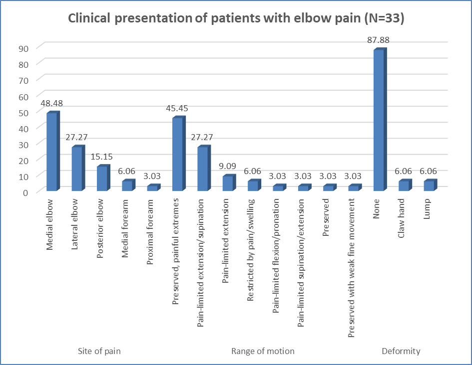
Figure 1 Clinical presentation of patients with elbow pain (N=33)
Table 3. Associated clinical findings among the study participants (N=33)
|
Finding |
n |
% |
|
Swelling |
||
|
Rare |
15 |
45.45 |
|
Minimal |
10 |
30.30 |
|
Mild |
3 |
9.09 |
|
Absent |
3 |
9.09 |
|
Obvious |
2 |
6.06 |
|
Associated symptoms |
||
|
Paresthesia in ulnar distribution |
15 |
45.45 |
|
Weak grip strength |
9 |
27.27 |
|
Weak push-off strength |
3 |
9.09 |
|
Warmth/redness |
2 |
6.06 |
|
Weak finger extension |
1 |
3.03 |
|
Tingling/numbness |
1 |
3.03 |
|
Numbness/tingling of 4th–5th digits |
1 |
3.03 |
|
Grip weakness |
1 |
3.03 |
|
Immobilization requirement |
||
|
Not required |
19 |
57.58 |
|
Splint sometimes |
10 |
30.30 |
|
Sometimes |
3 |
9.09 |
|
Rare |
1 |
3.03 |
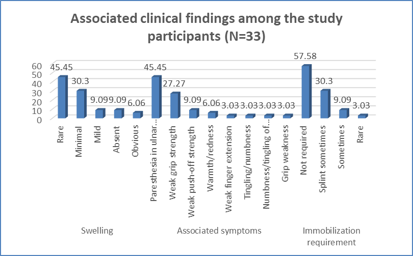
Figure 2 Associated clinical findings among the study participants (N=33)
Table 4. Distribution of clinical diagnoses according to history of trauma/fracture (N=33)
|
Clinical diagnosis |
Trauma absent, n |
Trauma present, n |
Total, n (%) |
|
Cubital tunnel syndrome |
1 |
0 |
1 (3.03) |
|
Lateral epicondylitis |
9 |
0 |
9 (27.27) |
|
Medial epicondylitis |
1 |
0 |
1 (3.03) |
|
Olecranon bursitis |
2 |
0 |
2 (6.06) |
|
Radial tunnel syndrome |
1 |
0 |
1 (3.03) |
|
Triceps tendinitis |
2 |
1 |
3 (9.09) |
|
Ulnar nerve compression neuropathy |
1 |
0 |
1 (3.03) |
|
Ulnar neuritis |
9 |
6 |
15 (45.45) |
|
Total |
26 (78.79) |
7 (21.21) |
33 (100.00) |
Table 5. Major MRI signal and sequence findings among the study participants (N=33)
|
MRI sequence |
Finding |
n |
% |
|
T2 FSPD axial |
Hyperintense nerve |
18 |
54.55 |
|
Hyperintense tendon |
13 |
39.39 |
|
|
Fluid collection |
2 |
6.06 |
|
|
T2 FSPD sagittal |
Nerve swelling |
18 |
54.55 |
|
Thickened tendon |
12 |
36.36 |
|
|
Fluid |
2 |
6.06 |
|
|
Edema |
1 |
3.03 |
|
|
T1 axial |
Isointense |
27 |
81.82 |
|
Hypointense |
4 |
12.12 |
|
|
Iso/hypointense fluid |
2 |
6.06 |
|
|
T2 STIR coronal |
Hyperintense signal |
28 |
84.85 |
|
Edema |
5 |
15.15 |
|
|
DWI |
Restricted diffusion |
26 |
78.79 |
|
Normal |
7 |
21.21 |
|
|
Post-contrast |
Nerve enhancement |
17 |
51.52 |
|
Tendon enhancement |
8 |
24.24 |
|
|
Tendon and nerve enhancement |
4 |
12.12 |
|
|
Rim enhancement |
2 |
6.06 |
|
|
Enhancement |
1 |
3.03 |
|
|
Mild enhancement |
1 |
3.03 |
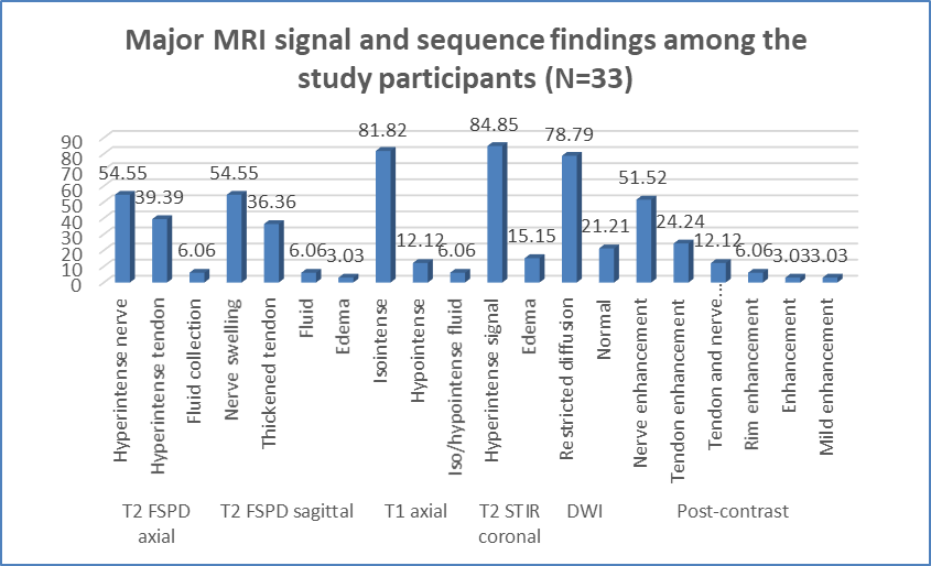
Figure 3 Major MRI signal and sequence findings among the study participants (N=33)
Table 6. MRI findings involving the ligamentous complexes and joint capsule (N=33)
|
Anatomical structure/MRI finding |
n |
% |
|
LCL complex |
||
|
Normal |
20 |
60.61 |
|
Mild annular ligament thickening |
4 |
12.12 |
|
No primary involvement |
3 |
9.09 |
|
Edema around RCL |
1 |
3.03 |
|
LUCL thickening/reactive edema |
1 |
3.03 |
|
Mild RCL thickening |
1 |
3.03 |
|
Partial-thickness RCL tear |
1 |
3.03 |
|
RCL, LUCL and annular ligament intact |
1 |
3.03 |
|
Thickened LUCL |
1 |
3.03 |
|
MCL/UCL complex |
||
|
Normal |
13 |
39.39 |
|
Mild anterior bundle thickening/posterior bundle signal increase |
7 |
21.21 |
|
Degenerative anterior bundle signal with thickened posterior bundle |
6 |
18.18 |
|
Intact |
2 |
6.06 |
|
No primary involvement |
2 |
6.06 |
|
Common flexor origin tendinosis |
1 |
3.03 |
|
Valgus instability-related MCL degeneration |
1 |
3.03 |
|
Anterior and posterior bundles intact |
1 |
3.03 |
|
Joint capsule |
||
|
Normal thickness |
10 |
30.30 |
|
Mild medial capsular thickening |
8 |
24.24 |
|
Medial capsular thickening with mild edema |
6 |
18.18 |
|
Lateral capsular thickening with edema |
5 |
15.15 |
|
Posterior capsular irritation |
3 |
9.09 |
|
Mild thickening near radiocapitellar joint |
1 |
3.03 |
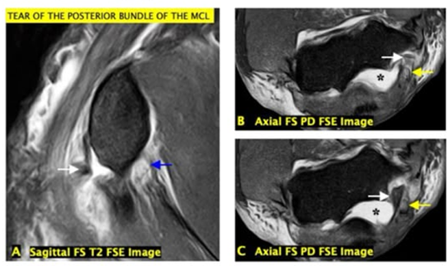
Figure 4: MRI findings of ulnar neuropathy secondary to posterior bundle MCL avulsion. (A–C) MR images demonstrate acute avulsion of the posterior bundle of the medial collateral ligament (MCL), with retraction of the osseous fragment (white arrow) causing mass effect and secondary subluxation of the ulnar nerve (yellow arrows). Associated findings include ulnohumeral incongruity with joint effusion (*) and a common flexor tendon full-thickness tear (blue arrow in A).
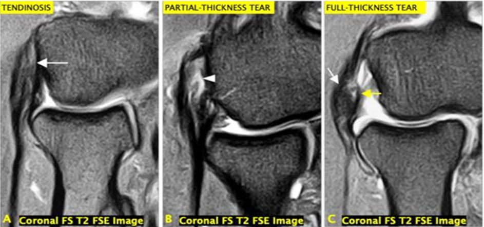
Figure 5: MRI spectrum of common extensor origin tendon injuries. Coronal fat-suppressed T2-weighted FSE images demonstrate (A) common extensor tendinosis (arrow), (B) partial-thickness tear of the common extensor tendon (arrowhead), and (C) full-thickness tear of the common extensor tendon (white arrow) with associated complete detachment of the proximal radial collateral ligament proper (yellow arrow).tear, showing focal fiber disruption with increased signal (arrowhead); and (C) full-thickness MCL tear, demonstrating complete disruption of ligament fibers with fluid signal traversing the defect (arrows).
DISCUSSION:
The present study evaluated the clinical and MRI findings in 33 patients with elbow pain. The largest proportion of patients belonged to the 31–45-year age group (39.39%), followed by 46–60 years (24.24%), with a slight male predominance (54.55%). This middle-aged distribution is comparable with Qi et al. [21], who evaluated 23 patients with lateral epicondylitis using 3-T MRI and reported a mean age of approximately 44 years. Clinically, medial elbow pain was the most common presentation (48.48%), followed by lateral elbow pain (27.27%). Ulnar-distribution paresthesia occurred in 45.45%, and weak grip strength in 27.27%. Correspondingly, ulnar neuritis was the most frequent clinical diagnosis (45.45%), followed by lateral epicondylitis (27.27%). Ayromlou et al. [22] studied 21 patients with ulnar neuropathy at the elbow and found increased ulnar nerve signal on MRI in 90.4%, nerve swelling in 42.8%, and nerve compression in 33.3%. Ulnar nerve hyperintensity demonstrated 90% sensitivity and 80% specificity for neuropathy. These observations support the prominent neural involvement observed in our patients. A history of trauma/fracture was present in only 21.21%, while 78.79% had no traumatic history. Among traumatic cases, 6/7 (85.71%) had ulnar neuritis. Thus, the overall pattern in our cohort was predominantly non-traumatic, with tendinous and neural abnormalities forming the major clinical spectrum. RI demonstrated hyperintense nerve signal in 54.55% and hyperintense tendon signal in 39.39% on T2 FSPD axial images. Nerve swelling was observed in 54.55%, while tendon thickening occurred in 36.36%. Similarly, Bäumer et al. [23] demonstrated excellent diagnostic performance of increased T2 signal for ulnar neuropathy (AUC 0.94), with qualitative MR neurography showing 83% sensitivity and 85% specificity. These findings reinforce the usefulness of T2-weighted MRI for detecting nerve pathology. Tendinous and ligamentous abnormalities were also evident. The LCL complex was normal in 60.61%, while mild annular ligament thickening occurred in 12.12%. In contrast, medial ligament abnormalities were more frequent: mild anterior bundle thickening/posterior bundle signal increase occurred in 21.21%, and degenerative anterior bundle signal with posterior bundle thickening in 18.18%. Qi et al. [21] demonstrated that elbow tendinopathy frequently represents multistructural disease; among 24 elbows with lateral epicondylitis, all had common extensor tendon abnormalities, while 22/24 (91.7%) had LUCL injury, 8/24 had RCL injury, and 3/24 had MCL injury. CET and LUCL injury severity were strongly correlated (r=0.877, p<0.01).
Capsular abnormalities were also frequent in our study, with medial capsular thickening in 24.24%, medial thickening with edema in 18.18%, and lateral thickening with edema in 15.15%. These findings further demonstrate that elbow pain may involve multiple periarticular structures rather than an isolated tendon or ligament. However, MRI abnormalities require clinical correlation. A 3-T MRI study of asymptomatic volunteers demonstrated common extensor tendon abnormalities in 37%, increasing from 10% at 35–44 years to 67% at 55–65 years. Another study identified LCL complex or common extensor tendon abnormalities in 5 of 22 asymptomatic individuals, all aged ≥65 years. Thus, the present findings highlight MRI as a valuable modality for comprehensive assessment of neural, tendinous, ligamentous, and capsular causes of elbow pain, while emphasizing the importance of correlating imaging abnormalities with clinical presentation.
CONCLUSION:
MRI provided comprehensive evaluation of the diverse causes of elbow pain by demonstrating neural, tendinous, ligamentous, and capsular abnormalities. Ulnar neuritis and lateral epicondylitis were the predominant diagnoses, with medial elbow pain and ulnar-distribution paresthesia being common clinical presentations. T2-weighted and STIR sequences were particularly useful for demonstrating nerve and tendon abnormalities. MRI also identified associated ligamentous and capsular changes that may not be evident clinically. Thus, MRI is a valuable imaging modality for accurate characterization of elbow pathology and should be interpreted in correlation with clinical findings.
LIMITATIONS
The study was limited by its small sample size and single-center, hospital-based design, which may limit the generalizability of the findings. The cross-sectional design did not permit assessment of disease progression or treatment outcomes. Histopathological, surgical, or electrophysiological confirmation of all MRI findings was not available. Larger multicenter studies with clinical follow-up and confirmatory investigations are warranted.
REFERENCES:
1. Sanders TL, Maradit Kremers H, Bryan AJ, Ransom JE, Smith J, Morrey BF, et al. The epidemiology and health care burden of tennis elbow: a population-based study. Am J Sports Med. 2015;43(5):1066-1071.
2. Kaur C, Kaur G, Bindra S, Singh P. Prevalence and risk factors of musculoskeletal disorders among housewives: a survey study. Int J Sci Healthc Res. 2024;9(2).
3. Kim H, Jang T. Epidemiology and etiology of elbow pain based on the Healthcare Bigdata Hub in Korea: a longitudinal observational study. Ewha Med J. 2023;46(4): e12.
4. Yasobant S, Mohanty S. Musculoskeletal disorders as a public health concern in India: a call for action. Physiother J Indian Assoc Physiother. 2018;12(1):46-47.
5. Sharma R, Sharma M, Sharma A, Dave J. Prevalence of elbow pain among agricultural farmers in North India: a cross-sectional study. Indian J Nat Sci. 2024;15(87).
6. Sathya D, Parasakthibala G. An overview: occupational health related musculoskeletal disorder among handloom weavers in India. Int J Creat Res Thoughts. 2025;13.
7. Bang AA, Bhojraj SY, Deshmukh M, Kalkotwar S, Joshi VR, Yarmal T, et al. Economic burden of pain in back and extremities in the adult population: a community-based estimate in rural Gadchiroli, India. J Glob Health Rep. 2021;5: e2021052.
8. Atkins N, Mukhida K. The relationship between patients' income and education and their access to pharmacological chronic pain management: a scoping review. Can J Pain. 2022;6(1):142-170.
9. Gadhavi B, Shukla Y. Prevalence of work-related musculoskeletal disorders in farmers of Gujarat. Int J Res Rev. 2019;6(11).
10. Husarik DB, Saupe N, Pfirrmann CWA, Jost B, Hodler J, Zanetti M, et al. Ligaments and plicae of the elbow: normal MR imaging variability in 60 asymptomatic subjects. Radiology. 2010;257(1):185-194.
11. Beltran LS, Bencardino JT, Beltran J. Imaging of sports ligamentous injuries of the elbow. Semin Musculoskelet Radiol. 2013;17(5):455-465.
12. Fliegel BE, Ekblad J, Varacallo MA. Anatomy, shoulder and upper limb, elbow annular ligament. In: StatPearls [Internet]. Treasure Island (FL): StatPearls Publishing; 2023.
13. Iyer RS, Thapa MM, Khanna PC, Chew FS. Pediatric bone imaging: imaging elbow trauma in children—a review of acute and chronic injuries. AJR Am J Roentgenol. 2012;198(5):1053-1068.
14. Wilps T, Kaufmann RA, Yamakawa S, Fowler JR. Elbow biomechanics: bony and dynamic stabilizers. J Hand Surg Am. 2020;45(6):528-535.
15. Liman MNP, Avva U, Ashurst JV, Butarbutar JC. Elbow trauma. In: StatPearls [Internet]. Treasure Island (FL): StatPearls Publishing; 2024.
16. Agins HJ, Chess JL, Hoekstra DV, Teitge RA. Rupture of the distal insertion of the biceps brachii tendon. Clin Orthop Relat Res. 1988;(234):34-38.
17. Acosta Batlle J, Cerezal L, López Parra MD, Alba B, Resano S, Blázquez Sánchez J, et al. The elbow: review of anatomy and common collateral ligament complex pathology using MRI. Insights Imaging. 2019;10(1):43.
18. Rosenberg ZS, Beltran J, Cheung YY, Ro SY, Green SM, Lenzo SR, et al. The elbow: MR features of nerve disorders. Radiology. 1993;188(1):235-240.
19. Kijowski R, Tuite M, Sanford M. Magnetic resonance imaging of the elbow. Part II: abnormalities of the ligaments, tendons, and nerves. Skeletal Radiol. 2005;34(1):1-18.
20. Walz DM, Newman JS, Konin GP, Ross G. Epicondylitis: pathogenesis, imaging, and treatment. Radiographics. 2010;30(1):167-184.
21. Qi L, Zhu ZF, Li F, Wang RF. MR imaging of patients with lateral epicondylitis of the elbow: is the common extensor tendon an isolated lesion? PLoS One. 2013;8(11):e79498.
22. Ayromlou H, Tarzamni MK, Daghighi MH, Pezeshki MZ, Yazdchi M, Sadeghi-Hokmabadi E, et al. Diagnostic value of ultrasonography and magnetic resonance imaging in ulnar neuropathy at the elbow. ISRN Neurol. 2012; 2012:491892.
23. Bäumer P, Dombert T, Staub F, Kaestel T, Bartsch AJ, Heiland S, et al. Ulnar neuropathy at the elbow: MR neurography—nerve T2 signal increase and caliber. Radiology. 2011;260(1):199-206.