Bronchial and Non-Bronchial Systemic Artery Embolization for Life-Threatening Hemoptysis: Single-Center Study.
- Rohit Yadav , Third-Year Resident, Department of Radiodiagnosis, Geetanjali Medical College and Hospital, Udaipur, Rajasthan, India.
- Brijesh Kumar Soni , Professor, Department of Radiodiagnosis, Geetanjali Medical College and Hospital, Udaipur, Rajasthan, India.
- Ravinder Kumar Kundu , Professor and Head, Department of Radiodiagnosis, Geetanjali Medical College and Hospital, Udaipur, Rajasthan, India.
Article Information:
Abstract:
Background: Life-threatening hemoptysis is a potentially fatal respiratory emergency requiring rapid identification and control of the bleeding source. Bronchial artery embolization (BAE), with embolization of non-bronchial systemic arteries when indicated, has emerged as an effective minimally invasive treatment. Objective: To evaluate the effectiveness and safety of bronchial and non-bronchial systemic artery embolization in patients presenting with life-threatening hemoptysis. Materials and Methods: This hospital-based observational study included 14 patients with life-threatening hemoptysis who underwent arterial embolization at the Department of Radio-Diagnosis, Geetanjali Medical College and Hospital, Udaipur. Clinical, radiological, angiographic, procedural, and follow-up data were recorded. Technical and clinical success, procedure-related complications, and rebleeding at three months were assessed. Data were summarized using descriptive statistics. Results: Of 14 patients, 9 (64.3%) were males and 5 (35.7%) were females. Tuberculosis was the predominant etiology (71.4%), followed by COPD (21.4%) and silicosis (7.1%). The mean baseline hemoglobin was 9.87 ± 1.34 g/dL. Bronchial arteries were the source of bleeding in 78.6% of patients, while internal mammary artery involvement was observed in 21.4%. PVA particles were used for embolization in all patients. Technical and immediate clinical success were achieved in 100% of cases, with no documented procedure-related complications. At three months, 13 patients (92.9%) remained free from rebleeding, while one (7.1%) experienced recurrence. Conclusion: Bronchial and non-bronchial systemic artery embolization provided excellent immediate control of life-threatening hemoptysis with a favorable short-term safety profile and low three-month recurrence.
Keywords:
Article :
INTRODUCTION:
Hemoptysis, defined as expectoration of blood originating from the lower respiratory tract, is an important clinical presentation ranging from blood-streaked sputum to life-threatening pulmonary hemorrhage[1]. Although most episodes are mild and self-limiting, massive or life-threatening hemoptysis constitutes a medical emergency because rapid accumulation of blood within the airways can result in airway obstruction, hypoxemia, respiratory failure, and death. Mortality in severe untreated hemoptysis has historically been reported to be high, with death occurring predominantly from asphyxiation rather than exsanguination [2]. Contemporary definitions therefore emphasize the physiological consequences of bleeding rather than an arbitrary volume threshold [3].The etiology of hemoptysis varies according to geographical region and underlying disease patterns. Bronchiectasis, pulmonary malignancy, and chronic inflammatory lung diseases are important causes worldwide, whereas pulmonary tuberculosis and its sequelae continue to constitute a major burden in developing countries, including India [4,5]. Other important causes include aspergillosis, pneumonia, pulmonary vascular abnormalities, and chronic obstructive pulmonary disease [6,7].
Chronic infection and inflammation promote neovascularization and hypertrophy of systemic arteries, producing fragile abnormal vessels susceptible to rupture and recurrent bleeding [8].The lungs have a dual arterial supply comprising the pulmonary and bronchial circulations. Although bronchial arteries account for only a small proportion of total pulmonary blood flow, they arise from the high-pressure systemic circulation and are responsible for approximately 90% of cases of life-threatening hemoptysis [9,10]. In chronic pulmonary diseases, bronchial arteries become enlarged, tortuous, and hypervascular. Additionally, non-bronchial systemic arteries, including intercostal, internal mammary, inferior phrenic, subclavian, and other systemic branches, may develop collateral supply to diseased lung through pleural adhesions and chronic inflammatory pathways [11]. Failure to recognize these vessels is an important cause of persistent or recurrent hemoptysis following treatment [12].
Imaging plays a central role in determining both the underlying etiology and vascular source of hemoptysis. Although chest radiography remains a useful initial investigation, computed tomography (CT) provides substantially greater information regarding pulmonary parenchymal abnormalities and the site and cause of bleeding [13]. CT bronchial angiography further enables detailed evaluation of hypertrophied bronchial arteries, ectopic bronchial arteries, and non-bronchial systemic collaterals and facilitates pre-procedural planning for embolization [14,15].Bronchial artery embolization (BAE), introduced by Rémy and colleagues, has become the preferred minimally invasive treatment for controlling life-threatening and recurrent hemoptysis [16]. Selective catheterization followed by embolization of abnormal vessels using polyvinyl alcohol particles, microspheres, coils, gelatin sponge, or liquid embolic agents provides rapid hemorrhage control while preserving functioning lung parenchyma . Contemporary studies have demonstrated high technical and immediate clinical success rates with BAE, with considerably lower morbidity than emergency surgical intervention [16]. Despite excellent initial control, recurrent hemoptysis remains an important limitation of embolization. Recurrence may result from recanalization, incomplete embolization, progression of the underlying pulmonary disease, or recruitment of previously unrecognized bronchial or non-bronchial systemic collaterals . Comprehensive identification and embolization of all pathological arterial supply may therefore improve sustained clinical success.
Hence, the present study was designedto evaluate the role of bronchial and non-bronchial systemic artery embolization in patients presenting with hemoptysis, with particular emphasis on vascular anatomy, angiographic findings, technical and clinical success, complications, and recurrence following embolization.
MATERIALS AND METHODS:
This hospital-based observational study was conducted in the Department of Radio-Diagnosis, Geetanjali Medical College and Hospital, Udaipur. The study evaluated the role of bronchial and non-bronchial systemic artery embolization in the management of patients presenting with life-threatening hemoptysis.The study was initiated after obtaining approval from the Institutional Ethics Committee and Institutional Research Review Committee. Written informed consent was obtained from all participants before enrolment and intervention.
Study Population
Patients presenting with massive or life-threatening hemoptysis who underwent bronchial artery embolization (BAE) during the study period were assessed for eligibility. All patients fulfilling the predefined selection criteria were included.
Sample Size and Sampling Technique
The study was time-bound, and a total of 14 patients fulfilling the eligibility criteria were enrolled. Participants were selected using a purposive sampling technique.
Inclusion Criteria
Patients were included if they:
· Presented with massive/life-threatening hemoptysis.
· Underwent bronchial artery embolization for control of hemoptysis.
Exclusion Criteria
Patients were excluded if they had:
· Pregnancy.
· Known allergy to iodinated contrast media.
· Chronic kidney disease.
· A common origin or close anatomical relationship of the target bronchial artery with a spinal artery that precluded safe embolization.
Clinical Evaluation
At presentation, all patients underwent detailed clinical assessment. A comprehensive history was obtained regarding the onset, duration, frequency, and severity of hemoptysis. Previous episodes of hemoptysis and histories of pulmonary tuberculosis, bronchiectasis, chronic obstructive pulmonary disease, fungal infection, lung malignancy, smoking, previous thoracic surgery, and treatment for respiratory illnesses were recorded.
Associated symptoms, including fever, cough, sputum production, weight loss, chest pain, and breathlessness, were documented. General physical examination included measurement of pulse rate, blood pressure, respiratory rate, oxygen saturation, and temperature, along with assessment of hemodynamic stability. A detailed respiratory system examination was performed to identify clinical evidence of underlying pulmonary disease.
Laboratory Investigations
Baseline laboratory investigations were performed before embolization. These included complete blood count, hemoglobin concentration, platelet count, coagulation profile, renal function tests, liver function tests, and blood grouping. Additional biochemical investigations were performed whenever clinically indicated.These investigations were used to assess the general clinical condition of the patients, identify abnormalities requiring correction, and determine suitability for angiography and embolization.
Imaging Evaluation
All patients underwent radiological evaluation to determine the underlying etiology, localize the probable site of bleeding, and identify abnormal vascular supply.
Chest Radiography
Chest radiography was performed as the initial imaging investigation. Radiographs were evaluated for pulmonary abnormalities such as cavitary lesions, consolidation, bronchiectatic changes, pulmonary masses, fibrosis, and pleural abnormalities.
Contrast-Enhanced Computed Tomography
Contrast-enhanced computed tomography (CECT) of the thorax was performed to characterize the underlying pulmonary pathology and localize the probable source of hemoptysis. Particular attention was given to tuberculosis and post-tubercular changes, bronchiectasis, cavitary lesions, aspergillosis, pulmonary masses, and other structural abnormalities.
CT Angiography
CT angiography was performed whenever indicated to evaluate the bronchial and systemic arterial anatomy. The images were assessed for hypertrophied bronchial arteries, ectopic bronchial arteries, non-bronchial systemic collateral vessels, vascular abnormalities, and the anatomical distribution of the underlying pulmonary disease. CT angiographic findings were used for planning subsequent catheter angiography and embolization.
Angiographic Evaluation
Following clinical and radiological evaluation, patients underwent catheter angiography in the angiography suite under strict aseptic precautions.Percutaneous arterial access was obtained through the common femoral artery under local anesthesia using the Seldinger technique. A vascular sheath was introduced, followed by thoracic aortography and selective bronchial artery angiography.
Angiographic features considered suggestive of abnormal or culprit vessels included:
· Bronchial artery hypertrophy and enlargement
· Arterial tortuosity
· Hypervascularity
· Neovascularization
· Systemic-to-pulmonary shunting
· Aneurysmal dilatation
· Contrast extravasation
Whenever non-bronchial systemic arterial supply was suspected on CT angiography or catheter angiography, selective angiography of potential collateral vessels was performed. These included the intercostal arteries, internal mammary arteries, inferior phrenic arteries, lateral thoracic arteries, subclavian artery branches, and other relevant systemic collateral vessels.
Embolization Procedure
After identification of the culprit artery, selective or super-selective catheterization was performed using appropriate angiographic catheters and microcatheters.
Before embolization, angiographic images were carefully evaluated to exclude significant spinal arterial supply arising from or in close proximity to the target vessel. Embolization was subsequently performed under fluoroscopic guidance.The target vessels included abnormal bronchial arteries and, whenever identified, non-bronchial systemic arteries contributing to the bleeding.
Embolic Materials
The embolic agents used during the procedures included:
· Polyvinyl alcohol (PVA) particles, 500–700 µm
· Gelatin sponge (Gelfoam)
· Coils
· N-butyl cyanoacrylate (glue), whenever indicated
The choice of embolic material was based on angiographic findings, vessel characteristics, vascular anatomy, and the interventional radiologist's discretion.Embolic material was administered carefully under fluoroscopic guidance until satisfactory occlusion of the abnormal vascular supply was achieved. Post-embolization angiography was subsequently performed to document reduction or disappearance of abnormal vascularity and satisfactory occlusion of the target vessel.
Assessment of Procedural Success
Technical Success
Technical success was defined as successful selective or super-selective catheterization and embolization of the identified abnormal vessel(s), with post-embolization angiography demonstrating satisfactory occlusion of the pathological vascular supply.
Clinical Success
Clinical success was defined as complete cessation or a clinically significant reduction in hemoptysis following embolization without the requirement for immediate additional intervention.
Post-Procedural Care
Following embolization, all patients were monitored for hemodynamic stability and control of hemoptysis. Vital parameters were monitored, and appropriate supportive treatment was provided whenever required.Patients were specifically assessed for procedure-related adverse events, including chest pain, dysphagia, fever, neurological deficits, vascular access-site complications, non-target embolization, and other immediate or delayed complications.
Outcome Measures
The principal outcome measures assessed were:
· Technical success of embolization
· Clinical success of embolization
· Procedure-related complications
· Recurrence or rebleeding following embolization
· Requirement for repeat intervention
· Clinical outcome at 3-month follow-up
Follow-Up
All patients were followed for three months after embolization. Follow-up information was obtained through outpatient visits, hospital records, or telephonic communication, whenever applicable.During follow-up, patients were assessed for recurrence of hemoptysis, procedure-related complications, requirement for repeat embolization or other interventions, and overall clinical outcome.
Data Collection
Demographic, clinical, laboratory, radiological, angiographic, procedural, and follow-up information was recorded using a structured data collection form.The variables collected included patient demographics, clinical presentation, underlying etiology of hemoptysis, CT findings, angiographic abnormalities, bronchial and non-bronchial systemic arteries involved, vessels embolized, embolic materials used, technical and clinical success, complications, recurrence of hemoptysis, repeat interventions, and 3-month clinical outcomes.
Statistical Analysis
The collected data were entered into Microsoft Excel and analyzed using descriptive statistical methods. Continuous variables were summarized as mean ± standard deviation (SD), while categorical variables were presented as frequencies and percentages. The findings were presented using appropriate tables, charts, and graphical representations.
RESULTS:
A total of 14 patients with hemoptysis were included. Most patients were middle-aged, with 28.6% each belonging to the 41–50 and 51–60-year age groups. Males constituted 64.3% of the study population, while females accounted for 35.7% (Table 1).Tuberculosis was the predominant etiology of hemoptysis, observed in 10 (71.4%) patients, followed by COPD in 3 (21.4%) and silicosis in 1 (7.1%). All patients were hemodynamically stable at presentation. The mean baseline hemoglobin level was 9.87 ± 1.34 g/dL (Table 2).Right-sided involvement was more frequent, occurring in 8 (57.1%) patients, compared with left-sided involvement in 6 (42.9%); no patient had bilateral involvement. The bronchial artery was the principal arterial source of bleeding in 11 (78.6%) patients, whereas a non-bronchial systemic source, specifically the internal mammary artery, was identified in 3 (21.4%) patients (Table 3; Figure 1).Polyvinyl alcohol (PVA) particles were used as the embolic material in all 14 (100%) procedures; none required Gelfoam, coils, or glue. Technical success was achieved in all patients (100%) (Table 4; Figure 2). Clinical success was also achieved in all 14 (100%) patients, with no documented procedure-related complications such as chest pain, dysphagia, or fever (Table 5).At the three-month follow-up, 13 (92.9%) patients remained free from recurrent hemoptysis, while rebleeding occurred in only 1 (7.1%) patient (Table 6; Figure 3). Thus, embolization demonstrated high immediate technical and clinical success, with sustained control of hemoptysis in the majority of patients during the three-month follow-up period.
Table 1. Demographic characteristics of the study participants (N=14)
|
Characteristic |
n (%) |
|
Age group (years) |
|
|
≤30 |
1 (7.1) |
|
31–40 |
1 (7.1) |
|
41–50 |
4 (28.6) |
|
51–60 |
4 (28.6) |
|
61–70 |
3 (21.4) |
|
>70 |
1 (7.1) |
|
Gender |
|
|
Male |
9 (64.3) |
|
Female |
5 (35.7) |
Table 2. Clinical characteristics and etiology of hemoptysis (N=14)
|
Characteristic |
n (%) / Mean ± SD |
|
Etiology |
|
|
Tuberculosis |
10 (71.4) |
|
COPD |
3 (21.4) |
|
Silicosis |
1 (7.1) |
|
Hemodynamic status |
|
|
Stable |
14 (100.0) |
|
Unstable |
0 (0.0) |
|
Baseline hemoglobin (g/dL) |
9.87 ± 1.34 |
Table 3. Distribution of side and arterial source of hemoptysis (N=14)
|
Variable |
n (%) |
|
Side of involvement |
|
|
Right |
8 (57.1) |
|
Left |
6 (42.9) |
|
Bilateral |
0 (0.0) |
|
Arterial source |
|
|
Bronchial artery |
11 (78.6) |
|
Non-bronchial systemic artery (internal mammary artery) |
3 (21.4) |
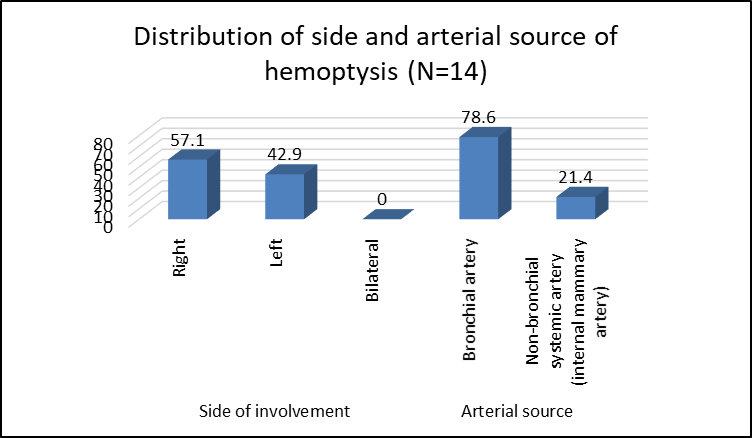
Figure 1 Distribution of side and arterial source of hemoptysis (N=14)
Table 4. Embolic material and technical outcome of embolization (N=14)
|
Procedural characteristic |
n (%) |
|
Embolic material |
|
|
PVA particles |
14 (100.0) |
|
Gelfoam |
0 (0.0) |
|
Coil |
0 (0.0) |
|
Glue |
0 (0.0) |
|
Technical success |
|
|
Successful |
14 (100.0) |
|
Unsuccessful |
0 (0.0) |
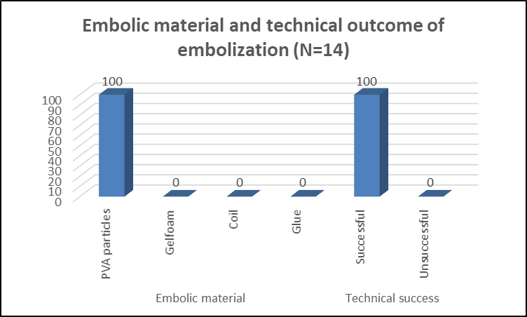
Figure 2 Embolic material and technical outcome of embolization (N=14)
Table 5. Clinical success and procedure-related complications (N=14)
|
Outcome |
n (%) |
|
Clinical success |
|
|
Successful |
14 (100.0) |
|
Unsuccessful |
0 (0.0) |
|
Procedure-related complications |
|
|
Chest pain |
0 (0.0) |
|
Dysphagia |
0 (0.0) |
|
Fever |
0 (0.0) |
|
No documented complication |
14 (100.0) |
Table 6. Three-month follow-up outcome after embolization (N=14)
|
Follow-up outcome |
n (%) |
|
No rebleeding |
13 (92.9) |
|
Rebleeding |
1 (7.1) |
|
Total |
14 (100.0) |
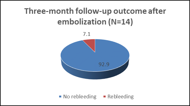
Figure 3 Three-month follow-up outcome after embolization (N=14)
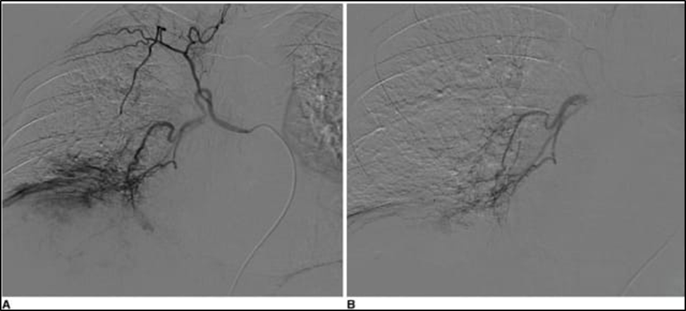
Fig. 4 Digital subtraction angiography before and after bronchial artery embolization. (A) Pre-embolization angiogram demonstrates abnormal hypertrophied bronchial arterial vascularity supplying the bleeding site. (B) Post-embolization angiogram shows marked reduction of abnormal vascularity with successful occlusion of the culprit vessel.
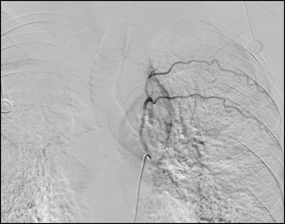
Fig. 5 Lt hypertrophied bronchial artery DSA image
DISCUSSION:
The present study evaluated the effectiveness and safety of bronchial and non-bronchial systemic artery embolization in 14 patients with life-threatening hemoptysis. Most patients were middle-aged, with 28.6% each belonging to the 41–50 and 51–60-year age groups, and males predominated (64.3%). Similarly, Bhalla et al.[13] studied 334 patients undergoing bronchial artery embolization (BAE), of whom 76.3% were males and 23.7% were females, with an age range of 5–81 years. Tuberculosis was the predominant etiology in our study, accounting for 10/14 (71.4%) patients, followed by COPD in 21.4% and silicosis in 7.1%. This finding was comparable to the Indian study by Bhalla et al.[13], in which post-tubercular changes constituted the predominant underlying pathology in 248/334 (74.3%) patients. Panda et al.[12], in their systematic review of 22 studies, also identified tuberculosis and its sequelae among the important indications for BAE, particularly in regions with a high burden of tuberculosis. Regarding the distribution of disease, 57.1% of our patients had right-sided and 42.9% had left-sided involvement, with no bilateral involvement.
Bhalla et al.[13] reported right-sided disease in 101/334 (30.2%), left-sided disease in 59/334 (17.7%), and bilateral involvement in 174/334 (52.1%) patients. The absence of bilateral disease in our series may be attributable to the relatively small sample size. In our study, the bronchial artery was the culprit arterial source in 11/14 (78.6%) patients, whereas non-bronchial systemic arterial supply through the internal mammary artery was observed in 3/14 (21.4%). The importance of non-bronchial systemic collateral vessels has also been emphasized in published literature. The CIRSE standards identify internal mammary arterial supply and recruitment of non-bronchial systemic collaterals as important considerations, particularly in recurrent hemoptysis. PVA particles were used in all 14 (100%) patients in our study. This was similar to Bhalla et al.[13], who used PVA in all 334 patients, although Gelfoam was additionally used in patients with significant shunting. PVA particles remain among the established embolic materials used for BAE. The technical success rate in our study was 100%, and immediate clinical success was also achieved in all 14 patients (100%). These findings were consistent with the CIRSE standards, which reported technical success rates of 90–100% and clinical success within 24 hours of 82–100%. Furthermore, the meta-analysis by Kettenbach et al.[17], involving 2,511 patients, demonstrated a pooled technical success rate of 99.9% and immediate bleeding control rate of 99.5%. A more recent meta-analysis involving 6,032 patients reported pooled technical and clinical success rates of 97.2% and 93.2%, respectively.
No procedure-related complications were documented in our patients. This finding supports the favorable safety profile of BAE, although the absence of complications should be interpreted cautiously because only 14 patients were studied. Panda et al.[12] reported a median major complication rate of only 0.1%, while the recent meta-analysis of 6,032 patients similarly reported a pooled major complication rate of 0.1%. At the 3-month follow-up, 13/14 patients (92.9%) remained free from rebleeding, while only one patient (7.1%) experienced recurrence. Panda et al.[12] reported recurrence rates ranging from approximately 10% to 57%, whereas Kettenbach et al.[17] found a pooled recurrence rate of 23.7%. The relatively lower recurrence of 7.1% in our study may be related to the short three-month follow-up period and small sample size. Nevertheless, the high immediate success, absence of documented complications, and low short-term recurrence observed in our series support bronchial and non-bronchial systemic artery embolization as an effective and relatively safe treatment for life-threatening hemoptysis.
CONCLUSION:
Bronchial and non-bronchial systemic artery embolization was highly effective in achieving immediate control of life-threatening hemoptysis, with 100% technical and clinical success in the present study. Tuberculosis was the predominant underlying etiology, and bronchial arteries were the most common source of bleeding. No procedure-related complications were observed, indicating a favorable short-term safety profile. At three months, 92.9% of patients remained free from rebleeding, supporting embolization as an effective minimally invasive treatment for life-threatening hemoptysis.
LIMITATIONS
The study was limited by its small sample size and single-center observational design, which may restrict the generalizability of the findings. The follow-up period of three months was relatively short and may not adequately capture late recurrence of hemoptysis. Larger multicenter studies with longer follow-up are required to confirm the long-term effectiveness and safety of embolization.
REFERENCES:
1. Ashraf O. Hemoptysis, a developing world perspective. BMC Pulm Med. 2006;6:1.
2. Hirshberg B, Biran I, Glazer M, Kramer MR. Hemoptysis: etiology, evaluation, and outcome in a tertiary referral hospital. Chest. 1997;112(2):440-444.
3. Ibrahim WH. Massive haemoptysis: the definition should be revised. Eur Respir J. 2008;32(4):1131-1132.
4. Larici AR, Franchi P, Occhipinti M, Contegiacomo A, del Ciello A, Calandriello L, et al. Diagnosis and management of hemoptysis. Diagn Interv Radiol. 2014;20(4):299-309.
5. Radchenko C, Alraiyes AH, Shojaee S. A systematic approach to the management of massive hemoptysis. J Thorac Dis. 2017;9(Suppl 10):S1069-S1086.
6. Davidson K, Shojaee S. Managing massive hemoptysis. Chest. 2020;157(1):77-88.
7. Issaka RB, Oommen S. Contemporary management of massive hemoptysis. Curr Opin Pulm Med. 2021;27(3):166-172.
8. Cahill BC, Ingbar DH. Massive hemoptysis: assessment and management. Clin Chest Med. 1994;15(1):147-167.
9. Ong TH, Eng P. Massive hemoptysis requiring intensive care. Intensive Care Med. 2003;29(3):317-320.
10. Corey R, Hla KM. Major and massive hemoptysis: reassessment of conservative management. Am J Med Sci. 1987;294(5):301-309.
11. Enting D, van der Werf TS, Prins TR, Zijlstra JG, Ligtenberg JJM, Tulleken JE, et al. Massive haemoptysis: primary care, diagnosis and treatment. Ned Tijdschr Geneeskd. 2004;148:1582-1586.
12. Panda A, Bhalla AS, Goyal A. Bronchial artery embolization in hemoptysis: a systematic review. Diagn Interv Radiol. 2017;23(4):307-317.
13. Bhalla A, Kandasamy D, Veedu P, Mohan A, Gamanagatti S. A retrospective analysis of 334 cases of hemoptysis treated by bronchial artery embolization. Oman Med J. 2015;30(2):119-128.
14. Takeda K, Kawashima M, Masuda K, Matsui H, Shinohara S, Kinoshita T, et al. Long-term outcomes of bronchial artery embolization for patients with bronchiectasis. Respiration. 2020;99:961-969.
15. Lee SH, Lee JH, Chang JH, Kim YS, Jeong BH, Shin B, et al. Hemoptysis requiring bronchial artery embolization in patients with nontuberculous mycobacterial lung disease. BMC Pulm Med. 2019;19:117.
16. Han K, Yoon KW, Kim JH, Kim GM. Bronchial artery embolization for hemoptysis in primary lung cancer. J Vasc Interv Radiol. 2019;30(3):428-434.
17. Kettenbach J, Ittrich H, Gaubert JY, Gebauer B, Vos JA. CIRSE Standards of Practice on Bronchial Artery Embolisation. Cardiovasc Intervent Radiol. 2022;45(6):721-732.