Flexible Bronchoscopy in Children: A Prospective Study.
- Abdul Samad Hashmi , Postgraduate Resident, Department of Pediatrics, Government Medical College, Srinagar, Jammu and Kashmir, India.
- Sheikh Quyoom Hussain , Assistant Professor, Department of Pediatrics, Government Medical College, Srinagar, Jammu and Kashmir, India.
- Khurshid Ahmad Wani , Professor, Department of Pediatrics, Government Medical College Srinagar, Jammu and Kashmir, India.
- Parvez Ahmad Dar , Professor, Department of Pediatrics, Government Medical College Srinagar, Jammu and Kashmir, India.
- Sheikh Mushtaq ahmad , Professor, Department of Pediatrics, Government Medical College Srinagar, Jammu and Kashmir, India.
Article Information:
Abstract:
Background: Respiratory diseases are a leading cause of morbidity and mortality in children, particularly in developing countries. Children with persistent, recurrent, or unexplained respiratory symptoms pose significant diagnostic challenges. Flexible bronchoscopy (FB) has emerged as an indispensable tool for direct airway visualization and microbiological sampling. However, prospective data from the Kashmir valley on the clinical profile and bronchoscopic findings in pediatric patients remain scarce. Material and Methods: This prospective hospital-based observational study was conducted over 18 months (September 2024–February 2026) at the 500-bedded Children Hospital, Government Medical College Srinagar. A total of 125 pediatric patients who underwent flexible bronchoscopy were enrolled. Clinical features, radiological findings, bronchoscopic findings, bronchoalveolar lavage (BAL) results, and procedural complications were systematically documented and analyzed using SPSS version 22. Results: The mean age was 4.2 ± 4.1 years; toddlers (1–3 years) were the largest group (30.4%). Male predominance was noted (59.2%). Persistent/recurrent pneumonia was the leading indication (25.6%), followed by chronic cough (21.6%), severe respiratory distress (18.4%), and stridor (17.6%). Chest radiograph showed consolidation in 38.4%. Bronchoscopy was abnormal in 71.2% of patients; foreign body aspiration was the most common specific finding (24.0%), followed by thick secretions/inflammation (16.0%) and laryngomalacia (12.8%). The overall specific diagnostic yield was 55.2%. BAL was performed in 16.0% of patients; culture positivity was 15.0%. The complication rate was 0.8% (one episode of transient desaturation). Conclusion: Flexible bronchoscopy is a safe, high-yield diagnostic modality in pediatric respiratory medicine. Foreign body aspiration and airway anomalies are the most frequent abnormal findings, especially in younger children. Early and judicious use of FB significantly improves diagnostic accuracy and facilitates timely management.
Keywords:
Article :
INTRODUCTION:
Respiratory diseases constitute one of the most significant causes of morbidity and mortality among the pediatric population worldwide, particularly in developing nations where infectious etiologies predominate.[1] Acute lower respiratory tract infections alone account for nearly two million deaths annually in children under five years of age and remain the single largest cause of childhood mortality globally.[2] In India, respiratory illnesses contribute substantially to pediatric hospital admissions, placing an enormous burden on the healthcare system.[3]
Children presenting with persistent, recurrent, or unexplained respiratory symptoms — including chronic cough, recurrent pneumonia, stridor, unexplained wheezing, or non-resolving radiological opacities — often pose considerable diagnostic challenges.[4] Conventional modalities including clinical examination, chest radiography, and computed tomography, while valuable, frequently fail to provide a definitive etiological diagnosis.[5] This diagnostic gap necessitates the use of advanced investigative procedures that allow direct visualization of the airways, of which flexible bronchoscopy (FB) has emerged as the most important.[6]
Bronchoscopy is defined as the endoscopic examination of the tracheobronchial tree, permitting direct visualization of the airway lumen from the larynx through the segmental and subsegmental bronchi.[7] The modern era of flexible bronchoscopy began in 1966 with Ikeda's development of the flexible fiberoptic bronchoscope.[10] Adaptation for pediatric use was pioneered by Wood and colleagues in the late 1970s, and subsequent miniaturization has now made the procedure feasible even in premature neonates.[12,13]
Diagnostic indications for flexible bronchoscopy in children include evaluation of stridor, persistent or recurrent wheezing, chronic cough, recurrent pneumonia, persistent atelectasis, hemoptysis, suspected foreign body aspiration, congenital airway anomalies, and microbiological sampling in immunocompromised patients.[18-20] Beyond diagnosis, flexible bronchoscopy serves therapeutic functions including removal of secretions, foreign body extraction, balloon dilatation of stenosis, and selective bronchial lavage.[17,18]
The diagnostic yield of flexible bronchoscopy in children consistently ranges from 70% to 89.3% in published literature, with alterations in clinical management reported in 23%–58% of cases.[22,23] The procedure carries an excellent safety record, with major complication rates below 1.7% in the largest prospective pediatric series.[21] Despite the growing utilization of pediatric flexible bronchoscopy in India, prospective data from the Kashmir valley on its comprehensive clinical and investigative profile remain scarce. The present study was therefore undertaken at the 500-bedded Children Hospital, Government Medical College Srinagar — the premier pediatric referral center in the Kashmir valley — to address this gap.
MATERIALS AND METHODS:
Study Design and Setting
This was a prospective hospital-based observational study conducted in the Department of Pediatrics, Government Medical College Srinagar, at a 500-bedded tertiary care children's hospital, Bemina, Srinagar, Jammu and Kashmir, India. The study was carried out over a period of 18 months, from September 2024 to February 2026, after obtaining prior ethical clearance from the Institutional Ethics Committee of Government Medical College Srinagar.
Study Population
All pediatric patients referred for flexible bronchoscopy during the study period and fulfilling the inclusion criteria were enrolled. Children were excluded if they were hemodynamically unstable, had bleeding diathesis or coagulation disorders, or if parents or guardians did not provide written informed consent. A total of 125 children were included.
Data Collection and Procedure
A comprehensive history emphasizing respiratory symptoms was obtained from caregivers. Thorough physical examination and anthropometric measurements were recorded in a pre-designed proforma. Baseline investigations included complete blood count, chest radiography, and CT/MRI of the chest in selected cases.
Flexible bronchoscopy was performed under appropriate monitoring and aseptic precautions in the bronchoscopy suite, PICU, or operation theatre. A pediatric flexible fiberoptic or video bronchoscope of appropriate size was introduced via the transnasal or transoral route. Standard pre-procedure fasting guidelines were followed. Continuous monitoring of heart rate, respiratory rate, oxygen saturation, and blood pressure was maintained. Sedation was administered using intravenous midazolam in weight-based doses, with topical 1–2% lignocaine for local anaesthesia. Supplemental oxygen was provided as required.
Bronchoalveolar lavage (BAL) was performed in clinically indicated cases by instilling sterile normal saline into the relevant bronchopulmonary segment, with subsequent gentle aspiration. BAL specimens were sent immediately for Gram staining, culture and sensitivity, and CBNAAT where indicated. Bronchoscopic findings, BAL results, and any procedural complications were systematically documented.
Statistical Analysis
Data were entered into Microsoft Excel and analyzed using SPSS version 22. Descriptive statistics were reported as frequencies, percentages, mean, and standard deviation. The chi-square test was used for categorical variables, Fisher's exact test where expected cell counts were below 5, and the independent t-test for normally distributed continuous variables. Univariate analysis assessed the influence of different variables on outcomes. A p-value <0.05 was considered statistically significant.
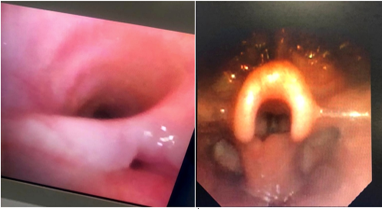
Tracheoesophageal fistula. Laryngomalacia.
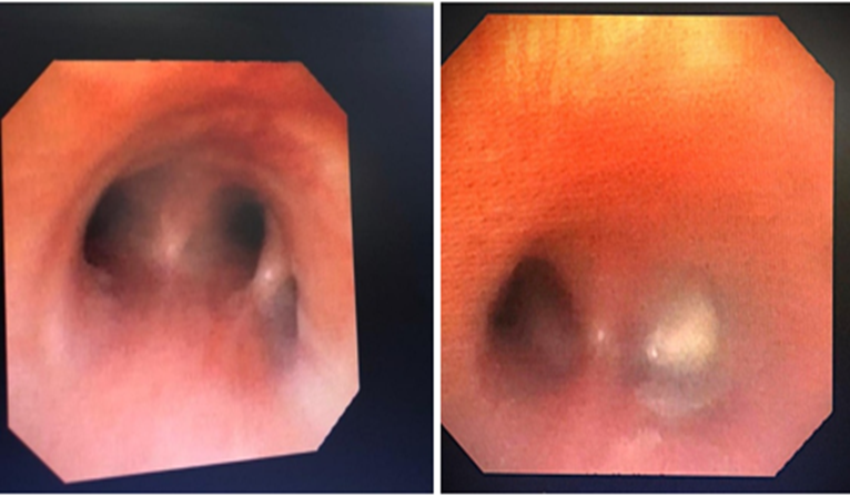
Tracheal bronchus( pig bronchus). Foreign body in right bronchus.
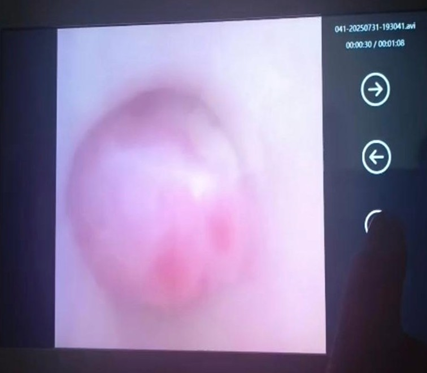
Carcinoid tumour
RESULTS:
Demographic and Clinical Profile
A total of 125 children underwent flexible bronchoscopy during the study period. The age distribution is shown in Table 1. The largest group comprised toddlers aged 1–3 years (30.4%), followed by infants aged 1–12 months (20.8%). Overall, 62.4% of patients were below three years of age. A male predominance was observed, with 74 males (59.2%) and 51 females (40.8%).
Table 1 - Age Distribution of Patients (n = 125)
|
Age Group |
Frequency |
Percentage (%) |
|
Neonates (<1 month) |
14 |
11.2 |
|
Infants (1–12 months) |
26 |
20.8 |
|
Toddlers (1–3 years) |
38 |
30.4 |
|
Preschool (3–6 years) |
17 |
13.6 |
|
School age (6–12 years) |
15 |
12.0 |
|
Adolescents (>12 years) |
15 |
12.0 |
|
Total |
125 |
100 |
The most common indication for bronchoscopy was persistent or recurrent pneumonia (25.6%), followed by persistent or chronic cough (21.6%), severe respiratory distress (18.4%), and noisy breathing or stridor (17.6%). Hemoptysis accounted for 10.4% and suspected foreign body aspiration for 6.4% of cases (Table 2). A statistically significant age-related variation in clinical indication was observed (p<0.05).
Table 2 – Clinical Indications for Bronchoscopy.
|
Indication |
Frequency |
Percentage (%) |
|
Persistent/Recurrent Pneumonia |
32 |
25.6 |
|
Persistent/Chronic Cough |
27 |
21.6 |
|
Severe Respiratory Distress |
23 |
18.4 |
|
Noisy Breathing/Stridor |
22 |
17.6 |
|
Hemoptysis |
13 |
10.4 |
|
Suspected Foreign Body |
8 |
6.4 |
|
Total |
125 |
100 |
Radiological Findings
Chest radiograph findings are shown in Table 3. Consolidation was the most common finding (38.4%), followed by hyperinflation (23.2%). Normal chest X-ray was seen in 21.6% of children despite persistent symptoms, illustrating the limitations of conventional radiology alone. CT or MRI was not performed in 67.2% of patients, reflecting its selective use; among those imaged, consolidation or collapse was found in 16.0%.
Table 3 – Radiological Findings on Chest X-ray
|
Finding |
Frequency |
Percentage (%) |
|
Consolidation |
48 |
38.4 |
|
Hyperinflation |
29 |
23.2 |
|
Normal |
27 |
21.6 |
|
Collapse |
21 |
16.8 |
|
Total |
125 |
100 |
Bronchoalveolar Lavage Results
BAL was performed selectively in 20 children (16.0%), predominantly in those with persistent or recurrent pneumonia (56.3% of such cases; p<0.05). Of 20 BAL specimens, 17 (85.0%) were culture-negative and 3 (15.0%) yielded positive results: Pseudomonas aeruginosa in one case and CBNAAT-positive findings consistent with Mycobacterium tuberculosis in two cases.
Bronchoscopic Findings
The spectrum of bronchoscopic findings is presented in Table 4. Normal bronchoscopy was seen in 36 children (28.8%). Among abnormal findings, foreign body aspiration was the most common (24.0%), followed by thick secretions or inflammation (16.0%), laryngomalacia (12.8%), and subglottic stenosis (9.6%). Tracheitis was present in 3.2% and tracheoesophageal fistula in 1.6%. Rare findings, each seen in one patient (0.8%), included aberrant tracheal bronchus (pig bronchus), laryngeal cleft, carcinoid tumour, nodule in the left bronchus, and vocal cord palsy.
Table 4 – Bronchoscopic Findings
|
Finding |
Frequency |
Percentage (%) |
|
Normal study |
36 |
28.8 |
|
Foreign body aspiration |
30 |
24.0 |
|
Thick secretions/inflammation |
20 |
16.0 |
|
Laryngomalacia |
16 |
12.8 |
|
Subglottic stenosis |
12 |
9.6 |
|
Tracheitis |
4 |
3.2 |
|
Tracheoesophageal fistula |
2 |
1.6 |
|
Aberrant tracheal bronchus |
1 |
0.8 |
|
Laryngeal cleft |
1 |
0.8 |
|
Carcinoid tumour |
1 |
0.8 |
|
Nodule in left bronchus |
1 |
0.8 |
|
Vocal cord palsy |
1 |
0.8 |
|
Total |
125 |
100 |
Diagnostic Yield and Associations
Positive findings yielding a specific diagnosis were observed in 69 children (55.2%), while 20 (16.0%) had thick secretions only (non-specific) and 36 (28.8%) had normal bronchoscopy. The diagnostic yield varied significantly by indication (p<0.05). The highest abnormal yield was seen in severe respiratory distress (91.3%), followed by persistent or chronic cough (74.1%) and noisy breathing/stridor (72.7%). All children presenting with hemoptysis had normal bronchoscopic findings. Abnormal chest X-ray findings were strongly associated with abnormal bronchoscopy (p<0.05): 72.9% of children with consolidation, 69.0% with hyperinflation, and 66.7% with collapse had abnormal findings, whereas children with normal radiographs uniformly had normal bronchoscopy. Foreign body aspiration was significantly more common in children below 3 years (32.1% vs 10.6% in those ≥3 years; p<0.05). Airway anomalies were detected in 40.0% of children below 1 year, compared to 10.5% in the 1–3 year group and 2.1% in those older than 3 years (p<0.05).
Complications
The procedure was highly safe. Only one complication was recorded — transient oxygen desaturation in a child below one year of age — giving an overall complication rate of 0.8%. No major adverse events including pneumothorax, significant haemorrhage, or procedure-related mortality were observed. The association between age and complications was not statistically significant (p>0.05).
DISCUSSION:
The present prospective study evaluated the clinical and investigative profile and bronchoscopic findings of 125 children undergoing flexible bronchoscopy at a tertiary pediatric centre in the Kashmir valley. The key findings confirm that flexible bronchoscopy is a safe and high-yield diagnostic tool in pediatric respiratory medicine.
The predominance of younger children — 62.4% below three years — reflects the higher vulnerability of infants and toddlers to respiratory illnesses, congenital airway abnormalities, and foreign body aspiration, consistent with published literature.[23,24] The male preponderance (59.2%) is also well established across published series.[20]
Persistent or recurrent pneumonia was the leading indication (25.6%), which is consistent with Özaslan et al. who reported recurrent lower respiratory tract infection as the most common indication in 26.8% of 410 procedures.[21] The statistically significant age-related variation in clinical presentation (p<0.05) — with stridor and severe respiratory distress predominating in infancy and recurrent pneumonia in older children — is well corroborated in the literature.[23,24]
The significant proportion of children with normal chest X-rays (21.6%) despite persistent symptoms underscores the well-recognized limitations of conventional radiology in airway pathology diagnosis, as Zerella et al. demonstrated normal radiographs in 41.5% of 265 children with confirmed foreign body aspiration.[25] Our finding of a significant correlation between radiological abnormality and positive bronchoscopic yield (p<0.05) provides clinicians with practical guidance for patient selection.
BAL positivity of 15.0% was lower than some series, possibly reflecting prior antibiotic exposure in this tertiary referral population. Bhat et al. reported BAL positivity in 52.3% of children with non-resolving pneumonia,[22] while Naguib et al. found 19.4% positivity in a larger multi-indication series. The selective approach to BAL in our study, concentrated in children with recurrent or persistent pneumonia, reflects judicious practice consistent with international guidelines.
Foreign body aspiration was the most common specific bronchoscopic diagnosis (24.0%), with a significantly higher prevalence in children below three years (32.1% vs 10.6%; p<0.05), consistent with Tang et al. who documented a median age of 17 months for foreign body aspiration in a series of 1,027 children.[24] The high proportion of airway anomalies in infants (40.0% in those <1 year), predominantly laryngomalacia and subglottic stenosis, is consistent with Yazan et al. and Pérez-Ruíz et al.[16,17]
The overall specific diagnostic yield of 55.2% in our series is consistent with published literature. Efrati et al. reported a diagnostic rate of 79% across 335 procedures, while Pérez-Ruíz et al. found the procedure diagnostically useful in 79.2% of 536 bronchoscopies.[19,17] The highest diagnostic yield in severe respiratory distress (91.3%) and chronic cough (74.1%) in our series aligns with Godfrey et al. who reported clinically meaningful contributions in 67.5% of 200 consecutive cases.[20]
The complication rate of 0.8% in our series is lower than the 6.9% reported in the landmark prospective study by de Blic et al. in 1,328 pediatric bronchoscopies.[21] The single complication — transient oxygen desaturation resolving spontaneously — is consistent with the self-limiting nature of minor complications described across the literature. The absence of a significant age-complication association (p>0.05) confirms that flexible bronchoscopy is safe across all pediatric age groups when performed by trained personnel with appropriate monitoring.
CONCLUSION:
Flexible bronchoscopy is a safe, effective, and indispensable diagnostic modality in pediatric respiratory medicine. In this prospective study of 125 children at a tertiary paediatric centre in the Kashmir valley, the procedure yielded a specific diagnosis in 55.2% of cases, with an overall abnormal finding rate of 71.2% and a complication rate of only 0.8%. Foreign body aspiration was the most common specific finding, particularly in children below three years, while airway anomalies predominated in infants. A statistically significant association between clinical indication, radiological abnormality, and bronchoscopic yield provides practical guidance for clinician decision-making. Early and judicious use of flexible bronchoscopy significantly improves diagnostic accuracy and facilitates timely clinical management in children with persistent or unexplained respiratory symptoms.
REFERENCES:
1. World Health Organization. Pneumonia in children. WHO Fact Sheet, 2022.
2. Liu L, Oza S, Hogan D, et al. Global, regional, and national causes of under-5 mortality in 2000-15: an updated systematic analysis. Lancet. 2016;388:3027-35.
3. Broor S, Pandey RM, Ghosh M, et al. Risk factors for severe acute lower respiratory tract infection in under-five children. Indian Pediatr. 2001;38:1361-69.
4. Owayed AF, Campbell DM, Wang EEL. Underlying causes of recurrent pneumonia in children. Arch Pediatr Adolesc Med. 2000;154:190-94.
5. Donnelly LF. Practical issues concerning imaging of pulmonary infection in children. J Thorac Imaging. 2001;16:238-50.
6. Midulla F, de Blic J, Barbato A, et al. Flexible endoscopy of paediatric airways. Eur Respir J. 2003;22:698-708.
7. Ernst A, Silvestri GA, Johnstone D. Interventional pulmonary procedures: guidelines from the American College of Chest Physicians. Chest. 2003;123:1693-717.
8. Ikeda S, Yanai N, Ishikawa S. Flexible bronchofiberscope. Keio J Med. 1968;17(1):1-16.
9. Wood RE, Fink RJ. Applications of flexible fiberoptic bronchoscopes in infants and children. Chest. 1978;73:737-40.
10. Sachdev A, Chhawchharia R. Flexible fiberoptic bronchoscopy in pediatric practice. Indian Pediatr. 2019;56:485-93.
11. Green CG. Assessment of the pediatric airway by flexible bronchoscopy. Respir Care. 1991;36:555-68.
12. Yin Y, Yuan S, Zhong W, Ding Y. Utility of pediatric flexible bronchoscopy in the diagnosis and treatment of congenital airway malformations. In: Endoscopy. InTech; 2012.
13. Daines C, DeBoer E. Indications and risks of flexible bronchoscopy in children. In: Pediatric Flexible Bronchoscopy. Springer; 2020.
14. Carroll C, Slattery D. Paediatric flexible bronchoscopy and its indications. Ir Med J. 2010;103:215-17.
15. Asha'ari ZA, Abdullah F, Yusof S, et al. Outcome from the combined physician-surgeon diagnostic flexible airway endoscopy protocol. Int J Pediatr Otorhinolaryngol. 2014;78:462-67.
16. Yazan H. Pediatric flexible bronchoscopy: a tertiary single center experience. North Clin Istanb. 2025;12:54-61.
17. Pérez-Ruíz E, Pérez-Frías J, Martínez-González B, et al. Pediatric fiberoptic bronchoscopy: analysis of a decade. An Esp Pediatr. 2001;55:421-28.
18. Wang LP, Chen SH, Huang YY, et al. Application of flexible bronchoscopy in children with respiratory diseases. Zhongguo Dang Dai Er Ke Za Zhi. 2017;19:1223-27.
19. Efrati O, Sadeh-Gornik U, Modan-Moses D, et al. Flexible bronchoscopy and bronchoalveolar lavage in pediatric patients with lung disease. Pediatr Crit Care Med. 2009;10:80-84.
20. Kretovics E, Schott O, Kendall O, et al. Aligning expectations with outcomes: a prospective study of pediatric bronchoscopy in the ICU setting. Am J Respir Crit Care Med. 2025;211:A2303.
21. de Blic J, Marchac V, Scheinmann P. Complications of flexible bronchoscopy in children: prospective study of 1,328 procedures. Eur Respir J. 2002;20:1271-76.
22. Bhat J, Wani W, Ahmad QI, et al. Flexible bronchoscopy in non-resolving pneumonia. Indian J Pediatr. 2017;84(9):681-84.
23. Huang YS, Tsai CK, Tsai C, et al. Age-dependent patterns in pediatric flexible bronchoscopy: a 5-year experience from a tertiary center in Southern Taiwan. Pediatr Respirol Crit Care Med. 2025.
24. Tang L, Xu Y, Wang YS, et al. Airway foreign body removal by flexible bronchoscopy: experience with 1027 children during 2000–2008. World J Pediatr. 2009;5(3):191-95.
25. Zerella JT, Dimler M, McGill LC, Pippus KJ. Foreign body aspiration in children: value of radiography and complications of bronchoscopy. J Pediatr Surg. 1998;33(11):1651-54.