Comparative Analysis of Needle Core Biopsy and Resected Specimens in Breast Carcinoma: An Evaluation of Diagnostic Concordance and Ki-67 Proliferative Activity.
- Dr. Krupa Kadarappa , Assistant Professor, Department of Pathology, Kidwai Memorial Institute of Oncology, Bengaluru, Karnataka.
- Dr. Vardendra Kulkarni , Professor and Head, Department of Pathology, Jagadguru Jayadeva Murugarajendra Medical College, Davangere, Karnataka.
- Dr. Shruthi , Assistant Professor, Department of Pathology, Navodaya Medical College hospital and Research centre, Raichur, Karnataka.
Article Information:
Abstract:
Background: Needle core biopsy (NCB) is an established pre-operative diagnostic modality for breast carcinoma. The concordance of NCB and RS for prognostic markers such as Ki-67 proliferation index is of clinical importance, particularly for therapeutic stratification. Methods: This was a prospective comparative study conducted over a period of two years at a tertiary care center. It consisted of 40 cases of breast carcinoma treated by modified radical mastectomy, 30 of which had matched NCB and RS pairs. The histologic grade, mitotic activity, and Ki-67 expression were assessed in the tissues, with a cut-off value of 20%. The Ki-67 index was assessed by manual counting and by Digital Image Analysis (DIA) using QuPath software. Results: There was 100% agreement for baseline histological diagnosis between NCB and RS. The concordance rate for Ki-67 index was 66.6% (κ = 0.5, moderate agreement) and there was no statistically significant difference between the means of the two sampling methods (p = 0.3461). High Ki-67 index was significantly related to mitotic activity (p = 0.0011) and histologic tumor grade (p = 0.0256). Furthermore, manual Ki-67 scoring showed nearly perfect agreement with DIA software in both NCB (κ = 0.84) and RS (κ = 0.79). Conclusion: NCB is a very reliable alternative to RS in determining histological diagnosis and in the estimation of Ki-67 proliferative activity. DIA offers a highly concordant and reproducible alternative to manual scoring for biomarker evaluation in standard clinical practice.
Keywords:
Article :
INTRODUCTION:
Breast cancer is the most frequently diagnosed cancer and the leading cause of cancer death among women and still constitutes a major health problem worldwide [1]. The management of early and advanced breast cancer has shifted from a purely surgical approach to a highly individualized, multimodal approach. This paradigm shift is highly dependent on the accurate preoperative assessment of prognostic and predictive factors, making Needle Core Biopsy (NCB) an indispensable foundation for the modern “triple assessment” diagnostic workup [2]. Currently, the selection of neoadjuvant and adjuvant systemic therapies is based on the tumor intrinsic molecular subtype, mainly estimated by immunohistochemical (IHC) evaluation of Estrogen Receptors (ER), Progesterone Receptors (PR), Human Epidermal Growth Factor Receptor 2 (HER2) and cellular proliferation markers [3]. One of these is the Ki-67 nuclear antigen, a protein expressed only during active cell cycle phases, which is a key surrogate marker of tumor proliferation [4]. High Ki-67 expression is an intrinsic feature of aggressive tumor behavior, increased recurrence risk and possibly increased sensitivity to cytotoxic chemotherapy [3].
The assessment of Ki-67 proliferation index is highly debated despite its clinical utility. The International Ki-67 in Breast Cancer Working Group has highlighted important issues such as technical variability with IHC staining, lack of universally accepted scoring protocols, and lack of agreement on definitive clinical cut-offs [5]. Furthermore, the diagnostic concordance of the Ki-67 index between preoperative NCB and definitive surgically resected specimens (RS) remains controversial. NCB is preferred due to rapid fixation and good preservation of antigens, but the small size of the sample may not represent the true biologic profile of highly heterogeneous tumors [6]. In contrast, RS provides a full representation of the tumor but is subject to cold ischemic degradation and uneven tissue fixation which can artificially reduce IHC staining intensity [7]. To overcome these pre-analytical and analytical limitations is critical for accurate patient stratification. Recently, Digital Image Analysis (DIA) using advanced machine learning algorithms has emerged as an attractive approach to mitigate the inter-observer and intra-observer variability associated with manual microscopic scoring [10]. For example, QuPath is an open source platform that provides advanced cellular segmentation and object detection, which promises highly reproducible and objective biomarker quantification [11]. Hence, this study is undertaken to evaluate the diagnostic utility and histological concordance of NCB and its corresponding RS for assessment of breast carcinoma. Secondary objectives include assessment of concordance rates for Ki-67 proliferation index using a 20% cut-off, assessment of intraobserver variability and comparison of traditional manual scoring with software-assisted DIA for validation in routine clinical practice.
MATERIALS AND METHODS:
Study Design and Setting: A prospective comparative study was conducted over a two-year June 2019 to May 2021 period at a tertiary care center (J.J.M. Medical College, Davangere). Ethical clearance was obtained from the Institutional Ethics Committee, and informed consent was secured from all participating patients.
Study Population: The study included 40 patients (39 female, 1 male) diagnosed with breast carcinoma who underwent a modified radical mastectomy (MRM). Out of this total cohort, 30 patients possessed matched preoperative NCB and postoperative RS available for rigorous comparative evaluation. Patients who received prior radiotherapy or neoadjuvant chemotherapy were excluded.
Histopathological Evaluation: Tissues derived from NCB and RS were fixed in 10% neutral buffered formalin, routinely processed, embedded in paraffin, and stained with Hematoxylin and Eosin (H&E). Tumors were classified histologically according to the 2019 WHO Classification of Breast Tumors. Histological grading was performed utilizing the Nottingham modification of the Bloom-Richardson system, assessing tubule formation, nuclear pleomorphism, and mitotic rate.
Immunohistochemistry and Ki-67 Scoring: Immunohistochemical staining for Ki-67 was performed utilizing a rabbit monoclonal antibody. Only clear nuclear staining was considered positive. For manual counting, a minimum of 200 invasive malignant cells in NCB and 1000 cells in RS were evaluated across multiple high-power fields (hotspots included). The Ki-67 index was expressed as the percentage of positively stained nuclei. A widely accepted 20% cut-off was applied to dichotomize the study population into high (≥20%) and low (<20%) proliferation categories.
Digital Image Analysis (DIA): Digital images of the Ki-67 IHC preparations were captured utilizing an Olympus BX41 microscope equipped with a color camera. These high-resolution images were subsequently subjected to automated DIA using the open-source QuPath software platform. The software algorithm segmented cells and quantified the percentage of positive nuclei to provide an objective Ki-67 index.
Statistical Analysis: Data were analyzed using MedCalc Statistical Software. Categorical variables were expressed as percentages. The Chi-square test evaluated correlations between clinicopathological parameters and the Ki-67 index (significance threshold: p < 0.05). Concordance and intra-observer agreement were analyzed using Cohen’s Kappa coefficient (κ) and Pearson correlation.
RESULTS:
The clinicopathological features of the 40 cases showed a mean age of 48.57 ± 9.36 years, with most of the diagnoses (95%) being Invasive Ductal Carcinoma, No Special Type (IDC-NST), and a 60% rate of positive axillary lymph node metastasis and moderately differentiated (Grade 2) tumors (Table 1). In 30 matched pairs, absolute concordance (100%) was observed between needle core biopsy (NCB) and resected specimens (RS) in the terms of primary histological diagnosis, and moderate categorical agreement (kappa = 0.50) and 66.6% concordance rate for the Ki-67 index (Table 2 and Figure 1). Notably, a high Ki-67 index (>20%) was strongly associated with increased mitotic counts (p=0.0011) and higher histological grade (p=0.0256). The automated digital image analysis (DIA) software (QuPath) demonstrated excellent reliability and nearly perfect agreement with the traditional manual scoring method for both specimen types, with no statistically significant difference in the mean proliferation estimates (Table 3 and Figure 2).
Table 1: Clinicopathological Characteristics of the Study Cohort (n=40)
|
Variable |
Category |
Number of Cases (n=40) |
Percentage (%)
|
|
Histological Type |
Invasive Ductal Carcinoma (NST) |
38 |
95.0 |
|
Medullary / Papillary Carcinoma |
2 |
5.0 |
|
|
Histological Grade |
Grade 1 (Well Differentiated) |
10 |
25.0 |
|
Grade 2 (Moderately Differentiated) |
24 |
60.0 |
|
|
Grade 3 (Poorly Differentiated) |
6 |
15.0 |
|
|
Lymph Node Status |
Positive (Metastasis Present) |
24 |
60.0 |
|
Negative / Not Assessed |
16 |
40.0 |
Table 2: Diagnostic Concordance Between Resected Specimens (RS) and Needle Core Biopsy (NCB)
|
Diagnostic Parameter |
Resected Specimen (RS) (n=30) |
Needle Core Biopsy (NCB) (n=30) |
Concordance Rate (%)
|
|
Histological Diagnosis |
30 |
30 |
100 |
|
Grade 2 Tumor Assessment |
17 |
15 |
88 |
|
High Ki-67 Proliferation (>20%) |
26 (out of 40 total) |
21 |
66.6 |
Table 3: Comparison of Manual Scoring vs. Digital Image Analysis (QuPath).
|
Specimen Type |
Scoring Method Comparison |
Mean Score (%) |
Kappa (κ) Value |
p-value
|
|
Needle Core Biopsy (n=30) |
Manual vs. QuPath Software |
33.05 vs 42.06 |
0.84 (Almost Perfect) |
0.2647 (NS) |
|
Resected Specimen (n=30) |
Manual vs. QuPath Software |
27.26 vs 33.65 |
0.79 (Almost Perfect) |
0.2774 (NS) |
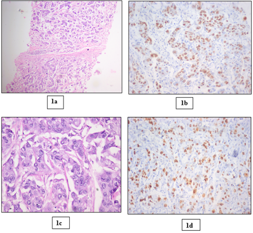
Figure 1: Well differentiated IDC NST showing high Ki-67 index in both NCB (1a & 1b) and RS (1c & 1d). 1a: NCB showing neoplastic cells arranged in tubules, cords and in clusters (100x H&E); 1b: NCB showing > 20% Ki-67 index – 100%;1c: High power image of RS showing tumor cells with mild pleomorphic vesicular nucleus and prominent nucleolus. (400x H&E); 1d: RS showing >20% Ki-67 index – 61.7%.
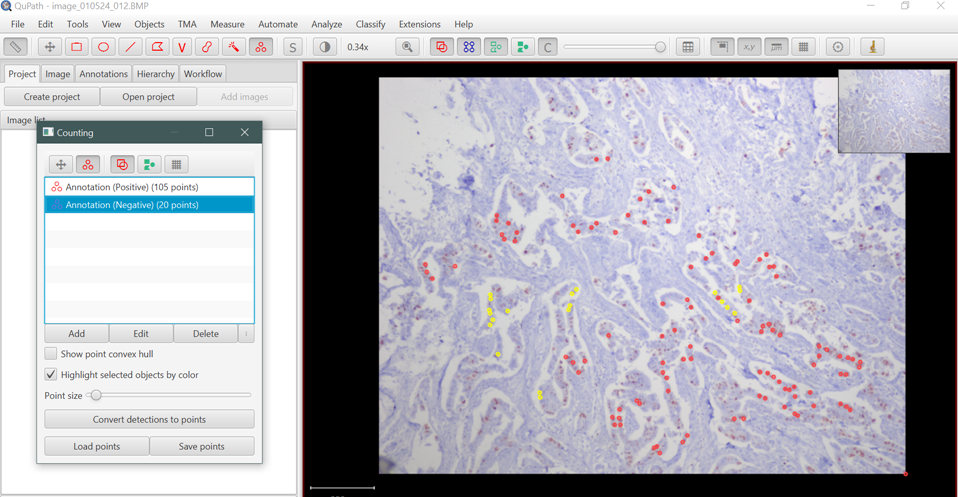
Figure 2 Digital image analysis of Ki67 by using QuPath software.
DISCUSSION:
Accurate preoperative characterization of breast carcinoma with NCB is critical to determine definitive surgical and oncological management. In the present study, 30 matched pairs of NCB and RS were compared and 100% concordance was found for the primary histological diagnosis. This reinforces the current literature supporting NCB as a highly reliable and definitive alternative to surgical excision for establishing baseline tissue diagnosis [2, 13]. The Ki-67 proliferation index is critical for distinguishing the luminal A from the luminal B molecular subtype with a direct impact on systemic therapy choices [8]. The present study demonstrated a moderate inter-specimen concordance rate of 66.6% (κ = 0.50) for the Ki-67 index between NCB and RS. This finding indicates a moderate agreement in line with, but slightly below, a number of comprehensive comparative studies. The concordance rates of Ahn et al. [8] and Ricci et al. [9] were higher at 82%. Ueno et al. [13] and Kombak et al. [12] reported concordance rates of 91.5% and 80%, respectively.
Our results are more similar to Focke et al. [7] who found a concordance rate of 75% (κ = 0.44) and Chen et al. [6] who found 79.5% concordance. The 33.4% rate of discordance observed in our cohort is likely due to a number of well documented, pre-analytical and biological factors. Intra-tumoral heterogeneity is the best example; the distribution of proliferating cells in breast carcinomas is often uneven, such that the limited volume of tissue obtained by a core needle may not be representative of the tumor biology in general [6, 9]. Surgical resection specimens are also subject to prolonged cold ischemic times and delayed formalin penetration, which are known to cause significant antigen degradation and false-negative IHC staining [8]. Because of these limitations, experts recommend that Ki-67 should ideally be assessed on both NCB and RS to avoid misclassification of intrinsic tumor subtypes [6]. We found strong biological associations between Ki-67 index and established markers of tumor aggressiveness.
High Ki-67 index (>20%) was found to be statistically significantly associated with high mitotic activity (p = 0.0011). This is in agreement with the work of Reddy et al. [15] and Soliman et al. [17] who both showed a strong positive correlation between high proliferation indices and a high mitotic score (Nottingham Score 3). In addition, a strong association was found between high Ki-67 index and higher histological tumor grades (p = 0.0256), a finding widely supported by Reddy et al [15] and Ragab et al [16]. These correlations confirm the utility of the Ki-67 index as a reliable marker of aggressive kinetic behavior of breast carcinomas and validate its prognostic utility in this cohort. Interestingly, we did not find any statistically significant correlation of the Ki-67 index with tumor size, lymph node status or lymphovascular invasion, which is in line with the specific findings of Reddy et al. [15] but not with the overall correlations observed by Elkablawy et al.
One major challenge with the clinical utility of Ki-67 index is the subjective nature of manual microscopic evaluation which often is associated with high inter-observer variability [5]. We thus compared conventional manual counting with software-assisted Digital Image Analysis using the QuPath platform. An almost perfect agreement was found between both methods for NCB (κ = 0.84) and RS (κ = 0.79). Furthermore, there was no statistically significant difference between the mean proliferation indices calculated by the manual and the software methods (p = 0.2647 for NCB; p = 0.2774 for RS).
These results provide strong support for incorporating DIA into routine pathology workflows. The results we obtained are similar to those of Bankhead et al. [11], who reported high reproducibility with the use of QuPath, and Shetty et al. [14], who found a high degree of concordance with other digital analysis platforms. The use of AI-assisted image analysis not only standardizes scoring, reducing human error and fatigue, but also improves turnaround times while maintaining diagnostic accuracy [10]. We conclude that digital image analysis is a highly competitive and objective alternative to the manual counting for the quantification of biomarkers in breast cancer pathology.
CONCLUSION:
Needle core biopsy is a very reliable alternative to surgical resection for histological diagnosis and estimation of Ki-67 proliferative activity when the whole tumor cannot be assessed. Despite moderate discordance due to tumor heterogeneity, NCB remains necessary for clinical therapeutic stratification. Furthermore, Digital Image Analysis provides a powerful and reproducible method for biomarker assessment that perfectly reflects the standards of manual pathology and overcomes the subjectivity of visual microscopic analysis.
REFERENCES:
1. Sung H, Ferlay J, Siegel RL, Laversanne M, Soerjomataram I, Jemal A, et al. Global Cancer Statistics 2020: GLOBOCAN Estimates of Incidence and Mortality Worldwide for 36 Cancers in 185 Countries. CA Cancer J Clin. 2021;71(3):209-249.
2. Rakha EA, Ellis IO. An Overview of assessment of Prognostic and Predictive factors in breast cancer needle core biopsy specimens. J Clin Pathol. 2007;60(12):1300-1306.
3. Ingolf JB, Russalina M, Simona M, Julia R, Gilde S, Boble RM, et al. Can Ki-67 Play a Role in Prediction of Breast Cancer Patient’s Response to Neoadjuvant Chemotherapy? Biomed Res Int. 2014;2014:1-7.
4. Romero Q, Bendahl PO, Klintman M, Loman N, Ingvar C, Ryden L, et al. Ki-67 Proliferation in Core biopsies versus Surgical Samples – A Model for Neo – Adjuvant Breast Cancer. BMC Cancer. 2011;11:1-12.
5. Dowsett M, Nielsen TO, Hern RA, Bartlett J, Coombes RC, Cuzick J, et al. Assessment of Ki-67 in Breast Cancer: Recommendations from the International Ki-67 in Breast Cancer Working Group. J Natl Cancer Inst. 2011;103(22):1656-1664.
6. Chen J, Wang Z, Lv Q, Du Z, Tan Q, Zhang D, et al. Comparison of Core Needle Biopsy and Excision Specimens for the Accurate Evaluation of Breast Cancer Molecular Markers: a Report of 1003 Cases. Pathol Oncol Res. 2017;23(4):769-775.
7. Focke CM, Decker T, van Diest PJ. Reliability of the Ki67-Labelling Index in Core Needle Biopsies of Luminal Breast Cancers is Unaffected by Biopsy Volume. Ann Surg Oncol. 2017;24(5):1251-1257.
8. Ahn S, Lee J, Suncho M, Park S, Sung SH. Evaluation of Ki-67 Index in Core Needle Biopsies and Matched Breast Cancer Surgical Specimens. Arch Pathol Lab Med. 2018;142(3):364-368.
9. Ricci MD, Calvano Filho CM, Oliveira Filho HR, Filassi JR, Pinotti JA, Baracat EC. Analysis of the concordance rates between core needle biopsy and surgical excision in patients with breast cancer. Rev Assoc Med Bras. 2012;58(5):532-536.
10. Acs B, Rantalainen M, Hartman J. Artificial intelligence as the next step towards precision pathology. J Intern Med. 2020;288(1):62-81.
11. Bankhead P, Loughrey MB, Fernandez JA, Dombrowski Y, McArt DG, Dunne PD, et al. QuPath: Opensource software for digital pathology image analysis. Sci Rep. 2017;7:1-7.
12. Kombak FE, Sahin H, Moollamemisoglu H, Onem I, Kaya H, Bugdayci O, et al. Concordance of Immunohistochemistry between Core needle biopsy and Surgical resection of breast carcinoma. Turk J Med Sci. 2017;47(6):1791-1796.
13. Ueno N, Kuroda H, Abe A, Kubota K, Kato H, Imai Y. The Reliability of Core-Needle Biopsy in Assessment of Hormone Receptor, HER2, and Ki-67 in Breast Carcinoma. Int Surg. 2017;102:522–529.
14. Shetty PV, Sandhya I, Prasad K, Rao PS, Rappai M. A comparison of Ki67 index in carcinoma breast by digital image analysis with manual method. Indian J Pathol Oncol. 2019;6(4):548–551.
15. Reddy P, Devi SM. Correlation of ER, PR, Her2neu and Ki67 with other Prognostic factors in Breast Carcinoma. Trop J Pathol Microbiol. 2020;6(5):349-361.
16. Ragab HM, Samy N, Afify M, El Maksoud NA, Shaaban HM. Assessment of Ki-67 as a potential biomarker in patients with breast cancer. J Genet Eng Biotechnol. 2018;16(2):479-484.
17. Soliman NA, Yussif SM. Ki-67 as a prognostic marker according to breast cancer molecular subtype. Cancer Biol Med. 2016;13(4):496-504.