Successful i-view™ Videolaryngoscopic Intubation in Ankylosing Spondylitis With Severe Cervical Rigidity and Fixed Spinal Posture: A Case Report.
- Akanksha , Assistant Professor, Department of Anaesthesiology, Pt. B.D. Sharma PGIMS, Rohtak, Haryana, India.
- Shilpa Popli , Associate Professor, Department of Anaesthesiology, Pt. B.D. Sharma PGIMS, Rohtak, Haryana, India.
- Manisha Manohar , Associate Professor, Department of Anaesthesiology, Pt. B.D. Sharma PGIMS, Rohtak, Haryana, India.
- Vritika Malik , Junior resident, Department of Anaesthesiology, Pt. B.D. Sharma PGIMS, Rohtak, Haryana, India.
- Preeti , Junior resident, Department of Anaesthesiology, Pt. B.D. Sharma PGIMS, Rohtak, Haryana, India.
- Mahima , Junior resident, Department of Anaesthesiology, Pt. B.D. Sharma PGIMS, Rohtak, Haryana, India.
Article Information:
Abstract:
Background: Ankylosing spondylitis (AS) is a chronic inflammatory disorder characterized by progressive ossification and rigidity of the axial skeleton. Advanced disease may result in severe cervical spine immobility and fixed kyphotic deformity, making conventional airway management particularly challenging. Forced manipulation of the cervical spine during laryngoscopy may potentially result in neurological injury, particularly in patients with a rigid and fragile spine. Video laryngoscopy may facilitate tracheal intubation while minimizing the need for alignment of the oral, pharyngeal, and laryngeal axes. A 57-year-old male patient with severe ankylosing spondylitis was scheduled for elective repair of a large umbilical hernia under general anaesthesia. Preoperative airway assessment revealed severe restriction of cervical spine movement with a fixed flexed posture. The patient was unable to lie with the head and neck on a conventional flat operating surface because of the fixed spinal deformity. The anticipated airway difficulty was discussed with the patient and the surgical team and a difficult-airway strategy was formulated. Following appropriate preoxygenation and positioning in the patient's natural alignment, tracheal intubation was successfully achieved using an i-view™ video laryngoscope without forceful cervical extension or manipulation. General anaesthesia was subsequently maintained uneventfully, and the patient underwent umbilical hernia repair. The tracheal tube was removed following adequate recovery from anaesthesia and confirmation of satisfactory respiratory and neurological status. Severe ankylosing spondylitis can present a complex airway challenge, particularly when fixed cervical deformity prevents conventional supine positioning. In appropriately selected patients, video laryngoscopy may provide an effective means of tracheal intubation while minimizing cervical movement. The i-view™ video laryngoscope, with its integrated screen and Macintosh-type blade, may be a useful addition to the difficult-airway armamentarium in such cases.
Keywords:
Article :
INTRODUCTION:
Ankylosing spondylitis is a chronic inflammatory rheumatological disorder characterized by progressive inflammation, ossification and fusion of the axial skeleton. Advanced disease can result in marked restriction of cervical spine movement, fixed flexion deformity and severe thoracolumbar kyphosis. These changes may substantially complicate airway management and positioning for anaesthesia. [1]
Airway management in patients with advanced ankylosing spondylitis requires particular caution because conventional positioning and cervical extension during direct laryngoscopy may be impossible or potentially hazardous. Previous reports have described difficult or failed direct laryngoscopy in such patients and have emphasized the role of awake fibreoptic intubation or alternative techniques that minimize cervical manipulation. [2]
More recently, video laryngoscopy has emerged as an important technique for managing anticipated difficult airways. Successful use of video laryngoscopes in patients with severe ankylosing spondylitis has been reported, including a series of four patients in whom GlideScope-assisted intubation was successful [3]. Video laryngoscopy has also been successfully employed in patients with severe cervical restriction when fibreoptic equipment is unavailable or impractical. [4]
The i-view™ video laryngoscope (Intersurgical, UK) is a single-use video laryngoscope incorporating a Macintosh-type blade and an integrated LCD screen, allowing indirect visualization of the glottis while retaining a familiar Macintosh insertion technique. The manufacturer describes its use in anaesthesia and difficult-airway management. [5]
We present a case of severe ankylosing spondylitis with fixed cervical deformity in which conventional positioning was not possible because the patient could not place his head and neck on a flat operating surface. Successful tracheal intubation was achieved using the i-view™ video laryngoscope, allowing general anaesthesia for repair of a large paraumbilical hernia.
CASE PRESENTATION:
A 57-year-old male patient, weighing 78kgs and with a height of 192 cm, was scheduled for elective repair of a large umbilical hernia posted for overlay meshplasty under general anaesthesia. The patient had a known history of ankylosing spondylitis for 30 years, with progressive restriction of spinal and cervical movements. [Add relevant treatment history, e.g. NSAIDs/biologics/steroids, if applicable.] There was no history of previous anaesthetic difficulty or any surgical history. On preoperative examination, the patient had a characteristic fixed kyphotic posture with marked restriction of cervical movement. The head and neck could not be brought into a conventional neutral or extended position, and the patient was unable to lie with the occiput comfortably supported on a flat operating table. The clinical appearance is shown in Figure 1.
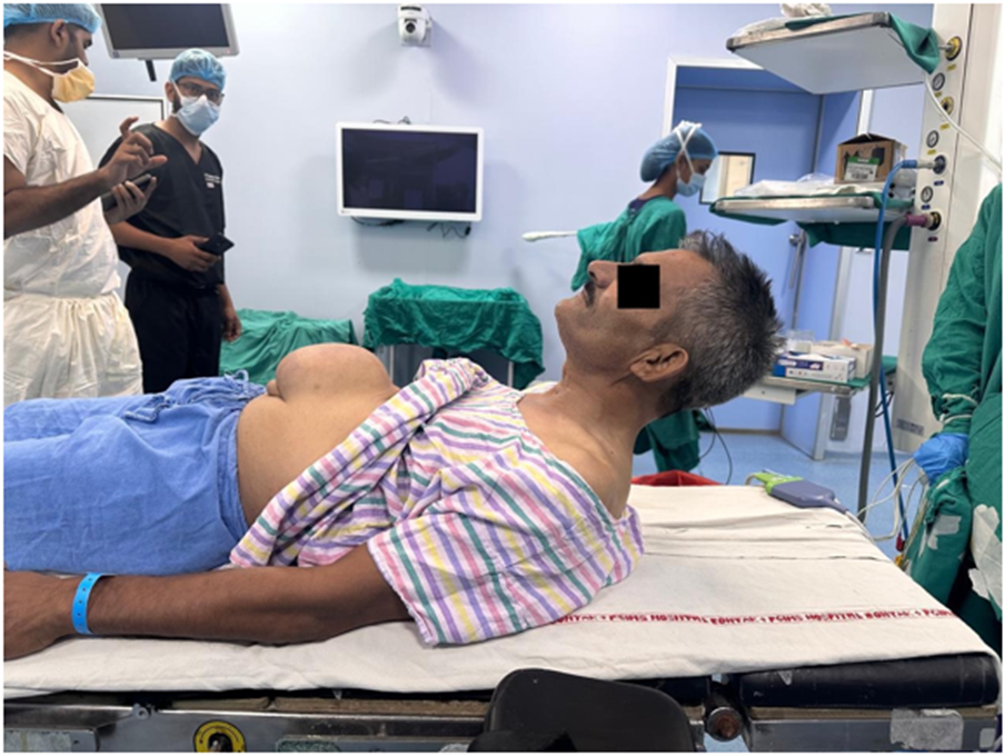
Figure 1: Preoperative positioning in severe ankylosing spondylitis. The patient was unable to comfortably place the head and neck on a conventional flat operating surface, necessitating individualized positioning and maintenance of the pre-existing spinal alignment during airway management.
Airway assessment revealed a mouth opening of approximately 4 cm, Mallampati class III, thyromental distance of 6.5 cm and severely restricted neck movement. The patient's respiratory examination revealed bilateral equal clear air entry. Cardiovascular examination was normal. Baseline oxygen saturation was 98-99% on room air. Preoperative investigations including [CBC, renal function, ECG, chest radiograph, pulmonary function tests] were [normal/relevant findings].
Given the severe cervical restriction and inability to achieve conventional supine positioning, the airway was considered anticipated difficult. The potential risks of airway manipulation, including difficult ventilation, failed intubation and cervical spine injury were discussed with the patient. A difficult-airway plan was formulated before induction of anaesthesia, with availability of alternative airway devices and equipment for emergency airway rescue.
The patient was transferred to the operating room and standard monitoring was established, including non-invasive blood pressure, electrocardiography and pulse oximetry. Particular attention was paid to maintaining the patient's pre-existing spinal alignment and RAMP [Rapid Airway Management Positioner] position was made [figure 2].
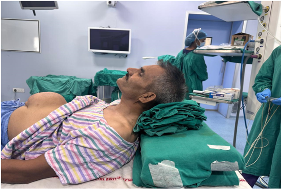
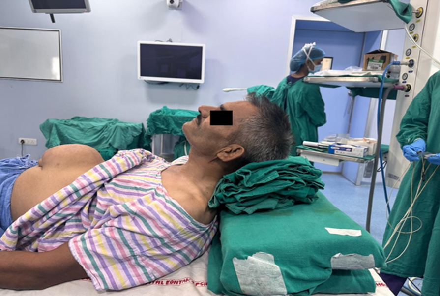
Figure 2: RAMP position to achieve adequate airway axis and alignment.
Attempts were made to make sure that no force was applied to the head and neck into a conventional neutral or sniffing position. The patient was positioned as comfortably as possible in his natural posture, using appropriate padding and support to accommodate the fixed spinal deformity. Preoxygenation was performed for 3–5 minutes with 100% oxygen. Induction was done using rapidly induced general anesthesia with IV boluses of fentanyl 100 µg, propofol 130 mg and rocuronium 50 mg. Mask ventilation was easily performed, after which tracheal intubation was completed without any issues using a i-view video laryngoscope with an 4 sized blade to avoid extending his neck[figure 3].
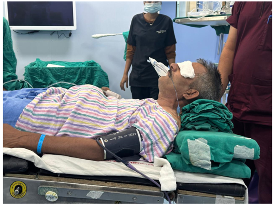
Figure 3: Intraoperative positioning of a patient with ankylosing spondylitis. The head and neck were carefully supported with folded sheets to accommodate the patient's fixed posture.
A 8.0-mm cuffed oral endotracheal tube was used for intubation. Correct tube placement was confirmed by bilateral chest excursion, auscultation and sustained end-tidal carbon dioxide waveform. No significant cervical manipulation was required during intubation. General anesthesia was maintained with sevoflurane 1% to 1.2%, oxygen 0.5 L/min, and air 1.5 L/min. A total of 39.6 mL of 2% lidocaine with epinephrine 1:160,000 was injected via infiltration for local anesthesia throughout the course of the surgery. The patient remained hemodynamically stable throughout the entire procedure. After completion of the surgery, we discontinued all the anesthetic maintenance agents. Neuromuscular blockade was reversed. After the patient was fully awake, maintaining adequate spontaneous ventilation with satisfactory oxygen saturation and demonstrating appropriate protective airway reflexes, the tracheal tube was removed in a controlled manner.
The patient was transferred to the PACU for postoperative monitoring. His postoperative course was uneventful and he was discharged from hospital on postoperative day 7 with advice to follow up in the surgery OPD.
DISCUSSION:
Patients with advanced ankylosing spondylitis pose a unique challenge to the anaesthesiologist because the disease can progressively transform a normally mobile vertebral column into a rigid, kyphotic structure. Cervical involvement may result in profound restriction of movement and a fixed flexed posture, substantially limiting the ability to achieve conventional airway positioning. The airway challenge in the present case was particularly significant because the patient's head could not be placed flat on the operating table. This is an important practical consideration that may be underestimated during routine airway assessment. Standard induction positioning assumes that the patient can tolerate supine positioning with the head and neck appropriately supported. In severe ankylosing spondylitis, attempting to impose such positioning may cause discomfort, excessive spinal stress or neurological complications.
Cervical spine manipulation during airway management is another major concern. Ankylosed spinal segments may be vulnerable to fracture and cervical movement can potentially result in neurological compromise. Consequently, airway management should aim to maintain the patient's established spinal alignment and minimize unnecessary cervical movement. Previous reports have highlighted the risks associated with cervical spine involvement and the difficulty of conventional direct laryngoscopy in these patients. [6] Awake fibreoptic intubation has traditionally been regarded as an important technique for anticipated difficult airway management in patients with severe ankylosing spondylitis. Early literature emphasized the advantages of awake intubation in these patients, particularly because spontaneous ventilation can be maintained while the airway is secured.[7] Current difficult-airway recommendations similarly emphasize consideration of awake tracheal intubation when difficult intubation is anticipated, particularly when other aspects of airway management may also be challenging. [8]
However, fibreoptic intubation is not always feasible. Equipment availability, operator experience, airway secretions, patient tolerance and the urgency or nature of surgery may influence the choice of technique. Video laryngoscopy provides an alternative by allowing indirect visualization of the glottis without requiring the same degree of alignment necessary for conventional direct laryngoscopy. Several reports have demonstrated successful video-laryngoscopic intubation in patients with ankylosing spondylitis. Saricicek et al. reported successful GlideScope intubation in four patients with severe ankylosing spondylitis and difficult airway features. More recent literature has similarly described successful videolaryngoscopy in patients with profound cervical restriction.
The i-view™ video laryngoscope used in our patient incorporates a Macintosh-type blade with an integrated LCD display. This design allows the operator to use a familiar Macintosh insertion technique while obtaining an indirect video view of the larynx. In the present case, this characteristic was particularly useful because significant cervical extension was neither possible nor desirable. An important lesson from this case is that airway management should begin with positioning rather than laryngoscopy. In patients with severe ankylosing spondylitis, the patient's natural spinal alignment should be identified preoperatively and reproduced on the operating table. Padding and positioning aids should be used to avoid pressure points and prevent inadvertent spinal movement.
A detailed rescue strategy is equally important. The airway plan should include preparation for difficult mask ventilation, supraglottic airway placement, alternative video/fibreoptic techniques and emergency front-of-neck access where appropriate. Current difficult-airway guidance emphasizes pre-induction planning, oxygenation, limiting repeated airway attempts and having an explicit rescue strategy.
CONSENT
Written informed consent was obtained from the patient for publication of this case report and any accompanying images.
Acknowledgement
We thank patient of our case for providing written informed consent and permission to publish the photos.
Financial or Other Competing Interests
None.
REFERENCES:
1. Sinclair JR, Mason RA. Ankylosing spondylitis. The case for awake intubation. Anaesthesia. 1984;39:3–11. PMID: 6696216.
2. Kamarkar US, Chaudhari LS, Hosalkar H, Budhi M, Venkataraghavan D. Difficult intubation in a case of ankylosing spondylitis: a case report. J Postgrad Med. 1998;44:43–46. PMID: 10703569.
3. Ahmad N, Channa AB, Mansoor A, Hussain A. Management of difficult intubation in a patient with ankylosing spondylitis—a case report. Middle East J Anaesthesiol. 2005;18:379–384. PMID: 16438012.
4. Saricicek V, Mizrak A, Gul R, Goksu S, Cesur M. GlideScope video laryngoscopy use tracheal intubation in patients with ankylosing spondylitis: a series of four cases and literature review. J Clin Monit Comput. 2014;28:169–172. doi:10.1007/s10877-013-9511-0.
5. Ahmad I, El-Boghdadly K, Bhagrath R, et al. Difficult Airway Society guidelines for awake tracheal intubation (ATI) in adults. Anaesthesia. 2020;75:509–528. doi:10.1111/anae.14904.
6. Apfelbaum JL, Hagberg CA, Connis RT, et al. 2022 American Society of Anesthesiologists Practice Guidelines for Management of the Difficult Airway. Anesthesiology. 2022;136:31–81. doi:10.1097/ALN.0000000000004002.
7. Chen WC, Lin S, He HF. Case Report: Double Visualization Intubation Strategy for Patients With Ankylosing Spondylitis. Front Med. 2022. doi:10.3389/fmed.2022.659624.
8. Intersurgical. i-view™ video laryngoscope. Manufacturer information and technical specifications.