Navigating the Difficult Airway in a Child With Pierre Robin Sequence: Anaesthetic Management During Cleft Palate and Hypospadias Surgery.
- Shilpa Popli , Associate Professor, Department of Anaesthesiology, Pt. B.D. Sharma PGIMS, Rohtak, Haryana, India.
- Akanksha , Assistant Professor, Department of Anaesthesiology, Pt. B.D. Sharma PGIMS, Rohtak, Haryana, India.
- Anshul , Associate Professor, Department of Anaesthesiology, Pt. B.D. Sharma PGIMS, Rohtak, Haryana, India.
- Neha goyal , senior resident, Department of Anaesthesiology, Pt. B.D. Sharma PGIMS, Rohtak, Haryana, India.
- Sunil Kumar , junior resident, Department of Anaesthesiology, Pt. B.D. Sharma PGIMS, Rohtak, Haryana, India.
- Divyansh Yadav , Senior resident, Department of Radiodiagnosis, Pt. B.D. Sharma PGIMS, Rohtak, Haryana, India.
Article Information:
Abstract:
Background: Pierre Robin sequence (PRS) is a congenital craniofacial condition characterized by micrognathia, glossoptosis, and upper-airway obstruction, frequently associated with cleft palate. These anatomical abnormalities can pose significant challenges during mask ventilation, tracheal intubation, maintenance of airway patency and postoperative extubation. Although airway obstruction is most pronounced during infancy, children with PRS may continue to present with difficult airway anatomy during subsequent surgical procedures. We report the perioperative anaesthetic management of a 5-year-old child diagnosed with Pierre Robin sequence and a large U-cleft palate at birth who presented for elective cleft palate repair and simultaneous correction of hypospadias under general anaesthesia. Children with Pierre Robin sequence remain challenging airway patients even beyond infancy. Careful preoperative airway assessment, preparation for difficult intubation, maintenance of oxygenation and a clearly defined extubation strategy are essential for safe anaesthetic management. The coexistence of another congenital anomaly, such as hypospadias, further emphasizes the importance of comprehensive preoperative evaluation and multidisciplinary perioperative planning.
Keywords:
Article :
INTRODUCTION:
Pierre Robin sequence (PRS) is a congenital craniofacial disorder characterized by the combination of micrognathia, glossoptosis, and upper-airway obstruction, with cleft palate occurring in a substantial proportion of affected children[1]. The sequence results from mandibular hypoplasia during fetal development, with posterior displacement of the tongue contributing to impaired palatal fusion and airway obstruction.
The anaesthetic management of children with PRS presents several challenges. Micrognathia and glossoptosis can result in difficult mask ventilation and tracheal intubation, while the presence of a cleft palate can further complicate airway instrumentation[2]. Although many children experience improvement in airway obstruction with growth, the abnormal craniofacial anatomy may persist into childhood and continue to influence airway management during subsequent surgical procedures.
Cleft palate repair itself presents additional anaesthetic considerations because the surgical field shares the upper airway, and postoperative airway obstruction, edema, bleeding, and impaired airway protective reflexes may complicate recovery. Recent literature continues to emphasize meticulous airway assessment and individualized airway planning in children with PRS undergoing palatal surgery.
The case highlights the importance of anticipating airway difficulties in an older child with PRS and developing a structured strategy for induction, tracheal intubation, intraoperative management, and extubation.
CASE PRESENTATION:
A 5-year-old male child weighing 14kg was scheduled for elective cleft palate repair and correction of hypospadias under general anaesthesia. The child had a history of feeding difficulty during infancy and recurrent respiratory infections with previous hospitalization history. There was no history of previous surgical intervention for airway obstruction such as tongue-lip adhesion or mandibular distraction osteogenesis. On preoperative examination, the child was conscious, cooperative and adequately nourished.
Preoperative assessment revealed characteristic craniofacial abnormalities, including micrognathia and glossoptosis. Preoperative assessment revealed child breathing spontaneously on room air. Examination demonstrated a wide cleft palate. Respiratory examination revealed bilateral equal air entry with no adventitious sounds. Cardiovascular examination was normal. Routine investigations, including complete blood count, renal and liver function tests, and coagulation profile, were within acceptable limits. The child had no clinical evidence of active respiratory infection at the time of surgery. Airway assessment suggested an anticipated difficult airway with Mallampati grade III, mouth opening two and a half fingers with normal neck movements and no previous significant airway history. A comprehensive difficult-airway plan was formulated, with preparation of conventional and advanced airway equipment and availability of experienced personnel. General anaesthesia was induced using inhalational technique, while maintaining spontaneous ventilation until adequate control of the airway was achieved.
Tracheal intubation was performed under controlled conditions, and a pediatric ENT surgeon remained on standby for emergency surgical airway access. Anesthesia was induced with 8% sevoflurane in 100% oxygen with maintenance of spontaneous ventilation. Eyes were taped. A 22-gauge IV was placed in the right saphenous vein. Glycopyrrolate 0.1 mg and fentanyl 5 mcg IVP (IV push) were given. Direct laryngoscopy with blade size 2 didot reveal visibilty of epiglottis and CL grade was found to be 4b. Video laryngoscopy using pediatric TASQscope with Miller 1 blade revealed a large U-cleft palate and a class IIb Cormack-Lehane view of the vocal cords. Spontaneous ventilation with vocal cord motion was observed. Tracheal intubation was accomplished using TASQscope guided bougie insertion followed by railroading of size 5 southpole endotracheal tube (ETT) and cuff was inflated with 0.5 ml of air and secured at 12 cm at the lips, with equal bilateral breath sounds confirming placement.
Anaesthesia was subsequently maintained with volatile agent and the child underwent cleft palate repair followed by hypospadias repair. Intraoperative haemodynamics and oxygenation remained stable. The procedure ensued uneventfully, with the patient receiving dexamethasone 3 mg, acetaminophen 110 mg, ketorolac 3.6 mg, and propofol 5 mg throughout the case. Local anesthesia was administered by the surgeon, lidocaine 0.5% with epinephrine and bupivacaine 0.25%. Total IV fluid was 350 ml.At the completion of surgery, the child was extubated fully awake and transferred to PICU for close postoperative monitoring. No significant perioperative airway or respiratory complications occurred.
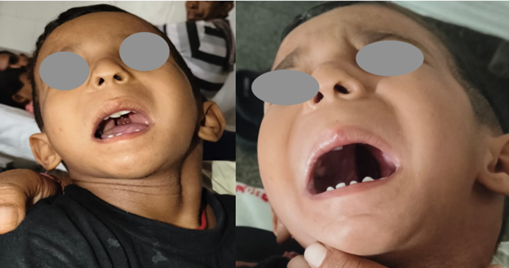
Figure 1: image depicts complete cleft palate in Pierre Robin Sequence syndrome patient
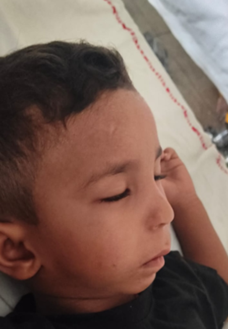
Figure 2: picture depicting micrognathia and retrognathia.
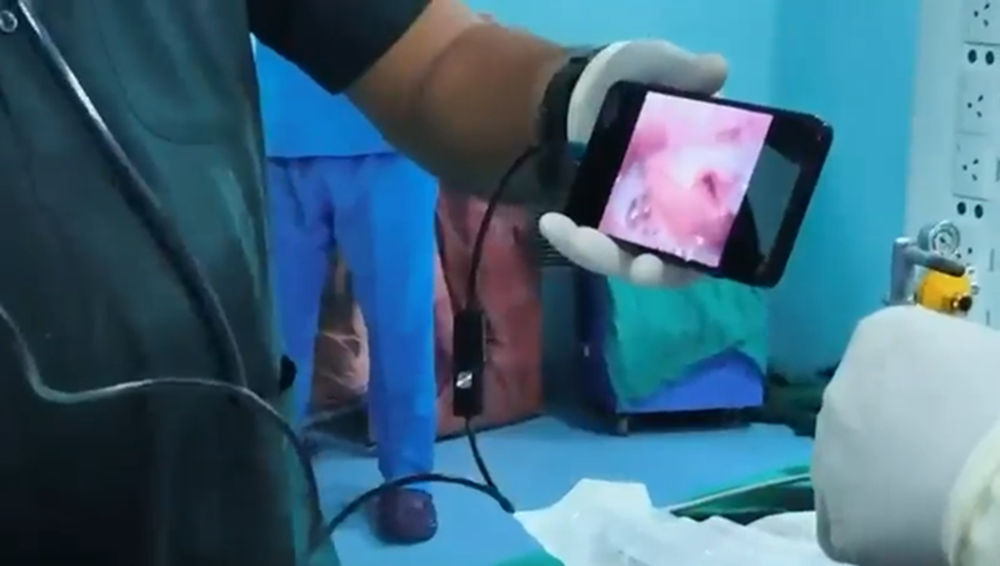
Figure 3: Video depicting Endotracheal tube being secured with the help of peadiatric TASQscope along with bougie.
DISCUSSION:
Pierre Robin sequence presents a distinctive challenge to the paediatric anaesthesiologist because the anatomical abnormalities responsible for the sequence can directly interfere with airway management. Micrognathia reduces the mandibular space available for the tongue, while glossoptosis contributes to posterior airway obstruction. These changes may make both mask ventilation and direct laryngoscopy difficult [3]. Although severe airway obstruction is most commonly recognized during infancy, airway-related difficulties can persist into later childhood. A series evaluating children with PRS found that more than half required some form of airway intervention, highlighting the variability and potential severity of airway involvement.
The first important step in managing these patients is identification of the anticipated difficult airway before induction. Previous anaesthetic records, history of airway interventions, symptoms suggestive of obstructive sleep apnoea, feeding and respiratory history, and a detailed craniofacial and airway examination should be considered. Where clinically indicated, additional assessment with sleep studies or endoscopic airway evaluation may provide useful information regarding the severity and anatomical level of obstruction.
Maintaining spontaneous ventilation during induction has traditionally been considered an important strategy in patients with severe PRS because loss of pharyngeal muscle tone may worsen upper-airway obstruction. However, the optimal induction technique should be individualized according to the child's age, severity of airway obstruction, previous airway history, institutional expertise and available equipment. Advanced techniques such as videolaryngoscopy and flexible fibreoptic intubation can facilitate tracheal intubation in selected patients [4]. Palatoplasty presents additional challenges because the airway is shared with the surgical team. The endotracheal tube must be securely positioned while providing unobstructed surgical access. Furthermore, postoperative airway obstruction may result from residual effects of anaesthetic drugs, airway edema, secretions, bleeding, altered tongue position and reduced airway tone. Therefore, extubation should be considered an elective airway procedure rather than simply the final step of anaesthesia [5].
In our patient, the additional diagnosis of hypospadias was noteworthy because it demonstrated the coexistence of another congenital anomaly. PRS may occur as an isolated condition or as part of a broader syndromic or genetic disorder, and associated congenital abnormalities warrant appropriate evaluation. The successful outcome in this case emphasizes the importance of anticipating rather than reacting to airway difficulty. Appropriate preparation, availability of advanced airway devices, experienced personnel, maintenance of oxygenation, and a carefully planned extubation strategy are central to safe perioperative management.
CONCLUSION:
Pierre Robin sequence should be regarded as an anticipated difficult-airway condition even when the child presents later in childhood without overt respiratory symptoms. In children undergoing cleft palate surgery, careful preoperative assessment and a structured airway plan are essential. Maintaining oxygenation, ensuring availability of advanced airway equipment, minimizing repeated airway instrumentation, and adopting a deliberate extubation and postoperative monitoring strategy can contribute to safe outcomes. The presence of associated congenital anomalies, such as hypospadias, further reinforces the need for comprehensive multidisciplinary evaluation.
REFERENCES:
1. Evans KN, Sie KC, Hopper RA, Glass RP, Hing AV, Cunningham ML: Robin sequence: from diagnosis to development of an effective management plan. Pediatrics. 2011, 127:936-48. 10.1542/peds.2010-2615.
2. Cladis F, Kumar A, Grunwaldt L, Otteson T, Ford M, Losee JE: Pierre Robin sequence: a perioperative review. Anesth Analg. 2014, 119:400-12. 10.1213/ANE.0000000000000301.
3. Cladis F, Kumar A, Grunwaldt L, Otteson T, Ford M, Losee JE: Pierre Robin sequence: a perioperative review. Anesth Analg. 2014, 119:400-12. 10.1213/ANE.0000000000000301.
4. Koerner IP, Brambrink AM: Fiberoptic techniques. Best Pract Res Clin Anaesthesiol. 2005, 19:611-21. 10.1016/j.bpa.2005.07.006.
5. Weiss M, Engelhardt T: Proposal for the management of the unexpected difficult pediatric airway. Paediatr Anaesth. 2010, 20:454-64.