Airway Management In Reinke’s Oedema Causing Airway Obstruction: Anaesthetic Challenge
- Nidhi Bangarwa , Department of Anaesthesiology, Pt. B.D. Sharma PGIMS, Rohtak, Haryana, India.
- Akanksha , Department of Anaesthesiology, Pt. B.D. Sharma PGIMS, Rohtak, Haryana, India.
- Susheela Taxak , Senior professor and Head Department of Anaesthesiology, Pt. B.D. Sharma PGIMS, Rohtak, Haryana, India.
- Rashmi rashmi , Senior resident Department of Anaesthesiology, Pt. B.D. Sharma PGIMS, Rohtak, Haryana, India.
Article Information:
Abstract:
Background: Reinke’s oedema is associated with the accumulation of fluid in the subepithelial area of vocal folds. It is caused by chronic tobacco consumption, vocal abuse and gastroesophageal reflux disorder. It may be associated with vocal cord polyp or laryngeal web formation. Narrowing of the glottic aperture leads to a difficult airway. We describe successful airway management in a patient with Reinke’s oedema via direct laryngoscopy with the backup of fiberoptic intubation and tracheostomy.
Keywords:
Article :
INTRODUCTION:
Reinke’s oedema is polypoidal degeneration of one or both vocal folds within Reinke’s space.1 Chronic irritation due to smoking, vocal abuse and laryngeal reflux are the potential contributory factors in the pathogenesis of Reinke’s oedema.2,3 Patients affected with Reinke’s oedema present with different levels of dysphonia, hoarseness of voice and some also report breathing difficulties, depending on the lesion size.4 We encountered a patient with Reinke’s oedema having hoarseness and stridor who was scheduled for surgical management under general anaesthesia.
CASE REPORT:
A 58 year old female presented with complaints of hoarseness of voice for the last 3 years which was progressively increasing in nature. The patient started experiencing difficulty in breathing and stridor in the last two months. She was a chronic smoker for the past ten yrs. Her breathing difficulties increased during supine positioning. During her opd visit, she was diagnosed with Reinke’s oedema. Indirect laryngoscopic examination showed swollen vocal cords with more involvement of the left side. The right side of vocal cord also had a polypoidal growth. The patient was posted for laryngeal cord stripping under general anaesthesia. During her pre-anaesthetic check-up, she was advised to quit smoking and nebulisation with budesonide. Informed written consent for tracheostomy, as well as awake fiberoptic intubation, was taken. Standard ASA monitors (NIBP, ECG and SPO2) were attached. Her heart rate was 90/min, BP was 140/78 mmhg and SP02 was 98%. She was premedicated with 0.2mcg glycopyrrolate. She was nebulised with 4% lignocaine in a semi-recumbent position and the posterior pharyngeal wall was sprayed with 10% lignocaine. After preoxygenation for 3 min induction was done with Inj. Fentanyl 120mcg and Inj. Propofol 120mg. Anaesthesia was maintained with (O2:N2O = 50:50) along with sevoflurane. The patient was planned for check laryngoscopy examination. During direct laryngoscopy, we found a very narrow glottic opening as most of the area was occluded by vocal cord oedema. The patient was given Inj. Atracurium 25 mg. Intubation was done with a 5 mm micro laryngeal tube without any airway trauma [Figure 1]. Polypoidal growth, as well as bilateral vocal cord lesions, were excised. Postoperatively there was no bleeding at the operative site [Figure 2]. After completion of surgery vocal cords were visualised endoscopically for assessment of any residual oedema. The patient was given Inj. Hydrocortisone 100mg postoperatively. Extubation was done after the reversal of neuromuscular blockade. There was no sign of upper airway obstruction in the postoperative period.
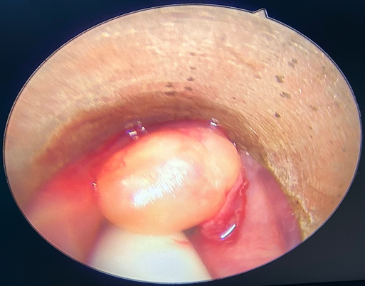
Fig 1. Endoscopic image showing edematous vocal cord along with polypoidal lesion after intubation
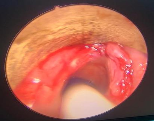
Fig 2. Endoscopic image showing glottic aperture post operatively
DISCUSSION:
Reinke’s oedema is a benign condition characterised by swelling of the superficial layer of lamina propria of the vocal fold. Hoarseness of voice is the chief clinical presentation.4 It is classified into four types.
1. Oedema of unilateral vocal fold
2. Oedema of bilateral vocal fold
3. Oedema of one vocal fold with associated polypoidal lesion
4. Oedema of both vocal fold with an associated polypoidal lesion in one or both vocal fold.5
5. Our patient was type 4 Reinke’s oedema.
The case described above had an anticipated difficult airway. There is limited space available for the passage of endotracheal tube in this condition so a smaller sized tube should be kept ready in advance. Airway management is of great concern specially in grade 4 Reinke’s oedema as the risk of airway obstruction is high. Patients having stridor are more prone to airway emergencies during tracheal intubation. Multiple attempts during intubation can further complicate the procedure. So careful handling of the airway by an expert anaesthesiologist should be done. Moreover, the risk of laryngospasm is also increased by repeated airway handling.
In our case, we preferred to do a check laryngoscopy under sedation to exactly assess the glottic opening. Posterior one-third of the glottic opening was visible so we decided to proceed with intubation under direct laryngoscopy. Glidescope provides better visualisation of the glottic opening.6,7 But we couldn’t use it because of non-availability. The use of supraglottic airway devices as a rescue measure is controversial in such cases as positive pressure ventilation through a supraglottic device may be obstructed by bowing of oedematous cords leading to ventilatory failure.8 So we kept the backup plan of fibreoptic intubation and tracheostomy ready.
Extubation is also a very crucial step in the management of such cases. Postoperative assessment of the degree of laryngeal oedema via direct or video laryngoscopy, and a quantitative cuff leak test should be done for better patient outcome.9,10 Post-extubation laryngeal oedema may lead to respiratory failure with need for reintubation. Airway exchange catheters (AECs) have been advocated in the Difficult Airway Society extubation guidelines for patients at risk for post-extubation oedema.11 Prophylactic use of corticosteroids help to prevent post-extubation oedema in high risk patients.12
, 19]
CONCLUSION:
We would like to conclude that collaborative efforts from otolaryngologists and anaesthesiologists in the anticipated difficult airway are most important for managing anticipated difficult airway (Reinke’s oedema in our case) with obstructive features.
Even in undiagnosed cases of Reinke’s care should be taken to properly evaluate and proceed for airway management. It is always prudent to keep backup plan for patient safety.
REFERENCES:
1. Tavaluc R, Tan-Geller M. Reinke's Edema. Otolaryngol Clin North Am. 2019;52(4):627-635.
2. Grill M, Lazzeri I, Kirsch A, Steurer N, Grossmann T, Karbiener M et al. Vocal Fold Fibroblasts in Reinke's Edema Show Alterations Involved in Extracellular Matrix Production, Cytokine Response and Cell Cycle Control. Biomedicines. 2021;9(7):735.
3. Marcotullio D, Magliulo G, Pezone T. Reinke's edema and risk factors: clinical and histopathologic aspects. Am J Otolaryngol. 2002;23(2):81-4.
4. Cortegiani A, Russotto V, Palmeri C, Raineri SM, Giarratano A. Previously undiagnosed Reinke edema as a cause of immediate postextubation inspiratory stridor. A A Case Rep. 2015;4(1):1-3.
5. De Vincentiis M, Ralli M, Cialente F, Greco A, Marcotullio D, Minni A et al. Reinke's edema: a proposal for a classification based on morphological characteristics. Eur Arch Otorhinolaryngol. 2020;277(8):2279-2283.
6. Griesdale DE, Liu D, McKinney J, Choi PT. Glidescope® video-laryngoscopy versus direct laryngoscopy for endotracheal intubation: a systematic review and meta-analysis. Can J Anaesth. 2012;59(1):41-52.
7. Sun DA, Warriner CB, Parsons DG, Klein R, Umedaly HS, Moult M. The GlideScope Video Laryngoscope: randomized clinical trial in 200 patients. Br J Anaesth. 2005;94(3):381-4.
8. D'Hulst D, Butterworth J, Dale S, Oaks T, Matthews B. Polypoid hyperplasia of the larynx misdiagnosed as a malpositioned laryngeal mask airway. Anesth Analg. 2004;99(5):1570-1572.
9. Newmark JL, Ahn YK, Adams MC, Bittner EA, Wilcox SR. Use of video laryngoscopy and camera phones to communicate progression of laryngeal edema in assessing for extubation: a case series. J Intensive Care Med. 2013;28(1):67-71.
10. Prinianakis G, Alexopoulou C, Mamidakis E, Kondili E, Georgopoulos D. Determinants of the cuff-leak test: a physiological study. Crit Care. 2005;9(1):24-31.
11. Difficult Airway Society Extubation Guidelines Group, Popat M, Mitchell V, Dravid R, Patel A, Swampillai C et al. Difficult Airway Society Guidelines for the management of tracheal extubation. Anaesthesia. 2012;67(3):318-40.
12. Kuriyama A, Umakoshi N, Sun R. Prophylactic Corticosteroids for Prevention of Postextubation Stridor and Reintubation in Adults: A Systematic Review and Meta-analysis. Chest. 2017;151(5):1002-1010.