Pterygium-Associated Corneal Astigmatism: Size-Dependent Burden and Surgical Reversibility After Conjunctival Autograft.
- Uzma Nausheen , KBN Medical College and Hospital, Kalaburgi
- Nandini H , Minto Ophthalmic Hospital, Regional Institiute of Ophthalmology, BMCRI, Bengaluru
- Bhavya Annadevula , Bhavya Hospital, Hyderabad
Article Information:
Abstract:
ground: Pterygium can alter corneal curvature and induce visually significant astigmatism. The magnitude of this refractive distortion may increase with lesion size and may improve after surgical excision. Objective: To evaluate the relationship between pterygium grade and corneal astigmatism and to assess the reversibility of refractive and keratometric astigmatism following pterygium excision with conjunctival autografting. Methods: This prospective interventional study included 100 eyes of 100 patients with primary nasal pterygium. Pterygia were graded according to the extent of corneal involvement. Preoperative assessment included visual acuity, autorefractometry, manifest refraction, slit-lamp examination, and keratometry. All patients underwent pterygium excision with conjunctival-limbal autografting and were followed postoperatively. Changes in refractive astigmatism, keratometric astigmatism, and best-corrected visual acuity were analyzed. Results: Grade II, III, and IV pterygia accounted for 68%, 23%, and 9% of eyes, respectively. Preoperative refractive astigmatism increased from 2.49±0.65 D in Grade II to 3.93±0.465 D in Grade III and 6.08±0.61 D in Grade IV. Corresponding keratometric astigmatism increased from 2.46±0.75 D to 3.71±0.63 D and 5.55±0.63 D. Overall refractive astigmatism decreased significantly from 3.15±1.258 D to 1.542±0.652 D, while keratometric astigmatism decreased from 3.03±1.189 D to 1.475±0.600 D (both P<0.001). BCVA also improved significantly. Conclusion: Increasing pterygium size was associated with progressively greater corneal astigmatism.
Keywords:
Article :
INTRODUCTION:
Pterygium is a common ocular surface disorder characterized by a triangular or wing-shaped fibrovascular proliferation of conjunctival tissue that progressively extends across the limbus onto the corneal surface. Although traditionally regarded as a degenerative condition, contemporary evidence supports a multifactorial proliferative process involving chronic ultraviolet (UV) radiation exposure, oxidative stress, inflammation, abnormal extracellular matrix remodeling, angiogenesis, and altered limbal epithelial homeostasis. Prolonged exposure to UV radiation, particularly UV-B, is considered one of the principal environmental factors involved in its development and progression. At the molecular level, UV-related injury can promote the expression of inflammatory cytokines, growth factors, matrix metalloproteinases, and angiogenic mediators, leading to epithelial alteration and fibrovascular proliferation across the cornea [1,2]. In addition to producing ocular irritation, foreign-body sensation, redness, and cosmetic concerns, progressive pterygium may have important optical consequences that can compromise visual function even before the lesion reaches the visual axis.
One of the most clinically relevant consequences of pterygium is its ability to alter corneal curvature and induce astigmatism. Pterygium-associated astigmatism results from distortion of the normally regular anterior corneal surface. Proposed mechanisms include mechanical traction exerted by the fibrovascular tissue on the cornea and alterations in the precorneal tear film, particularly localized tear pooling near the advancing pterygium head [3]. These changes commonly produce flattening of the horizontal corneal meridian and a corresponding predominance of with-the-rule astigmatism, although oblique and irregular astigmatic components may also occur. Importantly, pterygium-associated corneal distortion may be greater on computerized corneal topography than is apparent from manifest refraction, indicating that conventional refractive assessment alone may underestimate the optical burden of the disease [4].
The magnitude of corneal astigmatism appears to be strongly influenced by pterygium size. Lin and Stern demonstrated that progressive extension of the lesion toward the visual axis was associated with increasing central corneal astigmatism, with larger lesions producing clinically important asymmetric with-the-rule changes [4]. Subsequent studies have further quantified this relationship. Mohammad-Salih and Sharif reported that pterygium extension showed a stronger association with corneal astigmatism than lesion width and observed that approximately 2 diopters of astigmatism could occur when pterygium extension exceeded 2.2 mm, width exceeded 5 mm, or lesion area exceeded 6.25 mm² [5]. These observations are clinically important because they suggest that the refractive consequences of pterygium develop progressively and may become substantial well before direct pupillary or visual-axis involvement. Thus, lesion size may represent not merely an anatomical descriptor but also a practical surrogate for the functional burden imposed on the cornea.
The refractive influence of pterygium is not limited to regular cylinder. Increasing lesion size has also been associated with changes in corneal spherical power, surface asymmetry, and irregularity. In a prospective investigation, Tomidokoro et al. demonstrated a significant correlation between preoperative pterygium size and corneal astigmatism, as well as indices of surface regularity and asymmetry [6]. Larger lesions produced greater refractive distortion, and the magnitude of postoperative refractive change was likewise related to the preoperative extent of the pterygium. These findings are particularly relevant in patients undergoing refractive assessment, cataract evaluation, or intraocular lens power calculation, because unrecognized pterygium-induced corneal distortion may compromise keratometric measurements and affect subsequent refractive planning.
Surgical excision offers an opportunity not only to remove the fibrovascular lesion but also to reverse at least part of the pterygium-associated corneal distortion. Conjunctival autograft has become an established surgical approach because it covers the exposed scleral bed with autologous conjunctiva while offering favorable recurrence control. Prospective evaluation following excision with conjunctival autografting has demonstrated significant reductions in topographic astigmatism and improvement in visual parameters after surgery [7]. In an Indian prospective study, Gumber et al. reported a strong correlation between baseline pterygium size and preoperative astigmatism and observed a mean postoperative reduction of approximately 2.0 D following pterygium excision with limbal-conjunctival autografting [8]. Corneal curvature progressively stabilized during follow-up, highlighting that refractive assessment performed too early after surgery may not represent the final optical outcome.
The concept of surgical reversibility extends beyond conventional keratometric astigmatism. Pterygium may also increase higher-order optical aberrations, thereby reducing quality of vision even when Snellen visual acuity is relatively preserved. Gumus et al., in a prospective one-year study, demonstrated significant reductions in total ocular aberrations and several higher-order aberration components after pterygium excision with conjunctival autograft, indicating continued optical improvement beyond the immediate postoperative period [9]. Furthermore, systematic evidence supports conjunctival autografting as an effective reconstruction technique following excision, with lower recurrence rates than amniotic membrane transplantation at six months in pooled randomized evidence [10]. Reduction of recurrence is particularly important when evaluating long-term corneal refractive outcomes, because recurrent fibrovascular growth can reintroduce corneal traction and optical irregularity.
Despite substantial evidence demonstrating an association between pterygium and corneal astigmatism, clinically useful characterization of the size-dependent refractive burden together with the degree of surgical reversibility remains important. Evaluating astigmatism across different grades or dimensions of pterygium may help identify the stage at which corneal distortion becomes clinically significant and determine whether larger lesions demonstrate correspondingly greater postoperative improvement. Assessment of preoperative lesion size alongside serial postoperative keratometric or topographic measurements may therefore provide a practical basis for refining surgical indications and counseling patients regarding expected refractive outcomes. Accordingly, the present study aims to evaluate the relationship between pterygium size and the magnitude of corneal astigmatism and to determine the extent to which pterygium-associated astigmatic changes are reversible following surgical excision with conjunctival autograft.
MATERIALS AND METHODS:
Study design and setting
This prospective interventional study was conducted in the Department of Ophthalmology, Khaja Bandanawaz Teaching and General Hospital, Kalaburagi, over an 18-month period from November 2017 to June 2019. Patients were recruited consecutively from the ophthalmology outpatient department between November 2017 and April 2019. The study population comprised 100 eyes of 100 patients with primary nasal pterygium who fulfilled the eligibility criteria and underwent surgical excision with conjunctival autografting.
The study was designed to evaluate two principal aspects of pterygium-related refractive morbidity: the relationship between the extent of pterygium and preoperative corneal astigmatism, representing the size-dependent disease burden; and the change in refractive and keratometric astigmatism following surgical excision, representing surgical reversibility.
Participant selection
Consecutive patients with primary nasal pterygium extending across the limbus and scheduled for surgical treatment were considered eligible. Only one operated eye from each participant was included in the analytical cohort.
Patients were excluded if they had pseudopterygium, advanced cataract, glaucoma, keratoconus, corneal opacity or corneal dystrophy interfering with reliable keratometry, posterior-segment pathology capable of affecting visual acuity, previous ocular trauma, prior ocular surgery, conjunctival cicatrization or other significant ocular-surface disease, or a known autoimmune disorder. These criteria were intended to minimize alternative causes of corneal astigmatism or impaired visual function. The thesis specifies consecutive non-probability sampling and a final cohort of 100 eyes.
Preoperative assessment
All participants underwent a structured ophthalmic and systemic evaluation before surgery. Demographic characteristics, occupation, medical history, ocular history, previous trauma, and previous ocular procedures were recorded using a standardized clinical proforma. The fellow eye was also examined.
The ophthalmic evaluation included uncorrected visual acuity, best-corrected visual acuity using a Snellen chart, objective autorefractometry using a Topcon autorefractometer, manifest refraction, slit-lamp biomicroscopy, tonometry, anterior-segment examination, and fundus examination. Corneal curvature was measured using a Bausch and Lomb keratometer. The two principal keratometric readings, K1 and K2, and their respective axes were documented. Keratometric astigmatism was expressed in diopters and calculated as the absolute difference between K1 and K2.
For analysis of refractive astigmatism, the cylindrical component obtained during refraction was recorded in diopters together with its axis. Astigmatism was additionally categorized according to axis as with-the-rule, against-the-rule, or oblique where appropriate.
Assessment of pterygium size
Pterygium severity was classified according to the extent of corneal encroachment observed on slit-lamp examination. Grade I was defined as a pterygium crossing the limbus; Grade II as extension approximately midway between the limbus and the pupillary margin; Grade III as extension reaching the pupillary margin; and Grade IV as extension crossing the pupillary margin.
This ordinal grading system was used as the principal measure of pterygium size for assessing the size-dependent astigmatic burden. Preoperative refractive astigmatism, keratometric astigmatism, and visual acuity were subsequently evaluated according to pterygium grade.
Surgical procedure
All 100 eyes underwent primary pterygium excision followed by conjunctival-limbal autografting. Surgery was performed under local anesthesia. After topical 0.5% proparacaine, a peribulbar block was administered using 2% lignocaine with adrenaline 1:100,000 under aseptic conditions. Standard periocular preparation and draping were performed with povidone-iodine.
Following insertion of a wire speculum, a superior rectus bridle suture was placed. The conjunctiva was carefully separated from the underlying pterygium using Westcott scissors, with preservation of healthy conjunctival tissue. The corneal epithelium immediately anterior to the pterygium head was dissected, after which the pterygium head was separated from the corneal surface by blunt dissection and controlled traction. Residual fibrovascular tissue was removed using a No. 15 blade. The pterygium body, associated Tenon's tissue, and cicatricial tissue were excised while avoiding injury to the medial rectus muscle.
The residual abnormal limbal tissue was removed and the corneal-limbal surface was smoothened to provide an even recipient bed. Hemostasis was achieved conservatively, avoiding excessive cauterization.
The dimensions of the exposed scleral bed were measured with Castroviejo calipers. A thin conjunctival-limbal autograft of corresponding size was harvested from the superotemporal bulbar conjunctiva, with meticulous dissection of Tenon's tissue to obtain a thin graft. The graft was transferred to the recipient bed while preserving correct limbus-to-limbus orientation. It was secured with interrupted 10-0 nylon sutures, including episcleral anchoring at the corners. The operative technique documented in the thesis therefore represented a sutured conjunctival-limbal autograft procedure rather than bare-sclera excision alone.
Postoperative management and follow-up
At completion of surgery, topical antibiotic ointment was applied and the operated eye was patched. From the first postoperative day, patients received topical moxifloxacin 0.5% plus dexamethasone 0.1%, nepafenac 0.3%, and carboxymethylcellulose 0.5%, with postoperative therapy continued and tapered according to ocular inflammation over approximately 4–6 weeks.
Scheduled postoperative assessments were performed on days 7, 30, and 60. At every follow-up visit, patients underwent uncorrected and corrected visual-acuity assessment, autorefractometry, keratometry, manifest refraction, and slit-lamp examination.
Slit-lamp examination was additionally used to identify postoperative complications, including conjunctival congestion, chemosis, subconjunctival hemorrhage, graft displacement or wound dehiscence, graft necrosis, conjunctival cyst formation, Tenon's granuloma, and recurrence.
Outcome measures
The primary outcome was the change in corneal astigmatism following pterygium excision with conjunctival autografting. Surgical reversibility was evaluated by comparing preoperative and postoperative refractive astigmatism and keratometric astigmatism, with the 60-day postoperative assessment representing the principal postoperative endpoint.
The second principal outcome was the size-dependent burden of pterygium-associated astigmatism, assessed by examining differences in preoperative refractive and keratometric astigmatism across pterygium grades.
Secondary outcomes included change in best-corrected visual acuity, relationship between pterygium grade and visual acuity, pattern of astigmatism, and frequency of postoperative complications.
For clarity in the manuscript, absolute astigmatic reduction may be expressed as:
ΔAstigmatism=Astigmatismpreoperative−Astigmatismpostoperative\Delta Astigmatism = Astigmatism_{preoperative} - Astigmatism_{postoperative}
and percentage reversibility as:
% Reversibility=Astigmatismpreoperative−AstigmatismpostoperativeAstigmatismpreoperative×100\%\,Reversibility = \frac{Astigmatism_{preoperative}-Astigmatism_{postoperative}} {Astigmatism_{preoperative}}\times100
These derived measures would provide a more interpretable quantification of surgical reversibility without altering the original thesis data.
Statistical analysis
Continuous variables should be summarized as mean ± standard deviation when approximately normally distributed and as median with interquartile range when distributions are substantially skewed. Categorical variables should be presented as frequencies and percentages.
The original thesis used paired t-tests for comparison of preoperative and postoperative astigmatism within pterygium grades. For a journal-level analysis, the statistical framework can be strengthened while remaining consistent with the available data. Normality of continuous variables should first be examined using graphical assessment and the Shapiro-Wilk test. Paired t-tests should be used for normally distributed preoperative-postoperative comparisons, with the Wilcoxon signed-rank test used when normality assumptions are not satisfied.
Differences in baseline astigmatism among pterygium grades II, III, and IV should be analyzed using one-way analysis of variance with an appropriate post-hoc comparison when parametric assumptions are satisfied; otherwise, the Kruskal-Wallis test with corrected pairwise comparisons should be employed. Because pterygium grade is inherently ordinal, Spearman rank correlation should also be used to quantify the association between increasing pterygium grade and baseline astigmatism.
Where patient-level data are available, multivariable linear regression may additionally be performed with baseline keratometric or refractive astigmatism as the dependent variable and pterygium grade as the principal independent variable, adjusting for clinically relevant characteristics such as age and sex. Similarly, the magnitude of postoperative astigmatic reduction may be modeled to determine whether larger pterygia are associated with greater surgical reversibility.
For all principal comparisons, effect estimates should preferably be reported with 95% confidence intervals, rather than relying solely on P values. All statistical tests should be two-sided, with a P value <0.05 considered statistically significant.
RESULTS:
A total of 100 eyes of 100 patients with primary nasal pterygium underwent pterygium excision with conjunctival autografting and were included in the analysis. The mean age was 48.34 ± 10.27 years (range, 23–72 years); 51% were men and 49% were women. Most patients were outdoor workers (71%), 75% had unilateral disease, and 92% had a clinically progressive pterygium. The largest proportion of patients belonged to the 41–50-year age group (34%).
Table 1. Baseline demographic and clinical characteristics of the study population
|
Characteristic |
Category |
n (%) |
|
Age, years |
21–30 |
4 (4.0) |
|
31–40 |
24 (24.0) |
|
|
41–50 |
34 (34.0) |
|
|
51–60 |
31 (31.0) |
|
|
>60 |
7 (7.0) |
|
|
Sex |
Male |
51 (51.0) |
|
Female |
49 (49.0) |
|
|
Occupation/exposure |
Outdoor |
71 (71.0) |
|
Indoor |
29 (29.0) |
|
|
Pterygium laterality |
Unilateral |
75 (75.0) |
|
Bilateral |
25 (25.0) |
|
|
Clinical morphology |
Progressive |
92 (92.0) |
|
Atrophic |
8 (8.0) |
Grade II pterygium was the predominant presentation, accounting for 68 eyes, followed by Grade III in 23 eyes and Grade IV in 9 eyes. Preoperative astigmatism was predominantly with-the-rule (WTR), occurring in 77% of eyes; oblique and against-the-rule astigmatism accounted for 17% and 6%, respectively.
Table 2. Distribution according to pterygium grade and preoperative astigmatic axis
|
Variable |
Category |
n (%) |
|
Pterygium grade |
Grade II |
68 (68.0) |
|
Grade III |
23 (23.0) |
|
|
Grade IV |
9 (9.0) |
|
|
Preoperative astigmatism |
With-the-rule |
77 (77.0) |
|
Against-the-rule |
6 (6.0) |
|
|
Oblique |
17 (17.0) |
The increasing pterygium grade was accompanied by a marked increase in the magnitude of both refractive and keratometric astigmatism, demonstrating a clear size-dependent astigmatic burden.
Preoperative refractive astigmatism increased progressively from 2.49 ± 0.65 D in Grade II to 3.93 ± 0.465 D in Grade III and 6.08 ± 0.61 D in Grade IV. Following pterygium excision and conjunctival autografting, refractive astigmatism decreased significantly in every grade. The corresponding postoperative values were 1.27 ± 0.43 D, 1.84 ± 0.520 D, and 2.80 ± 0.496 D, respectively.
The absolute reduction was therefore progressively greater with increasing pterygium severity: 1.22 D in Grade II, 2.09 D in Grade III, and 3.28 D in Grade IV. Thus, although larger pterygia were associated with a substantially greater preoperative refractive burden, they also demonstrated a greater absolute reduction after surgery.
Table 3. Refractive astigmatism according to pterygium grade before and after surgery
|
Pterygium grade |
n |
Preoperative astigmatism, D, mean ± SD |
Postoperative astigmatism, D, mean ± SD |
Absolute reduction, D* |
P value† |
|
Grade II |
68 |
2.49 ± 0.65 |
1.27 ± 0.43 |
1.22 |
<0.001 |
|
Grade III |
23 |
3.93 ± 0.465 |
1.84 ± 0.520 |
2.09 |
<0.001 |
|
Grade IV |
9 |
6.08 ± 0.61 |
2.80 ± 0.496 |
3.28 |
<0.001 |
These findings demonstrate two complementary observations: increasing pterygium size was associated with increasing refractive astigmatism before surgery, and clinically substantial reversal occurred after excision across all grades.
A comparable pattern was observed for objectively measured keratometric astigmatism. Mean preoperative keratometric astigmatism increased from 2.46 ± 0.75 D in Grade II to 3.71 ± 0.63 D in Grade III and 5.55 ± 0.63 D in Grade IV. Following surgery, the corresponding values decreased to 1.23 ± 0.37 D, 1.73 ± 0.47 D, and 2.61 ± 0.73 D, respectively. Each within-grade reduction was statistically significant.
The absolute keratometric improvement was 1.23 D, 1.98 D, and 2.94 D for Grades II, III, and IV, respectively, again indicating that the magnitude of surgically reversible corneal distortion increased with disease severity.
Table 4. Keratometric astigmatism according to pterygium grade before and after surgery
|
Pterygium grade |
n |
Preoperative keratometric astigmatism, D, mean ± SD |
Postoperative keratometric astigmatism, D, mean ± SD |
Absolute reduction, D* |
P value† |
|
Grade II |
68 |
2.46 ± 0.75 |
1.23 ± 0.37 |
1.23 |
<0.001 |
|
Grade III |
23 |
3.71 ± 0.63 |
1.73 ± 0.47 |
1.98 |
<0.001 |
|
Grade IV |
9 |
5.55 ± 0.63 |
2.61 ± 0.73 |
2.94 |
<0.001 |
When the study population was considered collectively, mean refractive astigmatism decreased from 3.15 ± 1.258 D preoperatively to 1.542 ± 0.652 D postoperatively, representing an absolute mean reduction of 1.608 D (P<0.001). Similarly, mean keratometric astigmatism decreased from 3.03 ± 1.189 D to 1.475 ± 0.600 D, an absolute reduction of 1.555 D (P<0.001).
Best-corrected visual acuity also improved significantly after surgery. The mean BCVA increased from 0.412 ± 0.300 to 0.585 ± 0.273 (P<0.001). Grade-specific analysis showed improvement from 0.514 ± 0.293 to 0.70 ± 0.228 in Grade II, from 0.247 ± 0.181 to 0.41 ± 0.150 in Grade III, and from 0.058 ± 0.008 to 0.14 ± 0.072 in Grade IV.
Table 5. Overall preoperative and postoperative refractive and visual outcomes
|
Outcome |
Preoperative, mean ± SD |
Postoperative, mean ± SD |
Mean change* |
P value |
|
Refractive astigmatism, D |
3.15 ± 1.258 |
1.542 ± 0.652 |
−1.608 D |
<0.001 |
|
Keratometric astigmatism, D |
3.03 ± 1.189 |
1.475 ± 0.600 |
−1.555 D |
<0.001 |
|
BCVA |
0.412 ± 0.300 |
0.585 ± 0.273 |
+0.173 |
<0.001 |
Overall, 70% of patients demonstrated improvement in unaided visual acuity, whereas 30% remained unchanged and none showed deterioration in the thesis dataset.
The procedure had a favorable short-term safety profile. Subconjunctival hemorrhage was the most frequently documented complication (11%), followed by graft edema/chemosis (9%) and wound dehiscence (5%). Recurrence was observed in two eyes (2%), while one eye (1%) developed a conjunctival inclusion cyst. No graft necrosis or Tenon's granuloma was recorded.
Table 6. Postoperative complications following pterygium excision with conjunctival autograft
|
Postoperative event |
n (%) |
|
Subconjunctival hemorrhage |
11 (11.0) |
|
Chemosis/graft edema |
9 (9.0) |
|
Wound dehiscence |
5 (5.0) |
|
Recurrence |
2 (2.0) |
|
Conjunctival inclusion cyst |
1 (1.0) |
|
Graft necrosis |
0 (0) |
|
Tenon's granuloma |
0 (0) |
The principal finding was a pronounced size-dependent increase in pterygium-associated corneal astigmatism. Refractive astigmatism increased from 2.49 D in Grade II to 6.08 D in Grade IV, while keratometric astigmatism increased from 2.46 D to 5.55 D over the same severity range. Surgical excision with conjunctival autograft resulted in statistically significant reductions in both refractive and keratometric astigmatism across every grade. Importantly, the absolute reduction was greatest in Grade IV pterygium, supporting substantial surgical reversibility of the corneal distortion associated with larger lesions. Visual acuity improved concurrently, while postoperative complications were predominantly minor and recurrence during the available follow-up occurred in 2% of eyes.
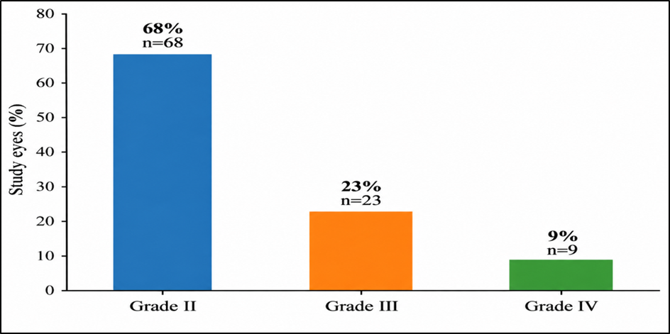
Figure 1. Distribution of study eyes according to pterygium grade
Figure 1 The majority of study eyes were classified as Grade II pterygium, accounting for 68% (n=68) of cases, followed by Grade III in 23% (n=23) and Grade IV in 9% (n=9). The distribution demonstrates that most patients presented with moderate corneal involvement, while advanced pterygium extending to or beyond the pupillary margin was less frequent.
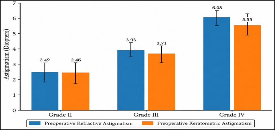
Figure 2. Size-dependent increase in preoperative refractive and keratometric astigmatism across pterygium grades
Figure 2 demonstrates a progressive rise in both preoperative refractive and keratometric astigmatism with increasing pterygium grade. Mean preoperative refractive astigmatism increased from 2.49 ± 0.65 D in Grade II to 3.93 ± 0.465 D in Grade III and 6.08 ± 0.61 D in Grade IV. Similarly, mean preoperative keratometric astigmatism increased from 2.46 ± 0.75 D in Grade II to 3.71 ± 0.63 D in Grade III and 5.55 ± 0.63 D in Grade IV. These findings indicate that larger and more advanced pterygia are associated with a greater corneal astigmatic burden, supporting a clear size-dependent effect on corneal optical distortion.
DISCUSSION:
The present study demonstrates a clear relationship between the extent of pterygium and the magnitude of corneal astigmatism, together with substantial reversibility of this optical distortion following pterygium excision with conjunctival autografting. The most important finding was the progressive increase in both refractive and keratometric astigmatism from Grade II through Grade IV disease. Preoperative refractive astigmatism increased from 2.49 ± 0.65 D in Grade II to 3.93 ± 0.465 D in Grade III and 6.08 ± 0.61 D in Grade IV, while corresponding keratometric astigmatism increased from 2.46 ± 0.75 D to 3.71 ± 0.63 D and 5.55 ± 0.63 D. Following surgery, both refractive and keratometric astigmatism decreased significantly in every grade. The overall refractive cylinder decreased from 3.15 ± 1.258 D to 1.542 ± 0.652 D, whereas keratometric astigmatism decreased from 3.03 ± 1.189 D to 1.475 ± 0.600 D. These findings support the concept that the optical effect of a pterygium is strongly size dependent and that a considerable component of the induced corneal distortion is surgically reversible.
The progressive increase in astigmatism with greater corneal encroachment is consistent with previous quantitative investigations. Fong et al. studied 123 eyes with primary pterygium and showed that larger lesions were associated with greater astigmatic alteration and that postoperative steepening of the previously flattened horizontal corneal meridian was related to pterygium size [11]. Similarly, Kampitak demonstrated a significant positive relationship between pterygium extension and corneal astigmatism in 243 eyes, with the probability of clinically relevant astigmatism increasing substantially when the lesion extended beyond approximately 2.25 mm onto the cornea [12]. These observations provide a physiological explanation for the grade-dependent pattern identified in the present study: as fibrovascular tissue advances centrally, mechanical traction and localized alterations of the tear–corneal interface increasingly distort the anterior corneal curvature. The present findings are also closely aligned with those of Maheshwari, who observed that refractive astigmatism increased significantly with increasing pterygium grade and reported a reduction in mean refractive cylinder from 4.60 ± 2.00 D preoperatively to 2.20 ± 2.04 D following excision [13]. The magnitude of improvement in our study was likewise clinically meaningful, although the exact values differ because of differences in baseline lesion severity, inclusion criteria, surgical technique, and timing of postoperative assessment. Importantly, the reduction was observed not only on subjective or objective refraction but also on keratometry, strengthening the interpretation that the postoperative refractive improvement reflected genuine restoration of corneal curvature rather than simply a change in subjective refraction.
A further notable observation was the predominance of with-the-rule astigmatism, which occurred in 77% of the study eyes, compared with 17% oblique and 6% against-the-rule astigmatism. This pattern is consistent with the characteristic effect of a nasal pterygium on the horizontal corneal meridian. Kampitak similarly reported a predominance of vertically oriented or with-the-rule astigmatic changes in eyes with pterygium [12]. The finding is clinically important because it reinforces the concept that pterygium produces a non-random and anatomically predictable alteration in corneal shape. Nevertheless, pre-existing age-related or idiopathic astigmatism may coexist with the pterygium; therefore, not all measured cylinder in an affected eye should automatically be interpreted as being caused exclusively by the lesion.
Surgical removal produced significant reductions in refractive astigmatism in all grades, but the absolute reduction was greatest in the more advanced lesions. Refractive astigmatism decreased by approximately 1.22 D in Grade II, 2.09 D in Grade III, and 3.28 D in Grade IV. A parallel pattern was observed for keratometric astigmatism, with reductions of approximately 1.23, 1.98, and 2.94 D, respectively. These findings suggest that the greater optical burden imposed by larger pterygia also provides greater potential for postoperative refractive recovery. Fong et al. similarly demonstrated that the magnitude of surgically induced refractive change was related to the preoperative extent of the lesion [11]. However, the persistence of postoperative astigmatism, particularly in Grade IV eyes, indicates that removal does not necessarily return every cornea to an optically normal state. Long-standing stromal remodeling, pre-existing corneal astigmatism, postoperative wound healing, and residual surface irregularity may all contribute to the remaining cylinder.
The keratometric improvement observed in the present study is supported by corneal-topography studies. Bahar et al. demonstrated improvement in corneal astigmatism and visual acuity following pterygium surgery and found that both preoperative and postoperative astigmatism were related to pterygium size [14]. Errais et al., studying patients undergoing pterygium excision with limbal-conjunctival autografting, reported a reduction in simulated keratometric astigmatism from 5.47 ± 3.45 D to 1.79 ± 1.52 D, together with significant improvements in corneal surface regularity and asymmetry indices [15]. Thus, the reduction in conventional keratometric cylinder observed in our study likely represents part of a broader restoration of corneal shape and optical regularity after removal of the fibrovascular tissue.
Modern tomographic studies have further shown that the influence of pterygium surgery is not confined to a single anterior keratometric measurement. Kheirkhah et al. demonstrated measurable postoperative alterations in anterior and posterior corneal surfaces and anterior-segment parameters after pterygium surgery [16]. A related study from the same group showed that excision can modify both anterior and posterior corneal astigmatism, indicating that the refractive consequences of surgery may be more complex than changes in anterior keratometry alone [17]. These findings are relevant when interpreting the present study because conventional keratometry, although clinically practical, does not fully characterize total corneal astigmatism. Incorporation of Scheimpflug tomography or modern corneal topography in future studies would permit more precise quantification of anterior, posterior, and total corneal refractive changes.
Visual acuity improved concurrently with the reduction in astigmatism. Mean decimal BCVA increased from 0.412 ± 0.300 preoperatively to 0.585 ± 0.273 postoperatively, and improvement was observed across all pterygium grades. Seventy percent of patients demonstrated improvement in unaided vision, whereas no eye was documented as having postoperative deterioration. This improvement is biologically plausible because excision reduces the optical distortion caused by corneal flattening and irregularity and, in advanced lesions, may also clear tissue encroaching toward or across the pupillary area. Bahar et al. found significant postoperative improvement in best-corrected visual acuity [14], while Errais et al. reported improvements in both uncorrected and spectacle-corrected visual acuity after successful excision [15]. Therefore, the visual benefit of surgery appears to extend beyond cosmetic or symptomatic relief and includes measurable optical rehabilitation.
The timing of postoperative assessment deserves particular attention. In the present study, final evaluation was performed at approximately 60 days. Although substantial improvement was already evident by this point, postoperative corneal remodeling may continue beyond the early follow-up period, particularly in larger lesions. Nejima et al., in a study of 562 eyes, reported that the time course of corneal topographic restoration depended on preoperative pterygium size and that lesions extending into the pupillary region could require 6–12 months for some topographic parameters to stabilize [18]. Conversely, Kam et al. observed relatively early stability of several keratometric and total corneal power measurements following primary pterygium excision, with values remaining stable during longer follow-up [19]. Taken together, these studies indicate that substantial optical improvement can occur early, but the exact interval required for complete stabilization may depend on lesion size, the parameter measured, and the imaging method used. Consequently, the present 60-day results should primarily be interpreted as evidence of early surgical reversibility, rather than definitive long-term refractive stabilization.
The surgical safety profile was also acceptable. The most frequent postoperative findings were subconjunctival hemorrhage (11%), graft edema or chemosis (9%), and wound dehiscence (5%); conjunctival inclusion cyst occurred in 1%, and recurrence was observed in 2% during the available follow-up. No graft necrosis or Tenon's granuloma was documented. Because the grafts in the present study were secured with sutures, graft-related inflammation and discomfort may differ from those encountered with tissue adhesives. Pan et al., in a meta-analysis comparing fibrin glue with sutures for conjunctival autograft fixation, found shorter operating times and lower recurrence with fibrin glue, although complication profiles varied across studies [20]. The 2% recurrence observed in the present series should nevertheless be interpreted cautiously because follow-up was limited to approximately two months; recurrence is a time-dependent outcome and longer surveillance is necessary before a definitive recurrence rate can be established.
The present study has several strengths. It prospectively evaluated a relatively large cohort of 100 operated eyes, applied a uniform conjunctival autograft technique, and assessed both refractive and keratometric astigmatism. Stratification by pterygium grade also permitted demonstration of a clinically meaningful size-dependent gradient in preoperative astigmatic burden and postoperative improvement. Nevertheless, several limitations should be acknowledged. First, pterygium size was categorized by ordinal grade rather than quantified continuously in millimeters, width, or surface area. This precluded determination of an exact size threshold at which clinically significant astigmatism develops. Second, conventional keratometry was used rather than computerized topography or tomography, preventing assessment of irregular astigmatism, higher-order aberrations, posterior corneal changes, and regional surface asymmetry. Third, astigmatism was analyzed predominantly by cylinder magnitude rather than formal vector analysis, which would provide a more rigorous characterization of changes in both magnitude and axis. Fourth, the relatively short postoperative follow-up restricts conclusions regarding long-term corneal stabilization and recurrence. Finally, the absence of an untreated or alternative-surgical-technique comparator limits causal comparisons between different management approaches.
Despite these limitations, the findings have practical clinical relevance. The progressive increase in refractive and keratometric astigmatism with advancing pterygium grade indicates that visual morbidity may occur before complete visual-axis obstruction. Surgical decision-making should therefore consider not only symptoms and proximity to the pupil but also the refractive consequences of increasing corneal encroachment. The marked postoperative reduction in astigmatism, particularly in Grade III and IV lesions, further suggests that a substantial proportion of pterygium-related corneal distortion is reversible after conjunctival autografting. From a refractive-planning perspective, pterygium should ideally be addressed and corneal measurements allowed to stabilize before definitive spectacle prescription, cataract biometry, toric intraocular lens planning, or corneal refractive surgery.
Overall, this study demonstrates a clear size–burden–reversibility relationship: larger pterygia were associated with greater refractive and keratometric astigmatism, while surgical removal produced significant reductions across all grades, with the largest absolute improvements occurring in the most advanced lesions. These results reinforce the importance of considering pterygium as a functional corneal refractive disorder rather than solely an ocular-surface lesion and support incorporation of lesion size and corneal astigmatism into decisions regarding the timing of surgical intervention.
CONCLUSION:
Pterygium was associated with a clear size-dependent increase in corneal astigmatism, with both refractive and keratometric astigmatism rising progressively from Grade II to Grade IV disease. With-the-rule astigmatism was the predominant preoperative pattern, supporting the characteristic effect of nasal pterygium on corneal curvature. Following pterygium excision with conjunctival autografting, significant reductions were observed in both refractive and keratometric astigmatism across all grades, with the greatest absolute improvement occurring in more advanced pterygia. Visual acuity also improved significantly after surgery, while postoperative complications were predominantly minor.
REFERENCES:
1. Zhou WP, Zhu YF, Zhang B, Qiu WY, Yao YF. The role of ultraviolet radiation in the pathogenesis of pterygia. Mol Med Rep. 2016;14(1):3-15. doi:10.3892/mmr.2016.5223.
2. Di Girolamo N, Chui J, Coroneo MT, Wakefield D. Pathogenesis of pterygia: role of cytokines, growth factors, and matrix metalloproteinases. Prog Retin Eye Res. 2004;23(2):195-228. doi:10.1016/j.preteyeres.2004.02.002.
3. Oldenburg JB, Garbus J, McDonnell JM, McDonnell PJ. Conjunctival pterygia: mechanism of corneal topographic changes. Cornea. 1990;9(3):200-204.
4. Lin A, Stern G. Correlation between pterygium size and induced corneal astigmatism. Cornea. 1998;17(1):28-30. doi:10.1097/00003226-199801000-00005.
5. Mohammad-Salih PAK, Sharif AFMD. Analysis of pterygium size and induced corneal astigmatism. Cornea. 2008;27(4):434-438. doi:10.1097/ICO.0b013e3181656448.
6. Tomidokoro A, Miyata K, Sakaguchi Y, Samejima T, Tokunaga T, Oshika T. Effects of pterygium on corneal spherical power and astigmatism. Ophthalmology. 2000;107(8):1568-1571. doi:10.1016/S0161-6420(00)00219-0.
7. Misra S, Craig JP, McGhee CNJ, Patel DV. A prospective study of pterygium excision and conjunctival autograft with human fibrin tissue adhesive: effects on vision, refraction, and corneal topography. Asia Pac J Ophthalmol (Phila). 2014;3(4):202-206. doi:10.1097/APO.0000000000000006.
8. Gumber A, Nishant P, Sinha S, Goel R, Malik KPS. Changes in corneal curvature and visual acuity in North Indian adults after pterygium excision with modified sutureless, glueless limbal-conjunctival autograft. Indian J Ophthalmol. 2021;69(9):2401-2405. doi:10.4103/ijo.IJO_2747_20.
9. Gumus K, Topaktas D, Goktas A, Karakucuk S, Oner A, Mirza GE. The change in ocular higher-order aberrations after pterygium excision with conjunctival autograft: a 1-year prospective clinical trial. Cornea. 2012;31(12):1428-1431. doi:10.1097/ICO.0b013e3182431465.
10. Clearfield E, Hawkins BS, Kuo IC. Conjunctival autograft versus amniotic membrane transplantation for treatment of pterygium: findings from a Cochrane systematic review. Am J Ophthalmol. 2017;182:8-17. doi:10.1016/j.ajo.2017.07.004.
11. Fong KS, Balakrishnan V, Chee SP, Tan DT. Refractive change following pterygium surgery. CLAO J. 1998;24(2):115-117.
12. Kampitak K. The effect of pterygium on corneal astigmatism. J Med Assoc Thai. 2003;86(1):16-23.
13. Maheshwari S. Effect of pterygium excision on pterygium induced astigmatism. Indian J Ophthalmol. 2003;51(2):187-188.
14. Bahar I, Loya N, Weinberger D, Avisar R. Effect of pterygium surgery on corneal topography: a prospective study. Cornea. 2004;23(2):113-117. doi:10.1097/00003226-200403000-00002.
15. Errais K, Bouden J, Mili-Boussen I, Anane R, Beltaif O, Meddeb Ouertani A. Effect of pterygium surgery on corneal topography. Eur J Ophthalmol. 2008;18(2):177-181. doi:10.1177/112067210801800203.
16. Kheirkhah A, Safi H, Nazari R, Kaghazkanani R, Hashemi H, Behrouz MJ. Effects of pterygium surgery on front and back corneal surfaces and anterior segment parameters. Int Ophthalmol. 2012;32(3):251-257. doi:10.1007/s10792-012-9560-2.
17. Kheirkhah A, Safi H, Molaei S, Nazari R, Behrouz MJ, Raju VK. Effects of pterygium surgery on front and back corneal astigmatism. Can J Ophthalmol. 2012;47(5):423-428. doi:10.1016/j.jcjo.2012.07.002.
18. Nejima R, Masuda A, Minami K, Mori Y, Hasegawa Y, Miyata K. Topographic changes after excision surgery of primary pterygia and the effect of pterygium size on topographic restoration. Eye Contact Lens. 2015;41(1):58-63. doi:10.1097/ICL.0000000000000065.
19. Kam KW, Kuan TA, Belin MW, Young AL. Long-term stability of keratometry, Scheimpflug-derived true net power, and total corneal refractive power after primary pterygium excision. Cornea. 2017;36(11):1358-1363. doi:10.1097/ICO.0000000000001341.
20. Pan HW, Zhong JX, Jing CX. Comparison of fibrin glue versus suture for conjunctival autografting in pterygium surgery: a meta-analysis. Ophthalmology. 2011;118(6):1049-1054. doi:10.1016/j.ophtha.2010.10.033.