Imaging Spectrum of Congenital and Acquired Craniovertebral Junction Abnormalities on 256 Slice Multidetector Computed Tomography.
- Ishita Saini , Post Graduate resident, Department of Radiodiagnosis, Geetanjali Medical College and Hospital, Geetanjali University, Udaipur
- Puneet Kumar Awasthi , Associate Professor, Department of Radiodiagnosis, Geetanjali Medical College and Hospital, Geetanjali University, Udaipur
- Ravinder Kumar Kundu , Professor & Head, Department of Radiodiagnosis, Geetanjali Medical College and Hospital, Geetanjali University, Udaipur
- Parthvi Jaiswal , Post Graduate resident, Department of Radiodiagnosis, Geetanjali Medical College and Hospital, Geetanjali University, Udaipur
Article Information:
Abstract:
Background: Disorders of the craniovertebral junction (CVJ), whether congenital or acquired, constitute a heterogeneous group. They have complex bony relationships and risk of cervicomedullary compromise requiring systematic cross-sectional imaging. Objective: To describe the imaging spectrum and etiology distribution of craniovertebral junction anomalies on 256-slice dual-energy multidetector computed tomography, and to evaluate the correlation of selected clinical and imaging features with neural involvement. Methods: This descriptive cross-sectional study was conducted on 64 eligible patients who were purposively recruited from a tertiary-care radiology department over a period of 18 months. Multiplanar and three-dimensional reconstructions were used to analyse the thin-section CT images. Magnetic resonance imaging was reviewed as clinically indicated. Frequencies and percentages were used to describe categorical variables, which were tested for associations by chi-square or Fisher exact test. Results: Mean age was 42.5±18.7 years; 35 patients (54.7%) were male. Congenital abnormalities and variants were identified in 44 cases (68.8%). Basilar invagination was the most common finding (28/64, 43.8%) followed by atlanto-occipital assimilation (23/64, 35.9%). Cord compression was seen in 18 patients (28.1%) and myelopathy or neural compression in 12 (18.8%). Acquired abnormalities were associated with cord compression (55.0% versus 15.9%, p=0.001) and myelopathy (35.0% versus 11.4%, p=0.038). Basilar invagination was associated with cord compression (53.6% vs 8.3%, p<0.001) and myelopathy (35.7% vs 5.6%, p=0.003). Limb weakness was linked to cord compression (76.2% vs 4.7%, p<0.001). A smaller clivus-canal angle was not independently associated with either outcome. Conclusion: Congenital anomalies were most prevalent but acquired disorders had a greater influence on the nervous system. MDCT provided detailed characterization of osseous morphology and alignment. MRI provided additional information regarding the cord and soft tissues. The pretreatment evaluation should include a combination of etiological classification, multiplanar anatomy, craniometry, clinical findings, and MRI when neural involvement is suspected.
Keywords:
Article :
INTRODUCTION:
The craniovertebral junction (CVJ) comprises the occiput, atlas, axis, supporting joints, and stabilising ligaments. It has to allow for a great range of motion while protecting the lower brainstem, upper cervical cord, cranial nerves and vertebral arteries. Congenital malformations, trauma, degeneration, inflammatory disease and infection may upset these relationships and produce a spectrum of symptoms from neck pain to progressive myelopathy. Diagnosis is often established and anatomical features relevant to management are defined by imaging as clinical manifestations may be delayed or nonspecific [1,3,4]. Due to embryological segmentation and the specialised biomechanics of the occiput-C1-C2 complex, developmental abnormalities frequently occur together and small changes in alignment can lead to neurological consequences [16-18].
Multidetector computed tomography (MDCT) offers thin section acquisition, high spatial resolution, multiplanar reformations and three-dimensional reconstructions. These capabilities are particularly useful for evaluating atlas and axis morphology, atlanto-occipital fusion, odontoid abnormalities, fractures, joint alignment, craniometric relationships [1,3]. Morphometric evaluation of the occipital condyles and foramen magnum is also relevant to surgical planning [19,20]. MRI is complementary to CT in demonstrating the spinal cord, brainstem, ligaments, inflammatory pannus and posterior fossa abnormalities [1,3,7]. Thus, combined approach is important in presence of neurological symptoms along with structural abnormalities.
The CVJ differs from the sub axial cervical spine in development and mechanics. The basiocciput, atlas and axis develop from closely related sclerotomal elements and failure of segmentation may affect several structures at the same time [16,17]. So basilar invagination may be associated with atlanto-occipital assimilation, odontoid dysplasia, atlas arch defects, block vertebrae or posterior fossa abnormalities. These combinations change the orientation of the foramen magnum, clivus, odontoid and upper cervical canal. A finding that seems trivial in isolation can be of clinical significance when it is part of a larger developmental constellation [3,16,18].
Basilar invagination is characterised by abnormal cranial migration of upper cervical spine especially of the odontoid process. In its imaging evaluation, it uses anatomical relationships such as the Chamberlain and McGregor lines, the clivus-canal angle and the relationship of the odontoid to the skull base [3,9,15]. The abnormality can cause ventral indentation of the cervicomedullary junction and is often associated with Chiari I malformation, syringomyelia or alteration in cervical alignment [9,10]. Atlanto-occipital assimilation may further restrict movement of the occiput-C1 articulation and transfer stress to the atlantoaxial joint. Os odontoideum and odontoid hypoplasia may predispose to instability despite preserved alignment on neutral imaging [7, 22, 33].
Acquired disease poses a different set of diagnostic problems. Trauma may disrupt the occipital condyles, atlas, axis or stabilising ligaments. Subtle malalignment can indicate potentially unstable injury [13,24]. Radiographic relationship measurements remain important in suspected occipitovertebral dissociation [26]. Destruction of bone and invasion of adjacent soft tissue may be caused by infectious or neoplastic processes, whereas rheumatoid arthritis may produce synovitis, erosions, ligamentous laxity, pannus and vertical migration. Degenerative change may result in narrow joint spaces, osteophytes, and worsening of symptoms in patients with pre-existing congenital anatomy [5,6]. Thus, accurate etiologic classification is not just descriptive: it influences the need for urgent stabilisation, further MRI, dynamic assessment, or operative planning.
Plain radiographs remain useful for assessment of alignment and dynamics, but the superimposition at the skull base limits characterisation of complex bony anatomy. MDCT overcomes this limitation by allowing isotropic thin-section review in the axial, coronal and sagittal planes. Bone-window images outline fusion, segmentation, fractures, erosions, joint incongruity and the direction of potential fixation corridors. Three-dimensional reconstructions can help the surgeon to understand spatial relationships but source images are still important for diagnosis [1,3,4]. MRI is the best modality to assess the intramedullary signal abnormality, brainstem or cord deformation, ligament injury, pannus, infection, tumour, and associated posterior fossa findings [1,7,13].
Congenital CVJ abnormalities like basilar invagination and atlanto-occipital assimilation have been reported to be a significant burden from published series of the Indian subcontinent [2,4,8]. The relationship of etiology, individual structural abnormalities, symptoms, and neural compromise is of clinical importance because these features affect the pretreatment evaluation. The objective of this study was to describe congenital and acquired CVJ abnormalities on 256-slice dual-energy MDCT and evaluate correlations between certain clinical and imaging variables and cord compression or myelopathy.
MATERIALS AND METHODS:
Study design and participants
A descriptive cross-sectional observational study was conducted over 18 months in the Department of Radiodiagnosis at Geetanjali Medical College and Hospital, Udaipur, a tertiary-care teaching hospital. Patients of any age or sex were eligible when they were clinically suspected of having a CVJ abnormality or were referred for CT evaluation because of neurological symptoms, trauma, suspected instability, or another congenital or acquired condition. Patients were excluded if consent was not provided, image quality precluded diagnostic assessment, or CT could not be performed because of clinical instability or contraindication. Purposive sampling yielded 64 participants, exceeding the prespecified minimum sample of 40.
Participants were recruited from outpatient, inpatient, and emergency referrals using the predefined eligibility criteria. The study population therefore represented a clinically referred cohort rather than a screening sample. Each participant was assigned to an etiological group after review of the clinical history and imaging findings. Congenital and congenital-variant categories included isolated developmental anomalies, congenital abnormalities with superimposed degeneration, and complex combinations of developmental findings. Acquired categories included degenerative, inflammatory, infective, traumatic, postoperative, and other nondegenerative disorders.
Clinical data and imaging protocol
Age, sex, presenting symptoms, and relevant history were recorded using a predefined proforma. CT was performed on a 256-slice dual-energy scanner (Revolution Frontier, GE Healthcare) with the patient supine and the head and neck immobilized. Thin-section axial images covered the occiput through the upper cervical spine. Sagittal and coronal multiplanar reformations were produced for all examinations, and three-dimensional volume-rendered images were generated when they improved delineation of complex anatomy or fractures. Bone windows were used for osseous assessment, with soft-tissue windows reviewed as required.
The field of view included the skull base, foramen magnum, occipital condyles, atlas, axis, and adjacent upper cervical levels. Axial source images were reviewed together with midline and parasagittal reformations for odontoid position, clival orientation, canal caliber, and posterior element morphology. Coronal reformations were used to assess lateral mass symmetry, atlanto-occipital relationships, occipital condylar anatomy, and asymmetric fusion or joint narrowing. Volume-rendered images were used selectively to display complex fusion patterns, congenital segmentation abnormalities, and fracture configuration; they were not used as a substitute for multiplanar source-image review.
MRI was obtained selectively when further evaluation of the cord, brainstem, ligaments, or other soft tissues was clinically required. Available MRI studies were assessed for cord compression, myelopathic signal change, syringomyelia, ligamentous abnormality, and posterior fossa anomalies. CT examinations were reviewed on dedicated workstations by radiologists experienced in musculoskeletal and neuroimaging; ambiguous findings were resolved by consensus.
Clinical and imaging observations were entered on the predefined study proforma. Reviewers identified the dominant abnormality and systematically assessed coexisting congenital or acquired findings. Particular attention was given to combinations that could affect stability or operative access, including basilar invagination with atlanto-occipital assimilation, odontoid abnormalities, block vertebrae, and degenerative change. When MRI was available, its neural and soft-tissue findings were correlated with the MDCT anatomy.
Imaging assessment and outcomes
Assessment included the morphology and alignment of the occiput, atlas, and axis; atlanto-occipital and atlantoaxial relationships; congenital anomalies; acquired traumatic, degenerative, inflammatory, infective, neoplastic, and postoperative changes; instability or vertical translocation; standard CVJ reference lines and angles; canal compromise; cord compression; and secondary changes. The primary outcome was the spectrum and frequency of congenital and acquired abnormalities on MDCT. Secondary outcomes were associated structural findings and neural involvement, supplemented by MRI where available.
Basilar invagination, atlanto-occipital assimilation, block vertebrae, os odontoideum, odontoid hypoplasia or aplasia, and Chiari malformation were recorded as separate imaging variables because more than one could occur in the same patient. Neural outcomes included canal compromise, cord compression, and myelopathy or other neural compression. Myelopathy was based on available MRI evidence of spinal cord signal abnormality in the appropriate anatomical and clinical setting. The analysis also examined whether etiology, basilar invagination, limb weakness, gait disturbance, and a reduced clivus-canal angle were associated with cord compression or myelopathy.
Statistical analysis and ethics
Data were analyzed using descriptive and inferential statistics. Continuous variables are reported as mean ± standard deviation and range; categorical variables as frequencies and percentages. Associations between categorical variables were examined with the chi-square test or Fisher exact test according to expected cell counts. A two-sided p value below 0.05 was considered statistically significant. The institutional ethics committee approved the protocol. Written informed consent was obtained from participants or legal guardians, and data were anonymized before analysis.
Percentages for symptoms, predisposing conditions, and individual imaging findings were calculated using the full cohort of 64 as the denominator. Because abnormalities and symptoms could coexist, percentages within those sets were not expected to sum to 100%. Etiological grouping was mutually exclusive at the broad congenital-versus-acquired level. Inferential comparisons used two-by-two contingency tables. Fisher exact testing was selected when expected cell frequencies were small; otherwise, the chi-square test was used.
RESULTS:
Participant characteristics and clinical presentation
The 64 participants ranged from 6 to 86 years of age (mean 42.5 ± 18.7 years; median 42 years). The two largest age groups were 31-40 and 41-50 years, each with 13 patients (20.3%). Thirty-five participants (54.7%) were male and 29 (45.3%) were female. Neck pain was the most frequent symptom (37/64, 57.8%), followed by limb weakness (21/64, 32.8%), sensory symptoms (14/64, 21.9%), gait disturbance (11/64, 17.2%), and headache (9/64, 14.1%). Trauma and rheumatoid arthritis were each recorded in two patients (3.1%), and tuberculosis in one (1.6%).
Eight patients (12.5%) were aged 20 years or younger, nine (14.1%) were 21-30 years, 13 (20.3%) were 31-40 years, 13 (20.3%) were 41-50 years, 10 (15.6%) were 51-60 years, six (9.4%) were 61-70 years, and five (7.8%) were older than 70 years. Thus, 26 patients (40.6%) presented between 31 and 50 years, while the presence of both pediatric and elderly participants reflected the mixture of developmental and acquired disease. No patient had a recorded cranial nerve symptom, and no participant had a documented history of ankylosing spondylitis.
Table 1: Participant characteristics and major imaging outcomes
|
Characteristic |
n |
% |
|
Male sex |
35 |
54.7 |
|
Neck pain |
37 |
57.8 |
|
Limb weakness |
21 |
32.8 |
|
Sensory symptoms |
14 |
21.9 |
|
Gait disturbance |
11 |
17.2 |
|
Headache |
9 |
14.1 |
|
Congenital abnormalities and variants |
44 |
68.8 |
|
Acquired abnormalities and variants |
20 |
31.2 |
|
Cord compression |
18 |
28.1 |
|
Myelopathy or neural compression |
12 |
18.8 |
|
Canal compromise |
4 |
6.2 |
Etiological and osseous imaging spectrum
Congenital abnormalities and variants accounted for 44 patients (68.8%): 34 isolated congenital cases, six congenital cases with degenerative change, and four complex congenital cases. The 20 acquired cases (31.2%) comprised degenerative, other nondegenerative, inflammatory, infective, traumatic, and postoperative disorders. Basilar invagination was the most common individual abnormality (28/64, 43.8%), followed by atlanto-occipital assimilation (23/64, 35.9%). Findings were not mutually exclusive, and the two leading congenital abnormalities frequently coexisted. Less common findings included block vertebrae, os odontoideum, odontoid hypoplasia or aplasia, and Chiari malformation (Figure 1).
Within the congenital group, isolated congenital abnormalities formed the single largest category (34/64, 53.1% of the full cohort). Congenital abnormalities with degenerative change accounted for six patients (9.4%), and complex congenital configurations for four (6.2%). Among acquired presentations, five patients (7.8%) had primarily degenerative disease, six (9.4%) had another nondegenerative acquired abnormality, three (4.7%) had inflammatory disease, two (3.1%) had infection, two (3.1%) had trauma, and two (3.1%) were postoperative. The broad congenital and acquired totals were used in the association analyses to avoid unstable estimates from these small subgroups.
Table 2: Etiological classification of CVJ abnormalities
|
Category or finding |
n |
% |
|
Congenital isolated |
34 |
53.1 |
|
Congenital with degeneration |
6 |
9.4 |
|
Complex congenital |
4 |
6.2 |
|
Acquired degenerative |
5 |
7.8 |
|
Acquired nondegenerative |
6 |
9.4 |
|
Inflammatory |
3 |
4.7 |
|
Infective |
2 |
3.1 |
|
Traumatic |
2 |
3.1 |
|
Postoperative |
2 |
3.1 |
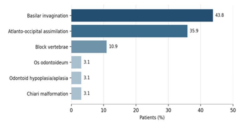
Figure 1: Major craniovertebral junction abnormalities detected in the study cohort. Categories are not mutually exclusive.
Associations with neural compromise
Cord compression was identified in 18 patients (28.1%); myelopathy or neural compression was identified in 12 (18.8%). Acquired etiology, basilar invagination, and limb weakness showed clinically and statistically important associations with neural compromise (Table 3). Acquired abnormalities were associated with a higher frequency of cord compression than congenital abnormalities (55.0% vs 15.9%, p=0.001) and a higher frequency of myelopathy (35.0% vs 11.4%, p=0.038).
Canal compromise without the broader cord-compression classification was documented in four patients (6.2%). No definite atlantoaxial instability, vertical translocation, or brainstem compression was recorded on the documented neutral-position examinations. These zero frequencies should be interpreted in light of selective MRI and the absence of routine dynamic imaging rather than as evidence that such findings are absent from the wider CVJ population.
Among patients with basilar invagination, 15 of 28 (53.6%) had cord compression compared with 3 of 36 patients (8.3%) without basilar invagination (p<0.001). Myelopathy was also more common with basilar invagination (35.7% vs 5.6%, p=0.003). Sixteen of 21 patients with limb weakness (76.2%) had cord compression compared with 2 of 43 without weakness (4.7%, p<0.001). Gait disturbance was not significantly associated with myelopathy (p=0.196). A reduced clivus-canal angle was not associated with cord compression (p=0.966) or myelopathy (p=0.438).
Table 3: Factors associated with cord compression and myelopathy
|
Comparison |
Outcome present |
Outcome absent |
Test |
p value |
|
Acquired etiology: cord compression |
11/20 (55.0%) |
9/20 (45.0%) |
Chi-square |
0.001 |
|
Congenital etiology: cord compression |
7/44 (15.9%) |
37/44 (84.1%) |
|
|
|
Acquired etiology: myelopathy |
7/20 (35.0%) |
13/20 (65.0%) |
Fisher exact |
0.038 |
|
Congenital etiology: myelopathy |
5/44 (11.4%) |
39/44 (88.6%) |
|
|
|
Limb weakness: cord compression |
16/21 (76.2%) |
5/21 (23.8%) |
Fisher exact |
<0.001 |
|
No limb weakness: cord compression |
2/43 (4.7%) |
41/43 (95.3%) |
|
|
|
Basilar invagination: cord compression |
15/28 (53.6%) |
13/28 (46.4%) |
Chi-square |
<0.001 |
|
No basilar invagination: cord compression |
3/36 (8.3%) |
33/36 (91.7%) |
|
|
|
Basilar invagination: myelopathy |
10/28 (35.7%) |
18/28 (64.3%) |
Fisher exact |
0.003 |
|
No basilar invagination: myelopathy |
2/36 (5.6%) |
34/36 (94.4%) |
|
|
|
Gait disturbance: myelopathy |
4/11 (36.4%) |
7/11 (63.6%) |
Fisher exact |
0.196 |
|
No gait disturbance: myelopathy |
8/53 (15.1%) |
45/53 (84.9%) |
|
|
|
Reduced clivus-canal angle: cord compression |
4/14 (28.6%) |
10/14 (71.4%) |
Chi-square |
0.966 |
|
Clivus-canal angle not reduced: cord compression |
14/50 (28.0%) |
36/50 (72.0%) |
|
|
|
Reduced clivus-canal angle: myelopathy |
4/14 (28.6%) |
10/14 (71.4%) |
Fisher exact |
0.438 |
|
Clivus-canal angle not reduced: myelopathy |
8/50 (16.0%) |
42/50 (84.0%) |
|
|
DISCUSSION:
This study achieved its goal of subclassifying CVJ disorders by etiology, delineating their MDCT spectrum and identifying findings relevant to pretreatment evaluation. Three results are of the greatest clinical importance. Congenital disorders were the predominant etiology, with basilar invagination and atlanto-occipital assimilation as the most prevalent osseous abnormalities, accounting for more than 2/3 of the cohort. Second, acquired disorders were less common, but were associated with significantly greater cord compression and myelopathy. Thirdly, the presence of basilar invagination and limb weakness were strongly suggestive of neural compromise, but not isolated reduction in the clivus-canal angle.
The predominance of congenital anomalies is similar to imaging series from India and other tropical locations where basilar invagination, atlanto-occipital assimilation, and segmentation anomalies constitute a large proportion of CVJ disease [2,8,21]. Congenital abnormalities may be asymptomatic until altered biomechanics, minor trauma or superimposed degeneration exhaust compensatory mechanisms. This pattern explains the wide age range and the concentration of the presentations in middle adulthood despite developmental origin [2, 16, 17]. The small male predominance found in the current cohort is in keeping with previous institutional series, although patterns of referral and access may play a part and a biological explanation has yet to be elucidated [1,4].
Basilar invagination was present in 43.8% of patients and was the main structural correlate of neural compromise. Upward migration of the odontoid can lead to narrowing of the ventral subarachnoid space, deformity of the cervicomedullary junction, and co-exist with abnormal cervical alignment or Chiari I malformation [9,10]. The strong associations with both cord compression and myelopathy in this cohort support careful evaluation of odontoid position, ventral compression, alignment and associated anomalies rather than recording basilar invagination as an isolated label. 35.9% have atlanto-occipital assimilation and it is often part of the same spectrum of development. Multiplanar MDCT is helpful as partial fusion and altered condylar or atlantal morphology may be difficult to appreciate on radiographs [3,6].
Occurrence of odontoideum and odontoid hypoplasia or aplasia were rare but noteworthy findings prior to treatment. Os odontoideum can be asymptomatic and allow for atlantoaxial instability and risk cord injury following minor trauma [11,12,22]. Pediatric series also emphasize the risk of instability and neurological injury [23]. Definite atlantoaxial instability was not reported in this work, although dynamic CT or MRI was not routinely done. Therefore, lack of instability on the neutral image should not preclude functional instability when there is a concern for symptoms and anatomy.
Acquired disorders accounted for 31.2% of cases, and were more common for cord compression and myelopathy than congenital disorders. Trauma may cause immediate loss of osseous and ligamentous stability and a rheumatoid or infective disease may produce erosions, pannus, laxity and progressive malalignment. MDCT is used as first-line imaging in the evaluation of fracture morphology and alignment in acute craniocervical trauma, with MRI performed when ligamentous injury, epidural disease, cord injury, and/or unexplained neurologic findings are suspected [13,14]. Surgical management principles for upper cervical and craniocervical injuries are described in additional clinical series [24,25]. The observed relationship between acquired disease and neural compromise, therefore, has a plausible structural underpinning, though with the heterogeneous acquired subgroup and small numbers of cases, inferences are made across conditions.
The most significant clinical variable correlated with cord compression was weakness of the limbs. This helps with rapid cross-sectional imaging if weakness is present with neck pain or a suspected CVJ disease. Gait disturbance, on the other hand, did not significantly associate with myelopathy. Gait impairment is a multi-factorial condition and may be due to pain, cerebellar dysfunction, peripheral disease or early cord dysfunction in which no signal change is seen. In addition, clinical evaluation should be used to guide, but not supplant, imaging. Likewise, the clivus-canal angle is of limited value in defining functional severity or neural compromise here, as it failed to have an independent association with either parameter.
Practical implications for pretreatment evaluation include a structured report that defines the abnormality and its etiology, describes occiput-C1-C2 morphology, outlines joint alignment, documents pertinent craniometric measurements, identifies canal or cord compromise, and emphasizes anatomy that may impact fixation, reduction, or decompression [27-29]. Biomechanical evidence also informs selection among occipito-atlanto-axial fixation constructs [30]. Three-dimensional reconstruction can enhance the communication of complex osseous relationships, but should not be used in place of viewing the thin-section source images and multiplanar reformations [3,5]. MRI is indicated if neurological deficits or suspected ligamentous or inflammatory disease, posterior fossa abnormalities or CT findings of possible neural compromise are present [1,7].
Normal population and age-related variation also need to be considered in interpretation of CVJ measures. Normative studies of bony dimensions and reference relationships based on CT are found to be different between age groups both in adults and children; thus, no single threshold would be applicable without anatomical context [31,32]. Dedicated imaging reviews of os odontoideum and basilar invagination also highlight the need to evaluate for associated anomalies and competing causes of neural compression and to not end the evaluation once the dominant osseous abnormality is identified, as there can be a number of rare co-occurring lesions that may be overlooked [33-35].
CONCLUSION:
Basilar invagination and atlanto-occipital assimilation were the most prevalent abnormalities for this group of MDCTs. Acquired disorders occurred less commonly but had a higher incidence of cord compression and myelopathy. A reduced clivus-canal angle alone did not identify patients at risk of neural compromise; in contrast, basilar invagination and limb weakness were associated with neural compromise. While MRI will continue to be used for the cord, ligaments and soft tissues, MDCT is central to defining osseous morphology, alignment and craniometric relationships. Systematic multimodal assessment gives anatomical information for accurate diagnosis and pretreatment planning.
REFERENCES:
1. Biswas A, Sarkar NR, Pandit A. Magnetic resonance imaging and multidetector computed tomography evaluation of craniovertebral junction abnormalities. J Evid Based Med Healthc. 2021;8(10):532-536. doi:10.18410/jebmh/2021/104.
2. Sankhe SS, Kumar SK. Neuroimaging in craniovertebral anomalies as seen in the tropics. Neuroimaging Clin N Am. 2011;21:879-895. doi:10.1016/j.nic.2011.07.014.
3. Smoker WR. Craniovertebral junction: normal anatomy, craniometry, and congenital anomalies. RadioGraphics. 1994;14(2):255-277. doi:10.1148/radiographics.14.2.8190952.
4. Dhadve RU, Garge SS, Vyas PD, Thakker NR, Shah SH, Jaggi ST, et al. Multidetector computed tomography and magnetic resonance imaging evaluation of craniovertebral junction abnormalities. North Am J Med Sci. 2015;7:362-367. doi:10.4103/1947-2714.163644.
5. Kumar M, Skandesh BM. Role of CT and MRI in evaluation of acquired disorders of craniovertebral junction in a rural based medical college. Int J Contemp Med Surg Radiol. 2018;3(3):C73-C76. doi:10.21276/ijcmsr.2018.3.3.16.
6. Abuzayed B. Computed tomography evaluation of craniovertebral junction. J Neurosci Rural Pract. 2020;11(3):363-364. doi:10.1055/s-0040-1713339.
7. Jain N, Verma R, Garga UC, Baruah BP, Jain SK, Bhaskar SN. CT and MR imaging of odontoid abnormalities: a pictorial review. Indian J Radiol Imaging. 2016;26(1):108-119. doi:10.4103/0971-3026.178358.
8. Talukdar R, Yalawar RS, Kumar M. Imaging in craniovertebral junction abnormalities. IOSR J Dent Med Sci. 2015;14(12):33-49. doi:10.9790/0853-141223349.
9. Brito JNPO, Santos BA, Nascimento IF, Martins LA, Tavares CB. Basilar invagination associated with Chiari malformation type I: a literature review. Clinics. 2019;74:e653. doi:10.6061/clinics/2019/e653.
10. Lin JY, Bao MG, Lin SY, et al. Cervical alignment of patients with basilar invagination: a radiological study. Orthop Surg. 2022;14(3):566-576. doi:10.1111/os.13212.
11. Arvin B, Fournier-Gosselin MP, Fehlings MG. Os odontoideum: etiology and surgical management. Neurosurgery. 2010;66(3 Suppl):A22-A31. doi:10.1227/01.NEU.0000366113.15248.07.
12. Cho S, Shlobin NA, Dahdaleh NS. Os odontoideum: a comprehensive review. J Craniovertebr Junction Spine. 2022;13(3):256-264. doi:10.4103/jcvjs.jcvjs_71_22.
13. Izzo R, Popolizio T, Balzano RF, et al. Imaging of cranio-cervical junction traumas. Eur J Radiol. 2020;129:108960. doi:10.1016/j.ejrad.2020.108960.
14. Joaquim AF, Patel AA. Craniocervical traumatic injuries: evaluation and surgical decision making. Global Spine J. 2011;1(1):37-42. doi:10.1055/s-0031-1296055.
15. Hou Z, Fan T, Fan W, Jian Q, Wang Y. Basilar invagination without atlantoaxial dislocation: treatment by correction of clivus canal angle with interfacet distraction and fixation. BMC Musculoskelet Disord. 2022;23:1138. doi:10.1186/s12891-022-06102-1.
16. Pang D, Thompson DN. Embryology and bony malformations of the craniovertebral junction. Childs Nerv Syst. 2011;27(4):523-564. doi:10.1007/s00381-010-1358-9.
17. Menezes AH. Craniocervical developmental anatomy and its implications. Childs Nerv Syst. 2008;24(10):1109-1122. doi:10.1007/s00381-008-0600-1.
18. Menezes AH, Traynelis VC. Anatomy and biomechanics of normal craniovertebral junction and biomechanics of stabilization. Childs Nerv Syst. 2008;24(10):1091-1100. doi:10.1007/s00381-008-0606-8.
19. Tubbs RS, Griessenauer CJ, Loukas M, Shoja MM, Cohen-Gadol AA. Morphometric analysis of the foramen magnum: an anatomic study. Neurosurgery. 2010;66(2):385-388. doi:10.1227/01.NEU.0000363407.78399.BA.
20. Naderi S, Korman E, Citak G, Guvencer M, Arman C, Senoglu M, et al. Morphometric analysis of human occipital condyle. Clin Neurol Neurosurg. 2005;107(3):191-199. doi:10.1016/j.clineuro.2004.06.007.
21. Ravikanth R, Majumdar P. Embryological considerations and evaluation of congenital anomalies of the craniovertebral junction: a single-center experience. Tzu Chi Med J. 2021;33(2):175-180. doi:10.4103/tcmj.tcmj_62_20.
22. Menezes AH. Os odontoideum: database analysis of 260 patients regarding etiology, associated abnormalities, and literature review. Front Surg. 2023;10:1291056. doi:10.3389/fsurg.2023.1291056.
23. Hedequist DJ, Mo AZ. Os odontoideum in children. J Am Acad Orthop Surg. 2020;28(3):e100-e107. doi:10.5435/JAAOS-D-18-00637.
24. Joaquim AF, Ghizoni E, Tedeschi H, Lawrence B, Brodke DS, Vaccaro AR, et al. Upper cervical injuries: a rational approach to guide surgical management. J Spinal Cord Med. 2014;37(2):139-151. doi:10.1179/2045772313Y.0000000158.
25. Bellabarba C, Mirza SK, West GA, Mann FA, Dailey AT, Newell DW, et al. Diagnosis and treatment of craniocervical dislocation in a series of 17 consecutive survivors during an 8-year period. J Neurosurg Spine. 2006;4(6):429-440. doi:10.3171/spi.2006.4.6.429.
26. Harris JH Jr, Carson GC, Wagner LK, Kerr N. Radiologic diagnosis of traumatic occipitovertebral dissociation: comparison of three methods of detecting occipitovertebral relationships on lateral radiographs of supine subjects. AJR Am J Roentgenol. 1994;162(4):887-892. doi:10.2214/ajr.162.4.8141013.
27. Chandra PS, Kumar A, Chauhan A, Ansari A, Mishra NK, Sharma BS. Distraction, compression, and extension reduction of basilar invagination and atlantoaxial dislocation: a novel pilot technique. Neurosurgery. 2013;72(6):1040-1053. doi:10.1227/NEU.0b013e31828bf342.
28. Goel A, Shah A. Atlantoaxial facet locking: treatment by facet manipulation and fixation. Experience in 14 cases. J Neurosurg Spine. 2011;14(1):3-9. doi:10.3171/2010.9.SPINE1010.
29. Goel A. Treatment of basilar invagination by atlantoaxial joint distraction and direct lateral mass fixation. J Neurosurg Spine. 2004;1(3):281-286. doi:10.3171/spi.2004.1.3.0281.
30. Oda I, Abumi K, Sell LC, Haggerty CJ, Cunningham BW, McAfee PC. Biomechanical evaluation of five occipito-atlanto-axial fixation techniques. Spine. 1999;24(22):2377-2382. doi:10.1097/00007632-199911150-00015.
31. Gupta PP, Dhok AM, Shaikh ST, Patil AS, Gupta D, Jagdhane NN. Computed tomography evaluation of craniovertebral junction in asymptomatic central rural Indian population. J Neurosci Rural Pract. 2020;11(3):442-447. doi:10.1055/s-0040-1712719.
32. Kaya M, Ceylan D, Kacira T, Keskin E, Celenk Y, Yaltirik Bilgin E, et al. Measuring the shape and dimensions of normal bony structures in the craniovertebral junction from computed tomography images of the pediatric age group. Ulus Travma Acil Cerrahi Derg. 2022;28(4):997-1007. doi:10.14744/tjtes.2022.45610.
33. Wang Q, Dong S, Wang F. Os odontoideum: diagnosis and role of imaging. Surg Radiol Anat. 2020;42(2):193-203. doi:10.1007/s00276-019-02351-3.
34. Shah A, Vutha R, Goel A. Basilar invagination associated with brainstem tumor opposite the odontoid process. J Craniovertebr Junction Spine. 2023;14(2):205-207. doi:10.4103/jcvjs.jcvjs_54_23.
35. Donnally CJ III, Munakomi S, Varacallo M. Basilar invagination. In: StatPearls. Treasure Island (FL): StatPearls Publishing; 2025.