Clinical, Radiological, Microbiological and Functional Profile of Bronchiectasis at a Tertiary Care Center: A Cross-Sectional Observational Study.
- Dr. Chebrolu Sai Atchyuth , Junior Resident-3, Department of Respiratory Medicine Dr. Shankarrao Chavan Government Medical College, Vishnupuri, Nanded. Dist. Nanded, Maharashtra 431606.
- Dr. Shrinivas Balajirao Barade , Assistant Professor, Department of Respiratory Medicine Dr. Shankarrao Chavan Government Medical College, Vishnupuri, Nanded. Dist. Nanded, Maharashtra 431606.
- Dr. Vijaykumar Ramappa Kapse , Professor and Head, Department of Respiratory Medicine Dr. Shankarrao Chavan Government Medical College, Vishnupuri, Nanded. Dist. Nanded, Maharashtra 431606.
- Dr. Sanjiv Vithalrao Zangde , Associate Professor, Department of Respiratory Medicine Dr. Shankarrao Chavan Government Medical College, Vishnupuri, Nanded. Dist. Nanded, Maharashtra 431606.
Article Information:
Abstract:
Background: Bronchiectasis is a heterogeneous chronic airway disease characterized by irreversible bronchial dilatation, recurrent respiratory symptoms, infection and progressive structural lung damage. Data describing the clinical, radiological, microbiological and functional phenotype of patients presenting to tertiary-care centers remain important for local service planning and risk stratification. Methods: We conducted a cross-sectional observational study of 127 adults with HRCT-confirmed bronchiectasis attending a tertiary-care respiratory medicine center. Demographic and clinical characteristics, exacerbation frequency, HRCT pattern and distribution, spirometric pattern, sputum culture, comorbidities, complications and disease severity were recorded. Severity was assessed using the FACED score; the Bronchiectasis Severity Index (BSI) was also reported. Categorical associations were assessed using the chi-square test, with two-sided p<0.05 considered statistically significant. Results: The mean age was 46.6±16.7 years and 79 (62.2%) participants were male. Chronic cough and sputum production were present in all participants; dyspnea occurred in 95 (74.8%), wheeze in 52 (40.9%) and hemoptysis in 38 (29.9%). Post-tubercular bronchiectasis was the most frequently reported etiology (60, 47.2%). Cylindrical bronchiectasis was the commonest HRCT pattern (48, 37.8%), while bilateral disease was present in 79 (62.2%) and upper-lobe predominance in 64 (50.4%). Obstructive ventilatory abnormality was the commonest spirometric pattern (42, 33.1%). No bacterial growth was reported in 39 (30.7%); Pseudomonas aeruginosa was the most frequently reported isolate (35, 27.6%). By FACED, 44 (34.6%) had mild, 51 (40.2%) moderate and 32 (25.2%) severe disease. Pulmonary hypertension, cor pulmonale and respiratory failure were significantly associated with FACED severity (chi-square 28.43, 31.84 and 19.87, respectively; all p<0.001). Conclusions: In this tertiary-care cohort, bronchiectasis was characterized by a high burden of chronic productive symptoms, frequent post-tubercular etiology, bilateral and predominantly cylindrical HRCT disease, frequent airflow obstruction, and clinically important cardiopulmonary complications. Severity stratification was associated with pulmonary hypertension, cor pulmonale and respiratory failure. These findings support systematic etiological evaluation, microbiological assessment, lung-function testing and multidimensional severity assessment in bronchiectasis care.
Keywords:
Article :
INTRODUCTION:
Bronchiectasis is a chronic structural airway disorder characterized by permanent bronchial dilatation and distortion resulting from recurrent infection, inflammation, impaired mucociliary clearance and airway injury. The interaction of mucus retention, microbial persistence and host inflammatory responses creates a self-perpetuating cycle that contributes to recurrent exacerbations and progressive respiratory impairment [1,2].
The clinical phenotype is heterogeneous. Patients may have chronic productive cough, dyspnea, wheeze, hemoptysis and recurrent exacerbations, while radiological disease may range from cylindrical to varicose or cystic bronchiectasis. Etiology also varies by population, with post-infectious disease and tuberculosis remaining important in settings with a high burden of previous pulmonary infection [3,4].
The Indian EMBARC/Respiratory Research Network of India registry demonstrated substantial heterogeneity and a significant burden of disease among Indian patients with bronchiectasis [5]. Microbiological colonization, particularly with Pseudomonas aeruginosa, is clinically relevant because chronic infection is associated with recurrent exacerbations and more severe disease phenotypes [6,7].
Multidimensional severity tools such as the FACED score and BSI incorporate clinical, radiological, microbiological and functional parameters to characterize disease severity and prognosis [8,9]. The present study was undertaken to describe the clinical, radiological, microbiological and pulmonary-function profile of patients with bronchiectasis attending a tertiary-care center and to examine the relationship between disease severity and major cardiopulmonary complications.
MATERIALS AND METHODS:
Study design and setting
This was a cross-sectional observational study conducted at a tertiary-care center in the Department of Respiratory Medicine. The thesis methodology states that both retrospective medical-record data and prospective clinical assessment data were used during the study period. Eligible patients were identified consecutively after institutional ethics approval, and written informed consent was obtained before enrollment.
Participants
Adults aged 18 years or older with bronchiectasis confirmed by high-resolution computed tomography (HRCT) of the chest and receiving care at the study center were eligible. The thesis lists cystic fibrosis or major respiratory comorbidity such as chronic obstructive pulmonary disease (COPD), recent major thoracic surgery, and absence of HRCT confirmation as exclusion criteria. The study enrolled 127 participants using consecutive sampling.
Sample size
The thesis calculated a minimum sample size of 115 using an assumed bronchiectasis prevalence of 1.2%, a 95% confidence level and 2% absolute precision, followed by a 10% allowance for incomplete data, yielding a target of 127 participants.
Data collection
Demographic and clinical variables included age, sex, cough, sputum production, dyspnea, wheeze, hemoptysis, chest pain, physical examination findings and annual exacerbation frequency. HRCT was reviewed for morphological pattern, distribution and predominant lobe involvement. Spirometry was recorded when feasible. Sputum samples underwent microbiological examination and culture. Comorbidities and complications were recorded. Disease severity was assessed using the FACED score; BSI categories were also reported in the thesis.
Statistical analysis
Data were entered into Microsoft Excel and analyzed using SPSS. Continuous variables were summarized as mean±standard deviation and categorical variables as frequencies and percentages. Associations between categorical variables were assessed using the chi-square test. One-way ANOVA was described as being used where applicable. All tests were two-sided and p<0.05 was considered statistically significant.
RESULTS:
Demographic and clinical profile
Among 127 participants, the mean age was 46.6±16.7 years. The largest age group was 41–50 years (34, 26.8%). Men constituted 79 (62.2%) and women 48 (37.8%). Chronic cough and sputum production were reported in all participants.
Dyspnea was present in 95 (74.8%), wheeze in 52 (40.9%), hemoptysis in 38 (29.9%) and chest pain in 31 (24.4%). Biphasic coarse crepitations were documented in 114 (89.8%) and clubbing in 24 (18.9%).
Dyspnea severity was most frequently mMRC grade 2 (51, 40.2%), followed by grade 3 (30, 23.6%). Annual exacerbation frequency was 2–3 episodes in 51 (40.2%), four or more in 32 (25.2%), and 0–1 in 44 (34.6%).
Table 1. Selected demographic and clinical characteristics (n=127).
|
Variable |
n (%) |
|
Male sex |
79 (62.2) |
|
Mean age, years |
46.6±16.7 |
|
Chronic cough |
127 (100) |
|
Sputum production |
127 (100) |
|
Dyspnea |
95 (74.8) |
|
Wheeze |
52 (40.9) |
|
Hemoptysis |
38 (29.9) |
|
2–3 exacerbations/year |
51 (40.2) |
|
≥4 exacerbations/year |
32 (25.2) |
Etiology and HRCT findings
Post-tubercular bronchiectasis was the most frequently reported etiology, accounting for 60 (47.2%) participants, followed by post-infectious disease in 28 (22.0%), idiopathic disease in 20 (15.7%), allergic bronchopulmonary aspergillosis in 12 (9.4%) and genetic causes in 7 (5.5%).
Cylindrical bronchiectasis was the most common HRCT morphology (48, 37.8%), followed by mixed (32, 25.2%), cystic (25, 19.7%) and varicose disease (22, 17.3%). Bilateral involvement was present in 79 (62.2%) participants. Upper-lobe predominance was reported in 64 (50.4%), lower-lobe predominance in 28 (22.0%) and multiple-lobe predominance in 35 (27.5%).
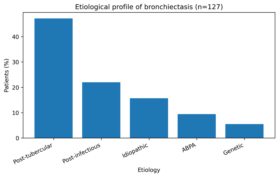
Figure 1. Etiological distribution of bronchiectasis as reported in the thesis (n=127).
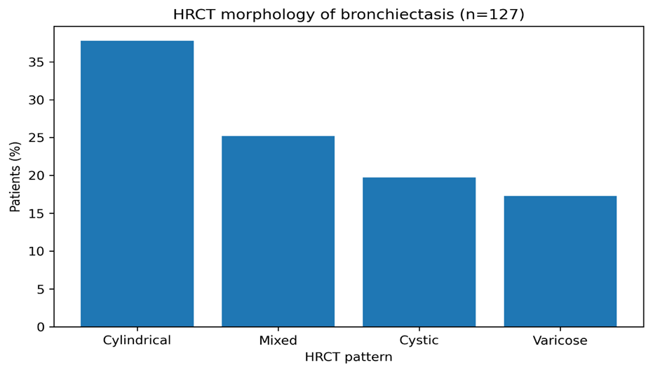
Figure 2. HRCT morphological pattern of bronchiectasis (n=127).
Table 2. Key radiological findings (n=127).
|
HRCT / distribution variable |
n (%) |
|
Cylindrical |
48 (37.8) |
|
Mixed |
32 (25.2) |
|
Cystic |
25 (19.7) |
|
Varicose |
22 (17.3) |
|
Bilateral distribution |
79 (62.2) |
|
Upper-lobe predominance |
64 (50.4) |
Pulmonary function and microbiology
Spirometry was normal in 26 (20.5%), obstructive in 42 (33.1%), restrictive in 21 (16.5%) and mixed in 31 (24.4%); 7 (5.5%) participants were unable to perform the test. Thus, obstruction was the most frequently reported spirometric abnormality.
Sputum culture showed no bacterial growth in 39 (30.7%) participants. Pseudomonas aeruginosa was the most frequently reported isolate (35, 27.6%), followed by Klebsiella species (25, 19.7%), Haemophilus influenzae (18, 14.2%) and budding yeast/fungal hyphae (10, 7.9%).
Table 3. Pulmonary function and sputum microbiology (n=127).
|
Finding |
n (%) |
|
Normal spirometry |
26 (20.5) |
|
Obstructive pattern |
42 (33.1) |
|
Restrictive pattern |
21 (16.5) |
|
Mixed pattern |
31 (24.4) |
|
Unable to perform spirometry |
7 (5.5) |
|
No bacterial growth |
39 (30.7) |
|
Pseudomonas aeruginosa |
35 (27.6) |
|
Klebsiella spp. |
25 (19.7) |
|
Haemophilus influenzae |
18 (14.2) |
Disease severity and complications
According to FACED, 44 (34.6%) participants had mild disease, 51 (40.2%) moderate disease and 32 (25.2%) severe disease; therefore, 65.4% were categorized as moderate-to-severe. By BSI, 30 (23.6%) were mild, 47 (37.0%) moderate and 50 (39.4%) severe.
Pulmonary hypertension was reported in 24 (18.9%), secondary respiratory infection in 20 (15.7%), massive hemoptysis in 18 (14.2%), cor pulmonale in 18 (14.2%) and respiratory failure in 12 (9.4%). Pulmonary hypertension increased from 4.5% in mild disease to 46.9% in severe disease (chi-square=28.43, p<0.001). Cor pulmonale increased from 2.3% in mild disease to 40.6% in severe disease (chi-square=31.84, p<0.001). Respiratory failure occurred in 0% of mild, 3.9% of moderate and 31.3% of severe disease (chi-square=19.87, p<0.001).
The thesis also reports a mean hospital stay of 8.7±3.9 days, with 58 (45.7%) participants hospitalized for 6–10 days. Five deaths (3.9%) were reported; because the study is cross-sectional and the definition and ascertainment period for this outcome require verification, mortality is not used as a principal endpoint in this manuscript.
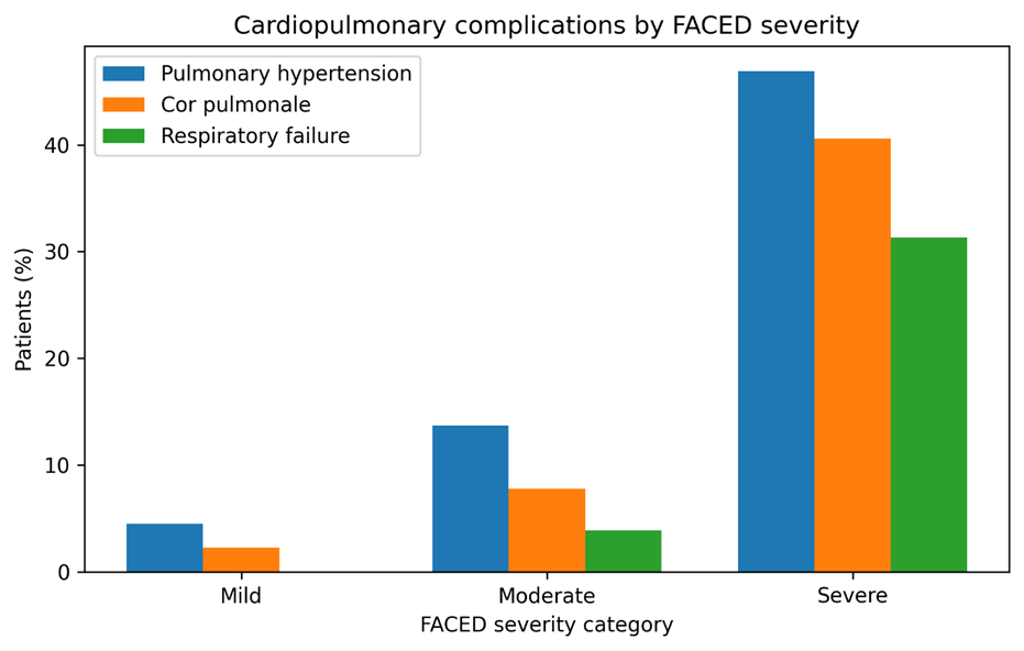
Figure 3. Prevalence of selected cardiopulmonary complications across FACED severity categories.
DISCUSSION:
This study describes a clinically heterogeneous tertiary-care bronchiectasis population with a substantial burden of symptoms, post-tubercular disease, structural lung involvement, airflow limitation and cardiopulmonary complications. The cohort was relatively young compared with many Western bronchiectasis populations, with a mean age of 46.6 years, and men constituted 62.2% of participants. The age and sex distribution is broadly compatible with reports from Indian tertiary-care cohorts, although population structure, referral patterns and local exposures can influence these characteristics [5,10,11].
Chronic cough and sputum production were universal in the reported cohort, while nearly three quarters had dyspnea. These findings are consistent with the clinical phenotype of bronchiectasis, in which impaired mucociliary clearance, mucus retention and chronic airway inflammation drive productive cough and recurrent respiratory symptoms [1,2]. The high prevalence of mMRC grade 2 or higher dyspnea also indicates a clinically important symptom burden in patients reaching tertiary care.
Post-tubercular bronchiectasis was the leading reported etiology, accounting for 47.2% of participants. This finding is particularly relevant in settings where pulmonary tuberculosis remains an important cause of permanent structural lung damage. The Indian bronchiectasis registry has demonstrated distinctive disease characteristics in Indian patients, including a substantial contribution of previous tuberculosis and more advanced disease compared with several international cohorts [5]. The present study therefore adds a single-center tertiary-care perspective to the broader Indian registry experience.
Cylindrical bronchiectasis was the most common radiological pattern, while bilateral disease and upper-lobe predominance were frequent. The upper-lobe distribution may plausibly reflect the high proportion of post-tubercular disease in this cohort, although this study was not designed to establish a causal relationship. HRCT remains central to confirming bronchiectasis, defining its morphology and distribution, and identifying structural consequences of previous infection [2,12].
Obstructive ventilatory abnormality was the most frequent spirometric pattern. This is biologically plausible because airway wall remodeling, mucus retention, chronic inflammation and airway distortion can increase airflow resistance. The presence of restrictive and mixed patterns in a substantial minority also illustrates the functional heterogeneity of bronchiectasis and may reflect coexisting parenchymal disease or extensive structural damage.
Pseudomonas aeruginosa was the most frequently reported organism in the thesis culture table. This is clinically relevant because Pseudomonas is a recognized marker of chronic airway infection and is associated with exacerbation-prone phenotypes and greater disease burden [6,7]. The absence of growth in approximately one third of participants may reflect prior antibiotic exposure, intermittent colonization, specimen quality or limitations of routine culture; these explanations are plausible but were not directly tested in this study.
The association between FACED severity and pulmonary hypertension, cor pulmonale and respiratory failure was one of the most clinically important findings. The prevalence of pulmonary hypertension rose from 4.5% in mild disease to 46.9% in severe disease, while cor pulmonale rose from 2.3% to 40.6%. Respiratory failure similarly clustered in the severe category. These findings support the clinical value of multidimensional severity assessment and are consistent with the established relationship between advanced bronchiectasis, impaired gas exchange and cardiopulmonary complications [8,9,13].
The findings should be interpreted in the context of the study design. A cross-sectional study can describe associations but cannot establish temporal relationships between bronchiectasis severity and complications. In addition, a tertiary-care cohort may over-represent patients with more advanced disease. Nevertheless, the combined clinical, radiological, microbiological and functional characterization provides a useful phenotype of patients encountered in specialized respiratory practice.
Strengths and limitations
Strengths include HRCT confirmation of bronchiectasis, consecutive recruitment, multidimensional clinical characterization, sputum microbiology, spirometry, and use of both FACED and BSI severity categories. The study also evaluates clinically important cardiopulmonary complications rather than limiting assessment to symptoms and imaging.
Limitations include the single-center setting, relatively modest sample size, cross-sectional design, incomplete feasibility of spirometry in some participants, reliance on routine sputum culture, and possible referral bias. The study therefore cannot establish longitudinal progression or treatment effectiveness. In addition, several internal inconsistencies between the thesis protocol, summary tables and the master chart require reconciliation before submission; these are listed separately in the accompanying verification document.
CONCLUSION:
In this tertiary-care cohort of 127 adults with HRCT-confirmed bronchiectasis, chronic cough and sputum production were universal, while dyspnea was common. Post-tubercular disease was the leading reported etiology, cylindrical morphology and bilateral involvement were frequent, and obstructive ventilatory abnormality was the commonest spirometric pattern. Pseudomonas aeruginosa was the most frequently reported sputum isolate. Moderate-to-severe disease constituted nearly two thirds of the cohort by FACED. Increasing FACED severity was significantly associated with pulmonary hypertension, cor pulmonale and respiratory failure. These findings support an integrated approach incorporating etiological assessment, HRCT characterization, microbiological testing, pulmonary function testing and multidimensional severity assessment in bronchiectasis.
Abbreviations
ABPA, allergic bronchopulmonary aspergillosis; BSI, Bronchiectasis Severity Index; COPD, chronic obstructive pulmonary disease; FACED, FEV1, Age, Chronic Colonization, Extension and Dyspnea; FEV1, forced expiratory volume in one second; HRCT, high-resolution computed tomography; mMRC, modified Medical Research Council; PHTN, pulmonary hypertension; PFT, pulmonary function test; TB, tuberculosis.
Declarations
Ethics approval and consent to participate: Institutional ethics approval was obtained and written informed consent was obtained from participants. [Insert the exact ethics committee name, approval/reference number and approval date before submission.
REFERENCES:
1. Chalmers JD, Chang AB, Chotirmall SH, Dhar R, McShane PJ. Bronchiectasis. Nat Rev Dis Primers. 2018;4(1):45. doi:10.1038/s41572-018-0042-3.
2. Polverino E, Goeminne PC, McDonnell MJ, Aliberti S, Marshall SE, Loebinger MR, et al. European Respiratory Society guidelines for the management of adult bronchiectasis. Eur Respir J. 2017;50(3):1700629. doi:10.1183/13993003.00629-2017.
3. Cole PJ. Inflammation: a two-edged sword—the model of bronchiectasis. Eur J Respir Dis Suppl. 1986;147:6-15.
4. Chotirmall SH, Chalmers JD. Bronchiectasis: an emerging global epidemic. BMC Pulm Med. 2018;18:76. doi:10.1186/s12890-018-0629-1.
5. Dhar R, Singh S, Talwar D, et al. Bronchiectasis in India: results from the European Multicentre Bronchiectasis Audit and Research Collaboration and Respiratory Research Network of India Registry. Lancet Glob Health. 2019;7(9):e1269-e1279. doi:10.1016/S2214-109X(19)30327-4.
6. Chalmers JD, Aliberti S, Filonenko A, et al. Characterization of the “Frequent Exacerbator Phenotype” in Bronchiectasis. Am J Respir Crit Care Med. 2018;197(11):1410-1420. doi:10.1164/rccm.201711-2202OC.
7. Aliberti S, Lonni S, Dore S, et al. Clinical phenotypes in adult patients with bronchiectasis. Eur Respir J. 2016;47(4):1113-1122. doi:10.1183/13993003.01899-2015.
8. McDonnell MJ, Aliberti S, Goeminne PC, et al. Multidimensional severity assessment in bronchiectasis: an analysis of seven European cohorts. Thorax. 2016;71(12):1110-1118. doi:10.1136/thoraxjnl-2016-208481.
9. Chalmers JD, Goeminne P, Aliberti S, et al. The Bronchiectasis Severity Index. An International Derivation and Validation Study. Am J Respir Crit Care Med. 2014;189(5):576-585. doi:10.1164/rccm.201309-1575OC.
10. Ramya VH, Sidharthan VH. A cross-sectional study on clinical, demographic, microbiological, and radiological profile of bronchiectasis patients attending a tertiary care teaching center. Indian J Tuberc. 2022;69(4):571-576. doi:10.1016/j.ijtb.2021.08.030.
11. Habesoglu MA, Ugurlu AO, Eyuboglu FO. Clinical, radiologic, and functional evaluation of 304 patients with bronchiectasis. Ann Thorac Med. 2011;6(3):131-136. doi:10.4103/1817-1737.82443.
12. Dimakou K, Triantafillidou C, Toumbis M, et al. Non-CF-bronchiectasis: aetiologic approach, clinical, radiological, microbiological and functional profile in 277 patients. Respir Med. 2016;116:1-7. doi:10.1016/j.rmed.2016.05.001.
13. Keistinen T, Säynäjäkangas O, Tuuponen T, Kivelä SL. Bronchiectasis: an orphan disease with a poorly-understood prognosis. Eur Respir J. 1997;10(12):2784-2787. doi:10.1183/09031936.97.10122784.
14. Martínez-García MA, de la Rosa Carrillo D, Soler-Cataluña JJ, et al. Prognostic value of bronchiectasis in patients with moderate-to-severe chronic obstructive pulmonary disease. Am J Respir Crit Care Med. 2013;187(8):823-831. doi:10.1164/rccm.201208-1518OC.
15. Chalmers JD, Metersky M, Aliberti S, et al. Neutrophilic inflammation in bronchiectasis. Eur Respir Rev. 2025;34(176):2400179. doi:10.1183/16000617.0179-2024.