Occult Malignancy in Non-Functional Kidney: Histopathological Insights From Routine Post- Nephrectomy Evaluations.
- Dr. Tazeen Jeelani , Assistant Professor, Department of Pathology Sher-i-Kashmir institute of Medical sciences soura Srinagar J&K, India.
- Dr. Nuzhat Samoon , Assistant Professor, Department of Pathology Government Medical College Srinagar J&K, India.
- Dr. Jubran Amain , Assistant Professor, Department of Pathology Government Medical College Anantnag J&K, India.
- Dr. Aiffa Aiman , Associate Professor, Department of Pathology Sher-i-Kashmir institute of Medical sciences Soura Srinagar J&K, India.
- Dr. Rukhsana Akhter , Associate Professor, Department of Pathology Sher-i-Kashmir institute of Medical sciences Soura Srinagar J&K, India.
- Dr. Faisal Guru , Associate professor, Department of Medical Oncology Sher-i-Kashmir institute of Medical Sciences Soura Srinagar J&K, India.
- Dr. Ather Hafiz Khan , Assistant Professor, Department of Transfusion Medicine Government Medical College Anantnag J&K, India.
Article Information:
Abstract:
Background: Objectives: The aim of the study was to evaluate the characteristics of patients with Renal tumors detected in the pathological examination performed on simple nephrectomy specimens due to a non-functioning kidney. Material and methods: The results of nephrectomy specimens from patients who underwent simple nephrectomy with the diagnosis of non-functioning kidney between January 2022 and December 2024 were retrospectively analysed. Data pertaining to demographic information, imaging methods, clinical and histopathological features of the patients were collected from the archival section of the Department of Pathology SKIMS. Results: A total of 185 patients with non-functioning kidney who underwent simple nephrectomy between January 2022 and December 2024 were included in the study. 98 (52.9%) patients were male and 87 (47.1%) patients were female. The most common complaint was flank pain (65.9%). The cause of the non-functioning kidney was urinary stones in 138 (74.6%) patients and ureteropelvic/ureterovesical junction stenosis and reflux in 47 (25.4%) patients. Renal tumors were detected in 19 (10.2%) patients. Urothelial cell carcinoma (UCC) was the most common tumor and was detected in 8 patients, 6 patients had renal cell carcinoma (RCC), 3 patients had oncocytic adenoma while as 2 patients had metanephric adenoma. 6 of the 8 patients with UCC had high grade invasive UCC while as 2 patients had low grade papillary UCC. Conclusion: In non-functioning kidneys, tumor may be overlooked due to radiological and clinical diagnostic difficulties. Therefore, careful examination and histopathological evaluation is necessary to detect these incidental renal tumors.
Keywords:
Article :
INTRODUCTION:
papery-thin renal parenchyma (parenchymal thickness <5 mm) on ultrasound or computed tomography (CT), non-visualization of collecting duct system on intravenous pyelography (IVP), less than 10% renal function in di-mercapto-succinic acid scan (DMSA) and a glomerular filtration rate of <10mL/min/1.73 m2 in the affected kidney (1). There are many causes of non-functional kidney such as urolithiasis, chronic pyelonephritis, adult polycystic kidney disease, acquired cystic kidney disease, unsuccessful transplantation and end-stage kidney disease due to medical conditions like diabetes, hypertension, etc (2).
A simple nephrectomy is usually performed in symptomatic patients with chronic infection, pyelonephritis, or obstruction. The advancements in minimally invasive surgical techniques has made simple laparoscopic nephrectomy as a viable alternative to open nephrectomy (3). Histopathological examination of non-functional kidney reveals various findings depending on the underlying cause, like inflammation, hyalinization of glomeruli, tubular atrophy, thyroidisation of tubules, nephrocalcinosis, chronic pyelonephritis and xantho-granulomatous pyelonephritis (XP) (1,2). Several cases have been reported in the literature about incidental detection of tumors of non-functioning kidneys (4,5). The pre-operative diagnosis of renal lesions in non-functioning kidneys with conditions such as chronic pyelonephritis, XP, and obstructing renal stones can be challenging using imaging modalities. In such cases, the diagnosis is often made through histopathological examination.
The main aim of this study was to assess the characteristics of patients who had undergone simple nephrectomy for a non-functioning kidney and incidentally identified to have renal tumors during pathological examination. Retrospective review of the histopathological features of these pat.
MATERIALS AND METHODS:
Patients who had undergone simple nephrectomy with the diagnosis of non-functioning kidney between January 2022 and December2024 were included in the study. The pathology reports of all the cases were reviewed, and the histopathological features and the histological types of the cases with kidney tumors were documented. Patients with a significant renal function or any suspicious lesion diagnosed preoperatively with computed tomography (CT) or magnetic resonance imaging (MRI) and patients <18 years old were excluded from the study.
Statistical analysis: Descriptive statistics to summarize the data, and the frequencies with percentages were reported for categorical variables, using SPSS software.
RESULTS:
A total of 185 patients with non-functioning kidneys who underwent simple nephrectomy between January 2022 and December 2024 were included in the study. Demographic data and clinical features are given in table 1. The median age of the patients was 49 (35-58) years. 98 (52.9%) patients were male and 87 (47.1%) patients were female. The most common complaint was flank pain and it was by 122 (65.9%) patients. Recurrent urinary tract infections and hematuria was reported by 28 (15.1%) and 18 (9.7%) patients respectively. Rest of the 17 patients had various complaints like abdominal swelling, urinary incontinence, nausea, and vomiting. Tumor in non-functioning kidneys was incidentally detected in 19 (10.2%) patients.
The main cause of non-functioning kidney was renal stones in 138 (74.6%) patients, followed by ureteropelvic/ureterovesical junction (UPJ/UVJ) stenosis in 38 (20.6%) cases and vesicoureteral reflux (VUR) in 9 (4.8%) patients. All patients underwent ultrasonography preoperatively. Additionally, 123 patients had non-contrast CT, 32 patients had contrast-enhanced CT, 10 patients had CT-urography, 2 patients had a non-contrast MRI, 5 patients had contrast-enhanced MRI, 3 patients had MR-urography and 10 patients had intravenous pyelography (IVP) before the operation. After careful pathological examination of nephrectomy specimens, chronic pyelonephritis was detected in 138 (74.6%) patients, granulomatous pyelonephritis(fig 2) in 20 (10.8%) patients, tumor in 19 (10.2%) patients, XP in 6 (3.3%) patients, and polycystic kidney in 2 (1.1%) patients. (Table 2)
Table 1. Demographic data and clinical features
|
Number of Patients (n) |
185
|
|
Gender Female, n (%) Male, n (%) |
87 (47.1%) 98 (52.9%) |
|
Median Age, years (IQR) |
49 (35-58) |
|
Laterality Right, n (%) Left, n (%) |
85 (46%) 100 (54%) |
|
Main Complaint Flank pain, n (%) Urinary tract infection, n (%) Hematuria, n (%) Others, n (%) |
122 (65.9%) 28 (15.1%) 18 (9.7%) 17 (9.2%) |
|
Etiology Renal stones, n (%) UPJ/UVJ stenosis, n (%) VUR, n (%) |
138 (74.6%) 38 (20.6%) 9 (4.8%) |
IQR: Interquartile range, UPJ: Ureteropelvic Junction, UVJ: Ureterovesical Junction, VUR: Vesicoureteral Reflux
Table 2. Histopathological characteristics of the patients
|
Chronic Pyelonephritis, n (%) |
138 (74.6%) |
|
Granulomatous pyelonephritis, n (%) |
20 (10.8%) |
|
Tumor, n (%) |
19 (10.2%) |
|
Xantho-granulomatous Pyelonephritis, n (%) |
6 (3.3%) |
|
Polycystic Kidney Disease, n (%) |
2(1.1%) |
Of the tumors detected histopathologically in 19 (10.2%) patients, Urothelial cell carcinoma (UCC) was the most common tumor in 8 patients (42.1%), 6 (31.5%) patients had renal cell renal cell carcinoma (fig 3), 3 (15.7%) patients had oncocytic adenoma while as 2 (10.5%) patients had metanephric adenoma. 6 (75%) of the patients with UCC had high grade invasive UCC (fig.1) while as 2(25%) patients had low grade papillary UCC. 5 (26.4%) cases were benign and 14 (73.6%) were malignant (Table 3). The median size of renal tumors in our study was 2.5cm (1.8-4.7cm).
Table 3. Classification of tumors (n=19)
|
Benign Tumors, n (%) Oncocytic adenoma Metanephric adenoma
|
5 (26.3%) 3 2 |
|
Malignant Tumors, n (%) UCC High grade invasive UCC Low grade papillary UCC RCC Clear cell RCC Papillary RCC Chromophobe RCC |
14 (73.7%)
6 2
3 2 1 |
RCC: Renal Cell Carcinoma; UCC: Urothelial Cell Carcinoma.
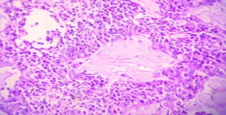
Fig 1: High grade urothelial cell carcinoma showing tumor arranged in irregular nests and trabeculae with tumor cells revealing marked cytological atypia.
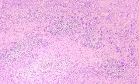
Fig 2: Shows renal parenchyma with chronic tubulointerstitial inflammatory infiltrate along with well formed granulomas and multinucleate giant cells. Tubules show focal thyroidization.
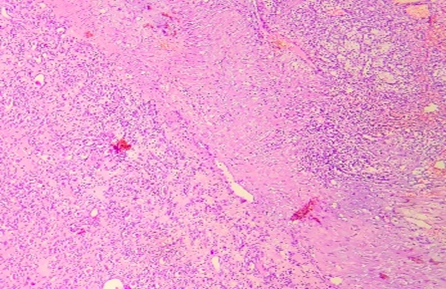
Fig 3: Shows features of chronic pyelonephritis in the left size of image and focus of clear cell RCC towards right upper corner.
DISCUSSION:
Owing to the advances in radiological imaging methods, the diagnosis of renal tumors has become easier with imaging modalities like ultrasonography, CT or MRI in individuals with non-functioning kidneys especially in those with severe inflammation, scarring and urolithiasis. These situations may obscure the presence of a malignant tumor. Simple nephrectomy is the surgical removal of the kidney through Gerota’s fascia and is performed in patients with benign diseases of the kidney. Radical nephrectomy is the removal of the kidney with Gerota’s fascia and is performed in malignant diseases of the kidney (6). Lin et al. evaluated the incidence of RCC in non-functioning kidneys in their study(2), they analysed 311 nephrectomy specimens which included 279 specimens obtained from patients with non-functioning kidneys. They found that 73 (23.5%) nephrectomy specimens had RCC and most of these cases had acquired cystic kidney disease or end-stage renal disease. Clear cell RCC was the most common subtype followed by papillary RCC. However, the type of nephrectomy (simple or radical) was not specified in their study, and it included patients who had a preoperative diagnosis of RCC.
Additionally, the study did not provide information on other types of renal tumors apart from RCC. Angerri et a(3) l. performed simple laparoscopic nephrectomy on 96 patients with urolithiasis and split renal function <15%they detected SCC in 1 (1.04%) patient. In our study, we detected renal tumors in 19 (10.2%) patients and 14 were malignant in the whole group. The number of patients who non-functioning kidney due to urolithiasis was 138; malignant tumors were detected through histopathological evaluation. This rate is lower than the value reported by Zengin et al. In the study conducted by Yeh et al., 47 patients with non-functioning kidneys due to urolithiasis who underwent nephrectomy were evaluated, they detected renal tumors in 24 patients. In 7 of these patients, tumors were detected preoperatively therefore the detection rate of incidental tumors was 36.2%. This is still a very high rate. They speculated that the inadequacy of USG, IVP and retrograde pyelography in detecting malignant lesions may have led to this result and they recommended performing an MRI before the operation in these patients. It is worth noting that the study by Yeh et al (7) is from 2007, and advancements in imaging techniques has since been made. Advancements may have improved the detection of malignancies in non-functioning kidneys.
The median tumor size of the malignant tumors in our study was 2.5 cm (1.8-4.7 cm), which is relatively a significant size. There are several reasons why these tumors cannot be detected in preoperative evaluations. Especially in cases with underlying stone disease and recurrent urinary infections, inflammatory and fibrotic changes in the kidney parenchyma may prevent the detection of the tumor. In cases with UPJ/UVJ stenosis, severe hydronephrosis of the kidney may mask cystic lesions. In our study, 7 (50%) of the patients with malignant lesions were evaluated with non-contrast CT before the operation. Non-contrast CT is mainly used for the evaluation of urinary stone disease and it is insufficient for malignancy evaluation. Four patients with malignant lesions were evaluated with contrast-enhanced CT, 2 patient was evaluated with contrast-enhanced MRI, and 1 patient was evaluated with MRI urography. Different radiological imaging methods were used during the evaluation of these patients with nonfunctioning kidneys depending on the underlying condition. Some of the patients were admitted to emergency service with acute complaints such as flank pain and fever so CT was the preferred imaging method.
For patients with normal kidney function contrast-enhanced CT was performed. In patients with coexisting kidney stones non-contrast CT was used. In patients with UPJ or UVJ obstruction, MRI or CT urography was used depending on the renal function. In cases with solid renal masses, the most important factor for differentiating malignant lesions from benign ones is the presence of contrast. We believe that contrast-enhanced imaging should be recommended to all patients with non-functioning kidneys before simple nephrectomy to minimize the risk of not diagnosing a malignancy. However, it should be kept in mind that even with contrast-enhanced CT and MRI, a concomitant malignant tumor may not be detected in these patients, since the blood flow to the kidney may be impaired in non-functioning kidneys. If a contrast-enhanced examination cannot be performed for any reason (kidney failure, allergy, etc.), a non-contrast MRI should be preferred instead of a non-contrast CT.
Yeh et al. reported that the most common preoperative complaints were flank pain and gross hematuria. In the case series of Jain et al., all patients presented with flank pain. The presenting complaint may also be related to the etiology of the non-functioning kidney (urolithiasis, ureteropelvic junction stenosis). There is no specific symptom that is associated with a specific tumor and malignant tumors can be detected in patients without any complaints. SCC of the kidney is a rare renal neoplasm and it constitutes about 0.8% of renal malignancies (9). Most of the information on renal SCC is obtained from case reports (10-12). It is believed that renal SCC is closely related to kidney stones, chronic infection, and inflammation(13). Jain et al. reported a case series of 4 patients with renal SCC who had staghorn calculi and non-functioning kidneys(4). There is no specific follow-up protocol defined for patients with malignant tumors detected after simple nephrectomy. We considered these patients at high risk for recurrence or metastasis and advice close follow-up. Similar to patients who underwent radical nephrectomy, with frequent contrast-enhanced CT or MRI.
A Metanephric adenoma is an unencapsulated circumscribed small-sized tumor with papillary or tubular architecture and low nuclear grade. Due to their small size, they are usually not detected by imaging methods and are most commonly found incidentally in nephrectomy specimens(14).
The most common histopathological diagnoses in nephrectomies performed for non-functioning kidneys are chronic pyelonephritis, granulomatous pyelonephritis, XP and polycystic kidney (3,15,16). In our study, chronic pyelonephritis was found in 74.6% and XP in 3.3%, which is consistent with the literature. Tuberculosis is a granulomatous disease and it is more commonly observed in developing and low-income countries like ours (17). Tuberculosis may affect the entire urinary system and the kidneys are the most frequently affected urogenital organs (18,19). We also had diagnosis of granulomatous pyelonephritis in 20 cases, and the cases were followed up for tuberculosis.
CONCLUSION:
In non-functioning kidneys, tumors may be overlooked due to radiological and clinical diagnostic difficulties. The possibility of detecting a tumor in histopathological examination after simple nephrectomy is low and most of these tumors are benign. However, malignant lesions like RCC and UCC can be missed preoperatively by imaging methods in some patients with non-functioning kidneys and detected after the pathological examination. Therefore, the possible presence of a concomitant tumor should be considered when evaluating non-functioning kidneys, and macroscopic and microscopic examinations should be done carefully.
REFERENCES:
1. Zengin K, Tanik S, Sener NC, Albayrak S, Ekici M, Bozkurt IH, et al. Incidence of renal carcinoma in non-functioning kidney due to renal pelvic stone disease. Mol Clin Oncol. 2015;3(4):941-943. doi: 10.3892/mco.2015.550.
2. Lin X, Goyal R, Yang XJ. Pathologic characterization of renal epithelial neoplasms arising in nonfunctioning kidneys. Hum Pathol. 2020;97:1-7. doi: 10.1016/j.humpath.2019.09.011.
3. Angerri O, López JM, Sánchez-Martin F, Millán-Rodriguez F, Rosales A, Villavicencio H. Simple Laparoscopic Nephrectomy in Stone Disease: Not Always Simple. J Endourol. 2016;30(10):1095-1098. doi: 10.1089/ end.2016.0281.
4. Jain A, Mittal D, Jindal A, Solanki R, Khatri S, Parikh A, et al. Incidentally detected squamous cell carcinoma of renal pelvis in patients with staghorn calculi: case series with review of the literature. ISRN Oncol. 2011;2011:620574. doi: 10.5402/2011/620574.
5. Akan S, Urkmez A, Tokuc E. Primary Squamous Cell Carcinoma of Renal Pelvis in Non-Functioning Kidneys. J Coll Physicians Surg Pak. 2018;28(6):S148-S150. doi: 10.29271/jcpsp.2018.06.S148.
6. Olumi AF, Blute ML. Open Surgery of Kidney. In: Partin AW, Dmochowski LR, Kavoussi LR, Peters CA, eds. Campbell-Walsh Wein Urology. 12th ed. Elsevier, 2021.
7. Yeh CC, Lin TH, Wu HC, Chang CH, Chen CC, Chen WC. A high association of upper urinary tract transitional cell carcinoma with nonfunctioning kidney caused by stone disease in Taiwan. Urol Int. 2007;79(1):19-23. doi: 10.1159/000102907.
8. Israel GM, Bosniak MA. How I do it: evaluating renal masses. Radiology. 2005;236(2):441-450. doi: 10.1148/radiol.2362040218.
9. Li MK, Cheung WL. Squamous cell carcinoma of the renal pelvis. J Urol. 1987;138(2):269-271. doi: 10.1016/s0022-5347(17)43116-8.
10. Chaurasia Z, Agarwal S, Gupta R, Khatana PS. Pelvicalyceal Squamous Cell Carcinoma: Incidental Diagnosis With Liver Metastasis on Follow Up. Cureus. 2021;13(9):e18345. doi: 10.7759/cureus.18345.
11. Mathur S, Rana P, Singh S, Goyal V, Sangwan M. Incidentally detected squamous cell carcinoma in non-functioning kidney presenting as multi-cystic mass. J Surg Case Rep. 2011;2011(9):8. doi: 10.1093/ jscr/2011.9.8.
12. Foss HE, Krewson BD, Simhal RK, Wang KR, Shah MS. Squamous cell carcinoma of the renal pelvis masquerading as xanthogranulomatous pyelonephritis. Can J Urol. 2023;30(2):11502-11504.
13. Erickson LA. Renal Pyelocalyceal Squamous Cell Carcinoma. Mayo Clin Proc. 2017;92(10):1603-1604. doi: 10.1016/j.mayocp.2017.08.006.
14. Moch H, Cubilla AL, Humphrey PA, Reuter VE, Ulbright TM. The 2016 WHO Classification of Tumours of the Urinary System and Male Genital Organs-Part A: Renal, Penile, and Testicular Tumours. Eur Urol. 2016;70(1):93-105. doi: 10.1016/j.eururo.2016.02.029.
15. Akmal M, Mirza ZI, Murtaza B. Are we performing a lot of simple nephrectomies? J Pak Med Assoc. 2017;67(3):438-441.
16. Peña PA, Torres-Castellanos L, Patiño G, Prada S, Villarraga LG, Fernández N. Minimally invasive nephrectomy for inflammatory renal disease. Asian J Urol. 2020;7(4):345-350. doi: 10.1016/ j.ajur. 2019.09.002.
17. WHO World Health Organization. Global tuberculosis report 2022. Available in: https://www.who.int/teams/global-tuberculosisprogramme/tb-reports/global-tuberculosis-report-2022.
18. Dias N, Pina-Vaz T, Abreu-Mendes P, Azeredo-Costa T, RodriguesPereira P, Silva C, et al. Review of 175 Cases of Tuberculosis Infections Affecting the Urogenital System. Turk J Urol. 2022;48(6):440 445. doi:10.5152/tud.2022.22148.
19. Kulchavenya E, Zhukova I, Kholtobin D. Spectrum of urogenital tuberculosis. J Infect Chemother. 2013;19(5):880-883. doi: 10.1007/ s10156-013-0586-9.