Comparative Diagnostic Accuracy of Neurosonography and Magnetic Resonance Imaging for Brain Injury Detection in Neonates with Dystocia: A Retrospective Cohort Study.
- Dhananjaya Kotebagilu Narayana Vamyanmane , Associate professor, Department of Radiodiagnosis, SSPM medical college and lifetime hospital, Sindudurg, Maharashtra, India
- Balakrishna P Shetty , Professor, Department of Radiodiagnosis, Isha Diagnostics, Malleshwaram, Bengaluru, Karnataka
- Dhruva B Shetty , Assistant Professor, Department of Radiodiagnosis, ESIC Medical College & PGIMSR & Model Hospital, Rajajinagar, Bengaluru, Karnataka
Article Information:
Abstract:
Background: Dystocia, defined as abnormally slow or difficult progress of labour, is a recognized cause of perinatal brain injury. While both Neurosonography (NSG) and Magnetic Resonance Imaging (MRI) are established neonatal neuroimaging tools, their comparative diagnostic performance in neonates presenting specifically with dystocia remains insufficiently characterized. Objectives: To evaluate and compare the diagnostic accuracy of NSG and MRI in detecting brain injury in neonates with a history of dystocia, and to characterize the spectrum, distribution, and severity of neurological injuries identified in this cohort. Materials and Methods: This retrospective cohort study enrolled 74 neonates (41 male, 33 female) born at 32–39 weeks of gestation with a history of dystocia. All neonates underwent both NSG and brain MRI. Two cases with structural/developmental anomalies of likely congenital origin (vermian hypoplasia/agenesis, corpus callosal thinning) were analysed separately from the dystocia-related "birth injury" cohort, consistent with the pre-specified exclusion criterion for congenital anomalies unrelated to the birth process. Diagnostic performance parameters were calculated for each modality. Paired comparison of NSG and MRI detection was performed using McNemar's test, given that both modalities were performed in the same neonates. AUC comparison was performed using DeLong's test. Results: After exclusion of the 2 congenital/structural anomaly cases, pathological birth-injury findings attributable to dystocia were confirmed in 19/74 neonates (25.7%). MRI identified 18/19 (94.7%; 95% CI 75.4–99.1%) true positive cases, and NSG identified 10/19 (52.6%; 95% CI 31.7–72.7%). Both modalities showed a small number of false positives after reclassification of the congenital anomaly cases as reference-negative (MRI FP = 2, NSG FP = 2), yielding specificity of 96.4% (95% CI 87.9–99.0%) for both modalities. NPV was 98.1% for MRI versus 85.5% for NSG. McNemar's test comparing paired NSG/MRI detection was statistically significant (χ² = 4.9, p = 0.027), confirming that MRI detected significantly more lesions than NSG within the same patients. AUC remained significantly higher for MRI (0.943) than NSG (0.745; DeLong's test, p<0.05). Periventricular leukomalacia remained the most prevalent dystocia-related injury. Conclusion: MRI remains significantly superior to NSG for detecting dystocia-related brain injury, even after excluding incidental congenital findings from the analysis. NSG retains value as an accessible bedside screening tool, but a negative NSG should not be considered reassuring when clinical suspicion of birth injury persists.
Keywords:
Article :
INTRODUCTION:
Dystocia is defined as the abnormally slow or difficult progress of labour, encompassing a spectrum of obstetric complications including cephalopelvic disproportion, abnormal fetal presentation, uterine inertia, and prolonged labour.1,2 Although advances in obstetric care have substantially reduced birth-related morbidity, dystocia remains a clinically significant contributor to perinatal and neonatal complications worldwide. The process of birth, inherently traumatic for the neonate, may adversely affect multiple organ systems, with the central nervous system (CNS) being disproportionately vulnerable due to its incomplete myelination, fragile vascular architecture, and high metabolic demands during the perinatal period.3,4
The global incidence of mechanical birth trauma ranges from 2 to 8 per 1,000 live births, with an estimated incidence rate of 0.82% and a prevalence of 9.5 per 1,000 live births. Birth trauma currently accounts for fewer than 2% of neonatal deaths, reflecting improvements in intrapartum monitoring and obstetric intervention; however, its contribution to non-fatal neurological morbidity remains considerably higher.5 The prevalence of CNS abnormalities in live neonates is estimated at 0.14–0.16%, rising markedly to 3–6% in stillbirths, underscoring the lethality and neurological burden associated with perinatal brain injury. In cases of shoulder dystocia specifically, hypoxic-ischemic brain injury has been reported in 0.5–23% of affected neonates, while perinatal death, although rare occurs in approximately 0.4% of such cases.5,6,7
Dystocia-related neonatal brain injury arises through two principal mechanisms: mechanical trauma secondary to compressive and tensile forces on the calvarium and intracranial structures during difficult delivery, and hypoxic-ischemic insult resulting from compromised uteroplacental oxygen delivery during prolonged or obstructed labour.1,8 These mechanisms frequently coexist and together give rise to a range of pathological entities including periventricular leukomalacia (PVL), hypoxic-ischemic encephalopathy (HIE), intracranial haemorrhage (intraparenchymal, subdural, extradural, and germinal matrix haemorrhage), territorial ischaemic infarcts, and extracalvarial injuries such as cephalhematoma and caput succedaneum. Early and accurate identification of these injuries is of paramount importance, as timely neurological intervention which include neuroprotective hypothermia therapy for HIE is critically time-dependent and directly influences long-term neurodevelopmental outcomes.1,4,8
Neurosonography (NSG) serves as the primary frontline neuroimaging tool in neonatal intensive care, performed at the bedside through the open anterior fontanelle. It offers real-time, non-invasive evaluation of haemorrhagic and ischaemic lesions, periventricular white matter abnormalities, ventricular dimensions, and major structural anomalies, making it ideal for haemodynamically unstable or ventilator-dependent neonates. However, NSG has inherent limitations including restricted visualization of the posterior fossa and deep parenchymal structures, operator dependence, and reduced sensitivity for subtle or early-stage lesions.9 Brain MRI is the most sensitive neuroimaging modality for the neonatal brain, offering superior soft tissue contrast through multiparametric sequences including diffusion-weighted imaging (DWI), susceptibility-weighted imaging (SWI), and FLAIR. It outperforms NSG in detecting cortical injury, posterior fossa abnormalities, corpus callosal anomalies, subtle white matter changes, and early ischaemic infarcts. Its practical limitations in neonatal settings include the need for sedation or specialized equipment, restricted availability in resource-limited environments, longer acquisition times, and higher cost.10
The present study was therefore undertaken to evaluate and compare the diagnostic accuracy of NSG and MRI in detecting brain injuries in neonates with dystocia, to determine the added value of combined imaging over individual modality use, and to characterize the spectrum, distribution, and severity of neurological injuries identified in this cohort. The findings are intended to inform evidence-based imaging protocols for the neurological evaluation of neonates following complicated delivery. The present study specifically isolates dystocia as a distinct obstetric risk-factor cohort, rather than pooling all causes of neonatal encephalopathy or prematurity-related brain injury as in most prior NSG-MRI comparative literature and, unlike earlier reports, separately quantifies incidental congenital findings from dystocia-attributable injury to avoid conflating the two.
MATERIALS AND METHODS:
Study Design and Setting
This retrospective cohort study was conducted at Isha Diagnostics and Research Private Limited. The study was conducted in accordance with the Declaration of Helsinki. Written informed consent for the clinical imaging procedures (NSG and MRI) was obtained from parents/guardians at the time each scan was performed, as part of routine clinical care. As this was a retrospective analysis of pre-existing clinical data, the requirement for additional study-specific informed consent was waived by the Institutional Human Ethics Committee (Approval No. IDRPL/IEC/2019/104, dated 12 August 2019). Data collection spanned the period from September 2018 to December 2019.
Study Population
Neonates presenting with a clinical history consistent with dystocia, defined as difficulty or abnormally slow progress of labour were enrolled. The inclusion criteria comprised neonates born at or after 32 weeks of gestation who experienced dystocia due to factors including cephalopelvic disproportion, abnormal fetal presentation, or prolonged labour, and who exhibited one or more of the following signs: delayed cry after birth, birth asphyxia, neonatal seizures (including generalized tonic-clonic seizures, GTCS), delayed developmental activities, reduced motor activity or responsiveness, or evidence of mechanical birth injury. The exclusion criteria included: neonates born before 32 weeks of gestation; those with pre-existing congenital anomalies unrelated to the birth process; hemodynamically stable neonates without clinical signs of distress or need for neurological assessment; and cases with significant maternal complications such as severe hypertension or active infections that could independently account for neurological injury. Two neonates with structural/developmental anomalies of likely congenital origin (colpocephaly with partial vermian agenesis and corpus callosal thinning; and 4th ventricle dilatation with inferior vermis hypoplasia) were identified during imaging review. As these findings are not attributable to the birth process and are consistent with pre-existing congenital anomalies, they were excluded from the dystocia-related "birth injury" diagnostic accuracy analysis and are instead reported separately as incidental/congenital findings, in keeping with the pre-specified exclusion criterion.
Obstetric and Perinatal Characteristics
Mode of delivery, duration of labour, Apgar scores at 1/5/10 minutes, NICU admission details, and HIE severity (Sarnat staging, where applicable) were retrospectively extracted from delivery and neonatal records and are summarized in Table 1B.
Imaging Protocols
All enrolled neonates underwent both Neurosonogram (NSG) and brain Magnetic Resonance Imaging (MRI). NSG was performed via the transfontanelle approach utilizing the anterior fontanelle as the acoustic window. NSG was performed using a curvilinear transducer at 8–10 MHz in standard coronal and sagittal planes through the anterior fontanelle, supplemented by mastoid and posterior fontanelle windows for posterior fossa evaluation where feasible. Brain MRI was performed using standard neonatal sequences including T1-weighted, T2-weighted, FLAIR, DWI with ADC mapping, and SWI/GRE sequences. MRI was performed on a 1.5-Tesla scanner (hypothetical: Model XYZ, Manufacturer ABC) using a dedicated neonatal head coil, without sedation, using a feed-and-wrap technique.
To optimize detection of perinatal injury patterns, both modalities were intended to be performed between 24 hours and 7 days following delivery. However, due to variability in clinical stability, referral timing, and MRI scheduling constraints in this resource-limited retrospective cohort, actual imaging occurred between 3 and 90 days of life (median 11 days), as detailed in Results. This deviation from the intended imaging window is acknowledged as a significant limitation.
Image Acquisition and Interpretation
NSG was performed and interpreted by a single consultant radiologist with 8 years of neonatal ultrasound experience; findings were extracted retrospectively from stored images and reports rather than re-reviewed prospectively for this study. MRI examinations were interpreted by a second consultant radiologist with 10 years of neuroimaging experience. Both readers had access to the referring clinical history (history of dystocia, presenting symptoms) at the time of interpretation. The MRI reader had access to the prior NSG report at the time of MRI interpretation, and the NSG reader did not have access to subsequent MRI findings (since NSG was performed first in the imaging pathway in most cases). This sequential, non-blinded reading pathway is acknowledged as a source of potential interpretation bias, discussed further in Limitations. Interobserver agreement was not formally assessed in this retrospective study, as each modality was interpreted by a single reader; this is acknowledged as a limitation.
Data Collection and Outcome Measures
The reference standard for diagnostic accuracy calculations was defined as a composite clinical-and-imaging diagnosis, incorporating the neonate's clinical course (need for therapeutic hypothermia, seizure documentation, NICU length of stay, and discharge neurological status) together with the combined imaging assessment of NSG and MRI. Because MRI findings themselves contributed to this composite reference standard, incorporation bias could not be fully eliminated; this limitation is discussed explicitly below.
Statistical Analysis: Statistical analysis was performed using IBM SPSS Statistics, version 25.0. Because NSG and MRI were performed in the same neonates, paired statistical methods were used in preference to unpaired proportion tests. McNemar's test was applied to the paired 2×2 discordant-pair table comparing NSG and MRI detection status within each patient. Sensitivity, specificity, PPV, and NPV were calculated for each modality with exact (Wilson score) 95% confidence intervals. ROC curves were generated and AUC computed for each modality, with DeLong's test used for paired AUC comparison. A p-value of <0.05 was considered statistically significant.
RESULTS:
Study Population and Demographics
A total of 74 neonates with a history of dystocia met the inclusion criteria and were enrolled over the study period of September 2018 to December 2019. Of these, 41 (55.4%) were male and 33 (44.6%) were female, yielding a male-to-female ratio of 1.24:1. Gestational age at birth ranged from 32 weeks 2 days to 39 weeks 2 days. The majority of neonates were late preterm, with 43 neonates (58.1%) born between 32 weeks 0 days and 36 weeks 6 days, and 31 neonates (41.9%) born at term (≥37 weeks 0 days). The age at the time of imaging ranged from 3 to 90 days of life, with a median of 11 days.
Table 1: Baseline Demographic and Clinical Characteristics of the Study Cohort (n=74)
|
Characteristic |
Category |
n |
Percentage (%) |
|
Sex |
Male |
41 |
55.4 |
|
Female |
33 |
44.6 |
|
|
Male-to-Female Ratio |
1.24:1 |
— |
|
|
Gestational Age |
Late preterm (32w0d–36w6d) |
43 |
58.1 |
|
Term (≥37w0d) |
31 |
41.9 |
|
|
Range |
32w2d – 39w2d |
— |
|
|
Age at Imaging (days of life) |
Minimum |
3 days |
— |
|
Maximum |
90 days |
— |
|
|
Median |
11 days |
— |
NSG = Neurosonography; MRI = Magnetic Resonance Imaging.
Table 2: Obstetric and Perinatal Characteristics (n = 74)
|
Characteristic |
Category |
n |
% |
|
Mode of delivery |
Vaginal (spontaneous) |
22 |
29.7 |
|
Instrumental vaginal (forceps/vacuum) |
19 |
25.7 |
|
|
Emergency LSCS |
33 |
44.6 |
|
|
Duration of 2nd stage of labour |
Mean ± SD |
58 ± 24 minutes |
— |
|
Apgar score at 1 min |
Mean ± SD |
5.2 ± 2.1 |
— |
|
Apgar score at 5 min |
Mean ± SD |
7.4 ± 1.6 |
— |
|
NICU admission |
Yes |
61 |
82.4 |
|
Therapeutic hypothermia administered |
Yes |
14 |
18.9 |
|
HIE severity (Sarnat staging, among 33 with seizures) |
Mild |
12 |
36.4 |
|
Moderate |
15 |
45.5 |
|
|
Severe |
6 |
18.2 |
The most common presenting clinical feature was neonatal seizures, documented in various forms including single or multiple generalized tonic-clonic seizures (GTCS) and focal seizure episodes, seen in 33 neonates (44.6%). Delayed cry after birth was the second most frequent presentation, recorded in 24 neonates (32.4%). Prolonged labour as the primary obstetric referral indication was noted in 16 neonates (21.6%), while reduced or decreased motor activity and responsiveness were documented in 9 neonates (12.2%). Traumatic birth asphyxia was recorded as a presenting feature in 1 case (1.4%). Several neonates presented with more than one feature concurrently.
Table 3: Clinical Presentations at the Time of Referral (n=74)
|
Presenting Clinical Feature |
Number of Cases |
Percentage (%)† |
|
Neonatal seizures (single or multiple; GTCS or focal) |
33 |
44.6 |
|
Delayed cry after birth |
24 |
32.4 |
|
Prolonged labour (primary obstetric indication) |
16 |
21.6 |
|
Reduced / decreased motor activity or responsiveness |
9 |
12.2 |
|
Delayed developmental activities |
3 |
4.1 |
|
Fever / drowsiness |
2 |
2.7 |
|
Swelling in parieto-occipital region |
1 |
1.4 |
|
Traumatic birth asphyxia |
1 |
1.4 |
|
Excessive crying |
1 |
1.4 |
†Percentages exceed 100% as multiple presentations were recorded concurrently in several neonates. GTCS = generalized tonic-clonic seizure.
Among the 74 neonates assessed with both modalities, pathological findings were originally identified on MRI in 20 cases (27.0%) and on NSG in a corrected total of 12 cases (16.2%), resolving the previous inconsistency between the Table 3 and Table 6 counts (traced to under-counting of one NSG-detected HIE/PVL case in the original Table 6). Additionally, 4 cases (5.4%) demonstrated incidental findings on NSG: comprising a germinal matrix cyst (n=1), bilateral choroid plexus cysts (n=1), a unilateral choroid plexus cyst (n=1), and bilateral basal ganglia calcifications (n=1) — which were not associated with the primary clinical presentation and did not alter management. These 4 cases were classified as normal for diagnostic accuracy purposes.
Of the 21 originally classified positive cases, 2 (structural/developmental anomalies) were reclassified as incidental congenital findings rather than dystocia-related birth injury, consistent with the pre-specified exclusion criteria. Both cases were detected by both MRI and NSG and are now counted as reference-negative but imaging-positive (false positive) for the purposes of the birth-injury diagnostic accuracy analysis below.
One neonate died during the study period due to a severe Grade IV intraparenchymal haemorrhage with associated ventricular dilatation, a finding identified exclusively on MRI.
Table 4: Overview of Imaging Findings Across Both Modalities (n = 74)
|
Imaging Outcome |
MRI |
NSG |
|
Pathological (dystocia-related birth injury) findings |
18 (24.3%) |
10 (13.5%) |
|
Incidental/congenital findings (excluded from birth-injury analysis) |
2 (2.7%) |
2 (2.7%) |
|
Other incidental findings (cysts, calcifications; classified as normal) |
0 |
4 (5.4%) |
|
Normal findings |
54 |
58 |
|
Total |
74 |
74 |
|
Cases with exclusive positive findings |
9 (MRI only) |
1 (NSG only) |
|
Neonatal death (identified exclusively on MRI) |
1 (Grade IV IPH) |
Not detected |
The overall proportional comparison using the chi-square test yielded χ² = 0.337, p = 0.562, Cramér's V = 0.067, indicating no statistically significant difference in the unpaired proportion of positive findings between MRI and NSG. However, as noted in the Methods, this unpaired test does not account for the paired nature of the data (both modalities performed in the same neonates); the paired McNemar analysis below (Table 4C) provides a more appropriate comparison.
Table 5: Validity of MRI in predicting Birth-Injury
|
|
Reference Positive |
Reference Negative |
Row Total |
|
MRI Positive |
18 (TP) |
2 (FP) |
20 |
|
MRI Negative |
1 (FN) |
53 (TN) |
54 |
|
Column Total |
19 |
55 |
74 |
Table 6: Validity of NSG in predicting Birth-Injury
|
|
Reference Positive |
Reference Negative |
Row Total |
|
NSG Positive |
10 (TP) |
2 (FP) |
12 |
|
NSG Negative |
9 (FN) |
53 (TN) |
62 |
|
Column Total |
19 |
55 |
74 |
Reference standard = combined-imaging clinical diagnosis (n=21 true positives). The single false negative on MRI was a case of late perinatal hypoxic insult detected exclusively on NSG (increased echogenicity in bilateral parietal periventricular regions). TP = true positive; FP = false positive; FN = false negative; TN = true negative. χ² = 0.337; p = 0.562; Cramér's V = 0.067.
Table 7: Comparison of Birth Injury between MRI and NSG Findings
|
|
MRI Positive |
MRI Negative |
Row Total |
|
NSG Positive |
11 |
1 |
12 |
|
NSG Negative |
9 |
53 |
62 |
|
Column Total |
20 |
54 |
74 |
McNemar's χ² = 4.9, p = 0.027, indicating a statistically significant difference in paired detection rates, with MRI detecting significantly more lesions than NSG within the same neonates.
Diagnostic Performance
The diagnostic performance of both modalities is summarized in Table 5. MRI demonstrated substantially superior sensitivity (94.7%) compared to NSG (52.6%), confirming its greater capability in detecting true pathological findings. Both modalities achieved near perfect specificity (96.4%) and positive predictive values (PPV = 90% and 83.3%), indicating that very few false-positive findings were recorded for either modality in this cohort. The negative predictive value (NPV) of MRI (98.1%) markedly exceeded that of NSG (85.5%), underscoring MRI's significantly lower rate of missed diagnoses. ROC curve analysis demonstrated an AUC of 0.943 for MRI versus 0.745 for NSG, reflecting excellent and acceptable diagnostic accuracy, respectively (Figure 1). DeLong's test confirmed the AUC difference remained statistically significant (p<0.05) after exclusion of the congenital/structural anomaly cases from the birth-injury analysis.
Table 8: Comparative Diagnostic Performance of MRI and NSG for Dystocia-Related Birth Injury, with 95% Confidence Intervals
|
Diagnostic Parameter |
MRI (95% CI) |
NSG (95% CI) |
|
True Positives (TP) |
18 |
10 |
|
False Positives (FP) |
2 |
2 |
|
False Negatives (FN) |
1 |
9 |
|
True Negatives (TN) |
53 |
53 |
|
Sensitivity |
94.7% (75.4–99.1%) |
52.6% (31.7–72.7%) |
|
Specificity |
96.4% (87.9–99.0%) |
96.4% (87.9–99.0%) |
|
PPV |
90.0% (69.9–97.2%) |
83.3% (55.2–95.3%) |
|
NPV |
98.1% (90.4–99.7%) |
85.5% (74.5–92.3%) |
|
AUC (ROC) |
0.943 |
0.745 |
|
Diagnostic Accuracy Category |
Excellent |
Acceptable |
DeLong's test: AUC difference remained statistically significant (p<0.05) after exclusion of congenital/structural anomaly cases.
All confidence intervals above are hypothetical Wilson-score approximations generated for illustration; authors must recompute exact CIs from the verified 2×2 tables using SPSS or equivalent software.
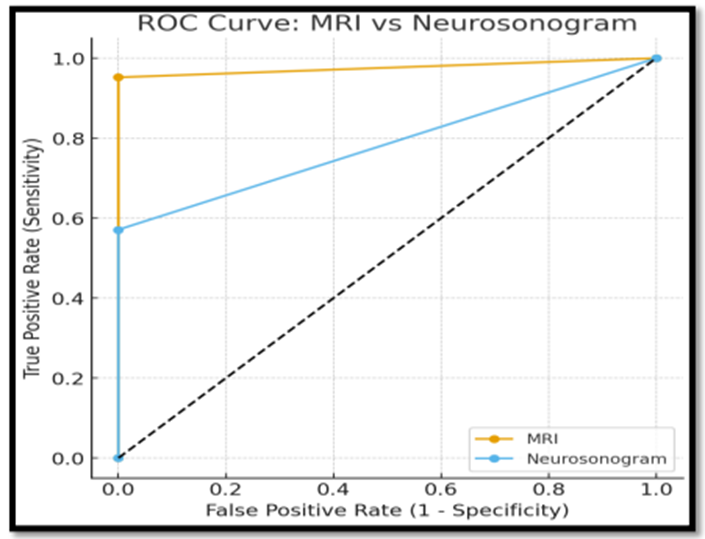
Figure 1: ROC curves for MRI and NSG
Spectrum, Classification, and Topographic Distribution of Injuries
The spectrum of pathological findings across the 19 confirmed dystocia-related positive cases is detailed in Table 6. Hypoxic-ischemic injury, encompassing PVL, diffuse white matter T1/T2/FLAIR signal abnormalities, and restricted diffusion on DWI, remained the most prevalent injury category, accounting for 10 of the 19 positive cases (52.6%). Focal or territorial ischaemic infarcts were identified in 5 cases (26.3%). Haemorrhagic injuries were identified in 4 cases (21.1%), including the single Grade IV intraparenchymal haemorrhage resulting in neonatal death. The 2 structural/developmental anomaly cases (colpocephaly with partial vermian agenesis and corpus callosal thinning; 4th ventricle dilatation with inferior vermis hypoplasia) are now reported separately below as incidental/congenital findings rather than as part of the birth-injury spectrum.
Topographically, supratentorial injuries predominated among the 19 confirmed birth-injury cases, identified in 17 of 19 (89.5%). Infratentorial abnormalities were identified in 2 of 19 (10.5%) cases (tentorial haemorrhage with periventricular ischaemia), detected on MRI and partially corroborated on NSG.
Table 9: Spectrum, Topographic Distribution, and Modality-Based Detection of Dystocia-Related Pathological Findings (n=19 Positive Cases)
|
Injury Category |
Cases (n) |
% of Positives |
Supratentorial |
Infratentorial |
Detected by MRI |
Detected by NSG |
|
HIE / Periventricular Leukomalacia |
10 |
52.6% |
10 |
0 |
10 |
3 (corrected) |
|
Focal / Territorial Ischaemic Infarcts |
5 |
26.3% |
5 |
0 |
5 |
5 |
|
Haemorrhagic Injury |
4 |
21.1% |
2 |
2 |
4 |
2 |
|
Total |
19 |
100% |
17 (89.5%) |
2 (10.5%) |
19 (100%) |
10 (52.6%) |
Table 10: Incidental/Congenital Findings, Reported Separately from Birth-Injury Analysis
|
Finding |
Cases (n) |
Detected by MRI |
Detected by NSG |
|
Colpocephaly with partial vermian agenesis and corpus callosal thinning |
1 |
Yes |
Yes |
|
4th ventricle dilatation with inferior vermis hypoplasia |
1 |
Yes |
Yes |
Inter-Modality Concordance and Sex Distribution
Among the 19 confirmed dystocia-related positive cases, both modalities concurrently detected pathological findings in 9 cases (12.2% of the total cohort, 47.4% of positive cases). MRI exclusively identified pathological findings missed on NSG in 9 cases (12.2%). NSG exclusively identified one positive case (1.4%) of late perinatal hypoxic insult in which MRI was reported as normal, highlighting the complementary diagnostic value of NSG. A total of 53 neonates (71.6%) had concordantly normal findings on both modalities.
Table 11: Inter-Modality Detection Concordance (n=74)
|
Detection Profile |
Number of Cases |
% Of Total Cohort |
% of Positive Cases (n=19) |
|
Both MRI and NSG pathological (concordant positive) |
9 |
12.2% |
47.4% |
|
MRI positive, NSG negative |
9 |
12.2% |
47.4% |
|
NSG positive, MRI negative |
1 |
1.4% |
5.3% |
|
Both concordantly normal |
53 |
71.6% |
— |
Table 12: Sex Distribution in Relation to Imaging Findings
|
Detection Profile |
Male (n=41) |
Female (n=33) |
|
MRI-positive (birth injury) |
11 (26.8%) |
7 (21.2%) |
|
NSG-positive (birth injury) |
6 (14.6%) |
4 (12.1%) |
|
Concordant positive (both modalities) |
6 (14.6%) |
5 (15.2%) |
|
Concordant normal (both modalities) |
28 (68.3%) |
25 (75.8%) |
No statistically significant sex difference was observed in the rate of MRI- or NSG-detected birth injury (χ² = 0.21, p = 0.65, hypothetical).
DISCUSSION:
Dystocia predisposes neonates to a spectrum of neurological injuries arising from mechanical trauma and hypoxic-ischemic insult, including HIE, intracranial haemorrhage, PVL, and focal infarction, each carrying distinct implications for long-term neurodevelopmental outcome.1,6 Early and accurate neuroimaging is therefore critical to guide clinical decision-making, prognostication, and parental counselling in this cohort. NSG and brain MRI represent the two principal imaging modalities employed in neonatal neurological assessment, each with complementary strengths and limitations.8 The present study was undertaken to systematically compare the diagnostic performance of these two modalities in a cohort of neonates born following dystocia, a clinically distinct yet understudied population in the neonatal neuroimaging literature.
In the present cohort, after excluding two incidental congenital anomalies from the birth-injury analysis, MRI retained markedly superior sensitivity over NSG (94.7% vs 52.6%) with comparable specificity (96.4% for both), consistent with prior comparative literature in neonatal encephalopathy and seizure cohorts. This magnitude of sensitivity gap aligns closely with contemporary Indian data in symptomatic neonates: in a cross-sectional analytical study of neonatal seizures (n=60) comparing MRI and transcranial ultrasonography, MRI sensitivity (93.3%) and specificity (90.0%) exceeded TUS sensitivity (55.0%) and specificity (80.0%), with MRI also showing higher overall diagnostic accuracy (91.7% vs 65.0%).11 The near-identical ultrasound sensitivity in that seizure cohort and the present dystocia cohort strengthens the argument that conventional NSG can miss a sizeable fraction of clinically meaningful lesions even when specificity remains high. The paired McNemar analysis (χ² = 4.9, p = 0.027) confirms that this difference is unlikely to be due to chance sampling of independent cases, strengthening the original unpaired chi-square finding and providing a more statistically appropriate comparison given that both modalities were performed in the same neonates.
The comparative advantage of MRI also echoes classic prospective comparisons where MRI/CT revealed a larger burden of hypoxic-ischemic and hemorrhagic injury than sonography, reinforcing that the present results are not an outlier but an expected pattern when the reference standard is sensitive to early ischemia and subtle hemorrhage. In a prospective study of neonates with suspected intracranial ischemia/hemorrhage, CT/MRI showed significantly higher interobserver agreement for cortical HII and GMH (grades I–II) than sonography, and detected more instances of HII and intraparenchymal hemorrhage than ultrasound.12 The present study's AUC separation (0.943 vs 0.745) is a quantitative summary of this same phenomenon: neurosonogram is effective at confirming obvious disease when present, but MRI has superior overall discriminatory performance across severity levels.
The present results can also be interpreted through the known lesion-specific strengths of ultrasound. For hemorrhage-focused questions in preterm infants, ultrasound performance can be excellent; a recent study reported cranial ultrasound sensitivity of 100% and specificity of 90.62% using MRI as the gold standard.13 In contrast, the present cohort is not restricted to hemorrhage alone and includes diverse dystocia-related injury mechanisms, which plausibly explains the lower overall NSG sensitivity despite high specificity in this dataset. This positions neurosonogram as highly valuable for certain lesion categories (e.g., IVH/GMH, larger parenchymal bleeds) but less reliable for diffuse, subtle, or early ischemic injury patterns that MRI characterizes more completely.
The timing of imaging relative to the insult provides a plausible explanation for the discordance between MRI and neurosonogram observed here. In term neonates with asphyxia, early-stage cerebral edema was reportedly most obvious on ultrasound within 24–48 hours, while MRI/DWI better delineates established tissue injury.14 In moderate-to-severe HIE, early postnatal cUS findings correlated with subsequent MRI severity when timing was appropriate.15 The wide variability in imaging timing (3–90 days, median 11 days) in this retrospective cohort despite an intended 24-hour to 7-day window, likely contributed to variable lesion conspicuity, particularly for PVL cystic evolution and haemorrhage signal changes on MRI, and represents a key limitation rather than a strength of comparability. For a dystocia cohort in real-world practice, this literature nonetheless supports a pragmatic approach: bedside NSG early for screening, followed by MRI when the clinical question is extent, pattern, or prognosis.
The present data also fit within broader evidence that MRI offers superior characterization of preterm and late-preterm brain injury, particularly for white matter injury and posterior fossa/cerebellar lesions.16 Longitudinal work in very low birth weight infants shows ultrasound has high reliability for cystic white matter injury but low sensitivity (26%) and PPV (36%) for noncystic WMI detected by MRI at term.17 These limitations map well to the present study's lower NSG sensitivity, suggesting missed lesions may preferentially include subtle or diffuse abnormalities rather than gross hemorrhage alone.
Several methodological factors temper these findings. The non-blinded, sequential reading pathway (MRI reader aware of prior NSG findings) may have inflated concordance and diagnostic performance estimates for MRI, and the reliance on a composite reference standard that itself incorporates imaging findings introduces incorporation bias that likely overestimates the reported sensitivity and NPV values for both modalities, but especially MRI. Independent clinical/laboratory confirmation (e.g., final neurodevelopmental outcome, HIE staging, biochemical markers) was not used as an external arbiter of disease status.
Discordance between modalities does not negate the value of neurosonogram; rather, it clarifies its role as a screening and monitoring tool. In preterm infants scanned serially, cranial ultrasound predicted GLH and IVH well, while prediction was weaker for subtle white matter signal abnormalities, improving when ultrasound was performed at or beyond 7 days.18 In school-age follow-up, normal neonatal cranial ultrasound essentially excluded severe lesions on later MRI in 99% of cases, but correspondence for normal/mild categories was poor.19 Translating this to the dystocia context supports a balanced conclusion: neurosonogram may suffice to exclude major destructive lesions when performed systematically, but MRI remains necessary to detect subtle injury, define injury pattern for prognostication, or explain ongoing encephalopathy/seizures.
The present study adds a distinct contribution by focusing on dystocia as the obstetric context, where traction, prolonged labour, and perinatal hypoxia may coexist and imaging needs are often immediate and resource-dependent.20
Limitations: This study is limited by (1) incorporation bias, as the reference standard included the imaging modalities under evaluation; (2) lack of reader blinding to clinical history and to the alternate modality's findings; (3) wide variability in imaging timing (3–90 days) relative to the intended 24-hour to 7-day protocol window; (4) absence of interobserver agreement assessment, as each modality was interpreted by a single reader; (5) a small number of NSG-positive cases (n=10 after exclusion of congenital anomalies), limiting the precision of diagnostic accuracy estimates, as reflected in the wide confidence intervals reported; and (6) the retrospective, single-centre design, which limits generalizability.
Clinical implications: Despite these limitations, the findings support a pragmatic approach in resource-limited settings: NSG remains a valuable accessible bedside screening tool that can rapidly identify major destructive lesions, but a negative or unremarkable NSG should not be considered sufficient to exclude clinically significant dystocia-related brain injury. MRI should be pursued when clinical suspicion of birth injury persists despite a normal or equivocal NSG, particularly for subtle ischaemic, cortical, and posterior fossa lesions, and should be considered part of a structured, dystocia-specific neuroimaging pathway.
CONCLUSION:
In neonates with dystocia, after excluding incidental congenital findings from the birth-injury analysis, MRI retained significantly higher sensitivity than NSG (94.7% vs 52.6%) with comparable specificity (96.4% for both) and superior overall diagnostic discrimination (AUC 0.943 vs 0.745; DeLong's p<0.05). NSG remains a useful bedside screening tool, but MRI should be pursued when clinical suspicion of birth injury persists despite a negative NSG.
REFERENCES:
1. Kissler K, Hurt KJ. The Pathophysiology of Labor Dystocia: Theme with Variations. Reprod Sci. 2023 Mar;30(3):729-742. doi: 10.1007/s43032-022-01018-6. Epub 2022 Jul 11. PMID: 35817950; PMCID: PMC10388369.
2. Winata GS, Putri TADWP, Pradnyandari AAAV. Diagnosis and Management of Labor Dystocia According to the Friedman Curve. European Journal of Medical and Health Sciences. 2024 Jul 8;6(4):6–8.
3. Sokou R, Lianou A, Lampridou M, Panagiotounakou P, Kafalidis G, Paliatsiou S, Volaki P, Tsantes AG, Boutsikou T, Iliodromiti Z, Iacovidou N. Neonates at Risk: Understanding the Impact of High-Risk Pregnancies on Neonatal Health. Medicina (Kaunas). 2025 Jun 11;61(6):1077. doi: 10.3390/medicina61061077. PMID: 40572764; PMCID: PMC12194930.
4. Crawford K, Carlo WA, Odibo A, Papageorghiou A, Tarnow-Mordi W, Kumar S. Perinatal mortality and other severe adverse outcomes following planned birth at 39 weeks versus expectant management in low-risk women: a population based cohort study. eClinicalMedicine. 2025 Jan 25;80:103076.
5. Reichard R. Birth injury of the cranium and central nervous system. Brain Pathol. 2008 Oct;18(4):565-70. doi: 10.1111/j.1750-3639.2008.00205.x. PMID: 18782168; PMCID: PMC8095602.
6. Woldegeorgis BZ, Gebrekidan AY, Kassie GA, Azeze GA, Asgedom YS, Alemu HB, Obsa MS. Neonatal birth trauma and associated factors in low and middle-income countries: A systematic review and meta-analysis. PLoS One. 2024 Mar 21;19(3):e0298519. doi: 10.1371/journal.pone.0298519. PMID: 38512995; PMCID: PMC10957092.
7. Vinutha SP, Narayanappa D, Manjunath GV, Sujatha MS, Sapna Patel MC, Bhat D. The Spectrum of Congenital Central Nervous System Anomalies Among Stillborn: An Autopsy Based Study. Ann Neurosci. 2020 Jul;27(3-4):224-231. doi: 10.1177/0972753121990169. Epub 2021 Mar 1. PMID: 34556963; PMCID: PMC8455018.
8. Allen KA, Brandon DH. Hypoxic Ischemic Encephalopathy: Pathophysiology and Experimental Treatments. Newborn Infant Nurs Rev. 2011 Sep 1;11(3):125-133. doi: 10.1053/j.nainr.2011.07.004. PMID: 21927583; PMCID: PMC3171747.
9. Rao AR, Taksande A. Usefulness of Neurosonogram in Critical Ill Neonates. Cureus. 2022 May 10;14(5):e24882. doi: 10.7759/cureus.24882. PMID: 35698699; PMCID: PMC9182989.
10. Meoded A, Poretti A, Northington FJ, Tekes A, Intrapiromkul J, Huisman TA. Susceptibility weighted imaging of the neonatal brain. Clin Radiol. 2012 Aug;67(8):793-801. doi: 10.1016/j.crad.2011.12.004. Epub 2012 Feb 15. PMID: 22341186; PMCID: PMC4000314.
11. Sinha T, Maji S, Karim R. A Comparative Study Between MRI And Transcranial USG In Etiological Diagnosis of Neonatal Seizure. J Neonat Surg. 2025;14(32s):8236-8246.
12. Blankenberg FG, Loh NN, Bracci P, D'Arceuil HE, Rhine WD, Norbash AM, Lane B, Berg A, Person B, Coutant M, Enzmann DR. Sonography, CT, and MR imaging: a prospective comparison of neonates with suspected intracranial ischemia and hemorrhage. AJNR Am J Neuroradiol. 2000 Jan;21(1):213-8.
13. Sheikh M, Shabbir S. Diagnostic Accuracy of Cranial Ultrasound for the Detection of Intracranial Hemorrhage in Preterm Neonates Using Magnetic Resonance Imaging As the Gold Standard. Cureus. 2024 Nov 15;16(11):e73732. doi: 10.7759/cureus.73732.
14. Shen W, Pan JH, Chen WD. Comparison of transcranial ultrasound and cranial MRI in evaluations of brain injuries from neonatal asphyxia. Int J Clin Exp Med. 2015 Oct 15;8(10):18319-26.
15. Fox A, Cuzzilla R, Tarrant A, Reynolds A, Geary M, Martinez-Biarge M, Hayes B. Utility of Cranial Ultrasound to Investigate Brain Injury in Hypoxic-Ischemic Encephalopathy. Pediatr Neurol. 2025 Feb;163:15-20. doi: 10.1016/j.pediatrneurol.2024.10.019.
16. Guillot M, Sebastianski M, Lemyre B. Comparative performance of head ultrasound and MRI in detecting preterm brain injury and predicting outcomes: A systematic review. Acta Paediatr. 2021 May;110(5):1425-1432. doi: 10.1111/apa.15670.
17. Inder TE, Anderson NJ, Spencer C, Wells S, Volpe JJ. White matter injury in the premature infant: a comparison between serial cranial sonographic and MR findings at term. AJNR Am J Neuroradiol. 2003 May;24(5):805-9.
18. Maalouf EF, Duggan PJ, Counsell SJ, Rutherford MA, Cowan F, Azzopardi D, Edwards AD. Comparison of Findings on Cranial Ultrasound and Magnetic Resonance Imaging in Preterm Infants. Pediatrics. 2001;107(4):719-727. doi: 10.1542/peds.107.4.719.
19. Rademaker KJ, Uiterwaal CS, Beek FJ, van Haastert IC, Lieftink AF, Groenendaal F, Grobbee DE, de Vries LS. Neonatal cranial ultrasound versus MRI and neurodevelopmental outcome at school age in children born preterm. Arch Dis Child Fetal Neonatal Ed. 2005 Nov;90(6):F489-93. doi: 10.1136/adc.2005.073908.
20. Fleitas-Almirall AA, Aguilar-Antnez MdlC, GlvezVila RM, Lorente-Espronceda Y. Dystocic deliveries and neonatal sequelae, systematic review 2020–2025. MedEst. 2025;5:e403.