Diagnostic Accuracy Of Mri In Rotator Cuff Injuries: Correlation With Arthroscopic Findings.
- Divyansh Yadav , Junior Resident, Department of Radiodiagnosis, Sarvodaya hospital, Faridabad
- Akanksha , Assistant Professor, Department of Anaesthesiology, Pt. B.D. Sharma PGIMS, Rohtak.
- Deepshiksha , Junior Resident, Department of Ophthalmology, Pt. B.D. Sharma PGIMS, Rohtak
- Ankit , Junior Resident, Department of Medicine, SMS medical college, Jaipur
- Amit Kumar , Assistant Professor, Department of Anaesthesiology, N.C Medical College, Panipat.
Article Information:
Abstract:
Background: Rotator cuff injuries are common musculoskeletal disorders that significantly impact shoulder function. Magnetic Resonance Imaging (MRI) has become a pivotal diagnostic tool for assessing rotator cuff injuries, providing detailed anatomical information. This study aims to investigate the correlation between preoperative MRI findings and arthroscopic observations in patients with rotator cuff injuries. Methods: A prospective cross-sectional, observational analysis was conducted on a cohort of 70 patients who underwent both preoperative MRI and subsequent arthroscopic evaluation for suspected rotator cuff injuries. MRI reports were independently reviewed and findings were compared with intraoperative arthroscopic observations. Parameters such as tear size, location, and degree of muscle involvement were analyzed for concordance between the two diagnostic modalities. Results: Preliminary findings reveal a substantial correlation between preoperative MRI assessments and arthroscopic findings in rotator cuff injuries. Sensitivity and specificity of MRI in identifying tear characteristics were determined, highlighting the accuracy of MRI in diagnosing the extent and nature of rotator cuff pathology. Additionally, discrepancies between the two methods were explored, shedding light on potential limitations and areas for improvement. Conclusion: This study underscores the significance of MRI as a reliable diagnostic tool for evaluating rotator cuff injuries, demonstrating a strong correlation with arthroscopic findings. Understanding the strengths and limitations of each modality is crucial for optimizing preoperative planning and enhancing overall patient care in the management of rotator cuff pathologies.
Keywords:
Article :
INTRODUCTION:
Shoulder pain and restriction of movements are common and a major health concern especially in elderly and post traumatic injuries. It is second most frequent acute musculoskeletal complaint in primary care and third most common cause of regional pain syndrome in the population.1,2
Of all the causes for shoulder pain in patients aged 40 years or above, rotator cuff musculotendinous unit injuries accounts for up to 85%. These symptoms results in significant morbidity including sleep disturbances and psychological distress resulting in increased health care utilization or reaching to chiropractors and undergoing investigations.3,4
In workup of patients with rotator cuff tears the role of imaging is to guide treatment decisions. Several radiological imaging techniques including ultrasound, MRI and arthrography are used to evaluate tears of rotator cuff. Ultrasound of rotator cuff is quick and relatively painless. Its accuracy for diagnosing both partial and full thickness tear is very high. The size of tear can be classified and the findings used as the basis for management decisions. The real time capability of ultrasound in conducting dynamic studies in the shoulder is a great asset. The simplicity, rapidity, low cost, and accuracy makes ultrasound the most effective imaging method for screening of rotator cuff injuries.5 Availability of high resolution ultrasound with higher frequency, better beam penetration for musculoskeletal parts and increase in expertise and experience has had a significant impact on reliability and sensitivity of ultrasound for detection of rotator cuff tears.6,7
At present, there is no universally recognized method for evaluation of the rotator cuff. MRI is considered superior for detecting both subtle and obvious internal derangement and assessing overall joint structure. MRI gives very accurate soft tissue details by providing information about rotator cuff tears such as tear dimensions, tear depth or thickness, shape of tear, muscle retraction, muscle atrophy involvement of adjacent structures, impingement, and provides alternate diagnosis when rotator cuff tears are not seen. Arthroscopy is the gold standard method for diagnosing rotator cuff injuries, but that benefit must be set against the invasiveness, use of general anaesthesia and potential discomfort to the patient.8 Arthroscopy is considered the gold standard in assessing shoulder pathologies because of direct 20 X magnified visualization of labrum, labroligamentous complex, rotator cuff tendon and articular surfaces and orthopaedician can manipulate these structures with a probe and does a dynamic evaluation.9
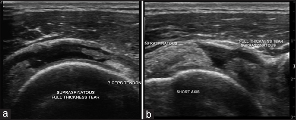
Figure1: Long (a) and shortaxis (b) examination reveals that there is a fluidfilled defect replacing the entire thickness and width of the right supraspinatus, suggesting a fullthickness tear of the supraspinatus tendon. The defect length or retraction is 30 mm. Reproduced from Patel, Radiopaedia.org, from the case of rotator cuff tear.
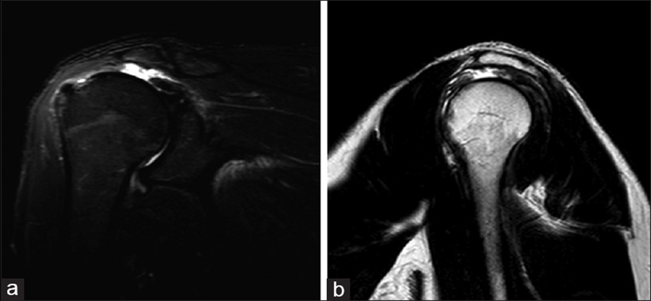
Figure 2: Coronal STIR image (a) and sagittal T2 image (b) showing a complete tear of the supraspinatus tendon, which is retracted medially, uncovering the humeral head, with subacromial bursal fluid collection.
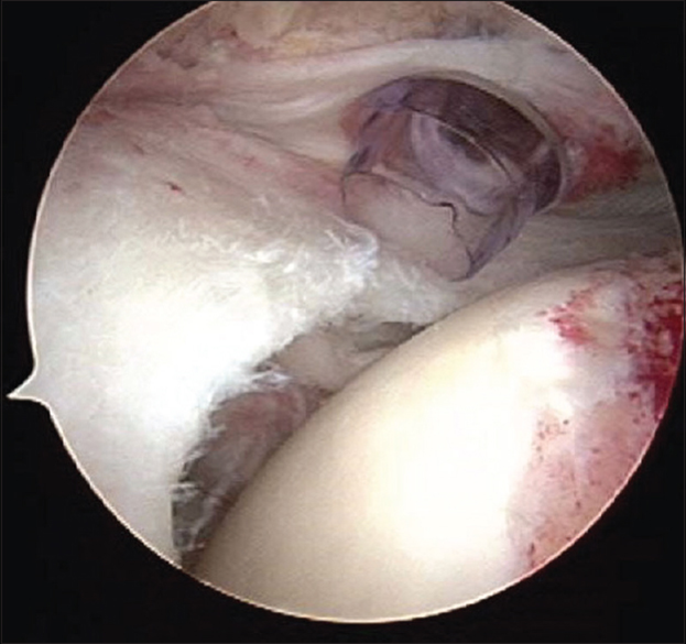
Figure 3: Arthroscopic view of the rotator cuff. The typical appearance of the torn cuff as viewed through the arthroscope. This view is from above the cuff, looking down the torn edge.
MATERIALS AND METHODS:
The present prospective cross-sectional, observational hospital based study was carried out from August 2018-December 2019 was conducted on 70 patients with suspected rotator cuff injuries patients of all age group referred to the department of Radio Diagnosis at Sarvodaya Hospital & Research centre, Faridabad, Haryana, 300 bedded tertiary care Centre during the time span of the study.
After taking consent from the patient and or his/her reliable attendant we saw different aspect as written in proforma.
Criteria of inclusion:
1. Patients with clinically suspected rotator cuff injuries who have undergone both MRI and arthroscopy at Sarvodaya Hospital & Research Centre.
2. Cases of all age group irrespective of sex.
3. Patients willing to take part in study and give written informed consent.
Criteria of exclusion:
1. Patients not willing to take part in the study.
2. Patients with active infection of shoulder joint.
3. Patients with osteoarthritis of shoulder joint.
4. Patients with tumor of the shoulder.
5. Patients with previous history of surgery or prosthesis in the shoulder.
6. Patients who had not undergone MRI and arthroscopy both at Sarvodaya Hospital & Research Centre.
7. Patients who were claustrophobic.
8. Patients having history of non-compatible metallic implant insertion, cardiac pacemaker and metallic foreign body in-situ.
9. Patients who required ventilation.
Statistical method used:
Sample size = z2pq
d2 = 1.96 x 1.96 x 0.21 x 0.79 0.1x 0.1 = 63.73
10% non-response taken, 63.73+6.37=70.1
A sample size of 70 patients was taken
Here,
z = is standard normal variate [at 5% type 1 error[p<0.05]it is 1.96 and at 1% type 1 error [p<0.01] it is 2.58] as in majority of studies p value are considered significant below 0.05 hence 1.96 used in formula.
P=expected proportion in population based on previous studies or pilot studies
q=1-p
d=absolute error or precision has to be decided by researcher
For the margin of error at 2% and confidence level of study at 95%, a minimum of 70 patients[calculated using raosoft sample size calculator and based on similar previous studies reported prevalence of rotator cuff injuries is around 21%].
After obtaining clearance and approval from the institutional ethical committee, 70 patients who had suspected rotator cuff injury fulfilling the inclusion and exclusion criteria who give informed consent were included in the study.
Technique:
Imaging was done with 1.5 Tesla Siemens Magnetom Essenza machine using shoulder coil the following sequences were selected as required:
1. Coronal oblique T1W/ proton density weighted (PDW) fast spin echo (FSE) sequence.
2. Coronal oblique fat suppressed (FS) PDW FSE/T2-W FSE sequence.
3. Sagittal oblique T2 W FSE sequence (with/without fat suppression).
4. Axial T2-W gradient echo (GE) sequence.
5. Axial PDW FSE (with/without fat suppression)
Field of view 14-16 cm, slice thickness 2-3mm and matrix 320x256.
Statistical analysis
The results are presented in frequencies and percentages. The Kappa statistic was calculated between MRI and arthroscopy. The sensitivity, specificity, positive predictive value (PPV) and negative predictive value (NPV) were calculated. The p-value<0.05 was considered significant. All the analysis was carried out on SPSS 16.0 version (Chicago, Inc., USA).
RESULTS:
A total of 70 patients were included in the study. Demographic characteristics were comparable between all age groups. Distribution of patients according to symptoms showed that more than half of patients had pain & restriction (54.3%) followed by history of trauma (20%), restriction of movements/difficulty in abduction (17.1%) and pain in shoulder (8.6%) Table-1 & Fig.4.
Table-1: Distribution of patients according to Symptoms
|
Symptoms |
No. (n=70) |
% |
|
Pain in shoulder |
6 |
8.6 |
|
Restriction of movements/difficulty in abduction |
12 |
17.1 |
|
Pain & restriction |
38 |
54.3 |
|
History of trauma |
14 |
20.0 |
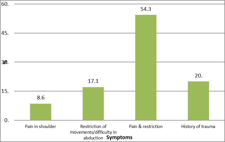
Fig. 4: Distribution of patients according to Symptoms
Table-2: Distribution of patients according to side
|
Side |
No. (n=70) |
% |
|
Right |
40 |
57.1 |
|
Left |
30 |
42.9 |
Table-2 & Fig.5 shows the distribution of patients according to side. More than half of patients had right side (57.1%).
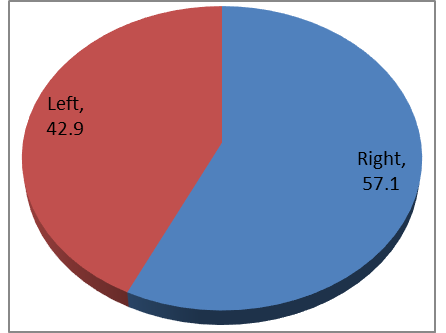
Figure 5: Distribution of patients according to side
Table-3 & Fig.6 shows the distribution of patients according to MRI findings. Tear supraspinatus on MRI was among more than half of patients (67.1%), Subscapularis (21.4%) and Infraspinatus (11.4%).
Table-3: Distribution of patients according to MRI findings
|
MRI findings |
No. (n=70) |
% |
|
Tear supraspinatus |
47 |
67.1 |
|
Subscapularis |
15 |
21.4 |
|
Infraspinatus |
8 |
11.4 |
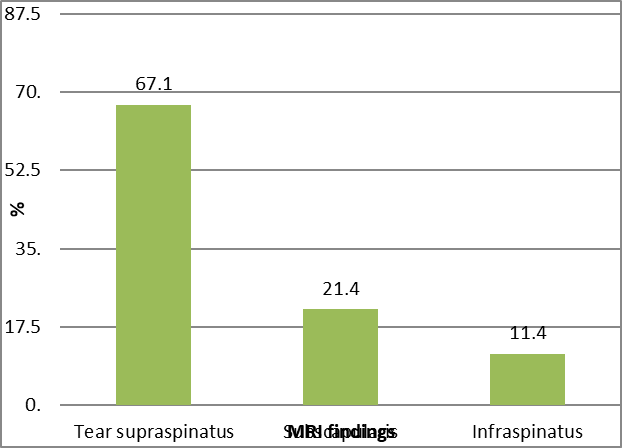
Fig. 5: Distribution of patients according to MRI findings
Table-4 & Fig. 6 shows the distribution of patients according to MRI findings and thickness. On MRI findings, supraspinatus tear was among more than half of patients (67.1%) followed by Subscapularis (21.4%) and Infraspinatus (11.4%). Full thickness tear was in 42.6% of supraspinatus tendon on MRI finding. Full thickness tear was in 40% of Subscapularis tendon on MRI finding.
Table-4: Distribution of patients according to MRI findings and thickness
|
MRI findings |
No. of patients (n=70) |
Full |
Partial |
|||
|
No. |
% |
No. |
% |
No. |
% |
|
|
Tear supraspinatus |
47 |
67.1 |
20 |
42.6 |
27 |
57.4 |
|
Subscapularis |
15 |
21.4 |
6 |
40.0 |
9 |
60.0 |
|
Infraspinatus |
8 |
11.4 |
5 |
62.5 |
3 |
37.5 |
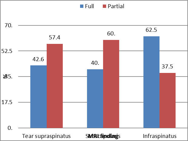
Fig. 6: Distribution of patients according to MRI findings and thickness
Table-5 & Fig.7 shows the distribution of patients according to MRI findings–partial thickness tear of supraspinatus tendon, articular surface was among more than half of patients (51.9%) followed by bursal (29.6%) and Intrasubstance (18.5%).
Table-5: Distribution of patients according to MRI findings in supraspinatus tendon- Partial tear
|
Supraspinatus tendon- Partial tear |
No. (n=27) |
% |
|
Articular |
14 |
51.9 |
|
Bursal |
8 |
29.6 |
|
Intrasubstance |
5 |
18.5 |
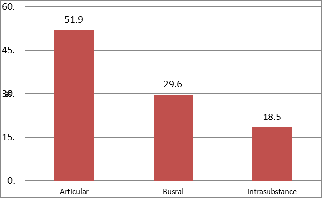
Fig. 7: Distribution of patients according to MRI findings in supraspinatus -Partial tear
Table-6 & Fig.8 shows the distribution of patients according to arthroscopy findings-partial thickness tear of supraspinatus tendon. on arthroscopy-partial thickness tear of supraspinatus tendon, articular surface was among more than one third of patients (48%) followed by Bursal (32%) and Intrasubstance (20%). 2 patients diagnosed as partial thickness tear on MRI were found to be full thickness tear on Arthroscopy.
Table-6: Distribution of patients according to arthroscopy findings in supraspinatus tendon- partial tear
|
Arthroscopy findings-Partial |
No. (n=25) |
% |
|
Articular |
12 |
48.0 |
|
Busral |
8 |
32.0 |
|
Intrasubstance |
5 |
20.0 |
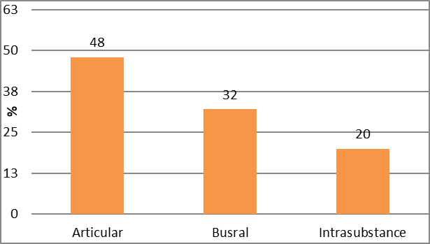
Fig.8: Distribution of patients according to arthroscopy findings-Partial
Table-7 & Fig.9 shows the distribution of patients according to ancillary MRI findings. SASD bursal effusion finding was the most common ancillary MRI finding (28.6%). SC bursal effusion finding was the second most common ancillary MRI finding (25.7%). Hill sach's and bankarts & Labral tear were least common ancillary MRI findings each constituted 2.9%.
Table-7: Distribution of patients according to ancillary MRI findings
|
Ancillary MRI findings |
No. (n=70) |
% |
|
SASD bursal effusion |
20 |
28.6 |
|
SC bursal effusion |
18 |
25.7 |
|
Joint effusion |
14 |
20.0 |
|
AC joint arthrosis |
10 |
14.3 |
|
Fracture greater tubercle |
4 |
5.7 |
|
Hill sach's and bankarts |
2 |
2.9 |
|
Labral tear |
2 |
2.9 |
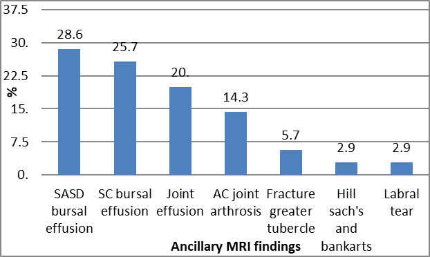
Fig. 9: Distribution of patients according to ancillary MRI findings
Table-8 & Fig.10 shows the accuracy and efficacy of MRI shoulder in comparison to arthroscopy. Full thickness tear of supraspinatus tendon was correctly detected by MRI among 42.6% patients with sensitivity and specificity of 90.9% and 100% respectively.
Table-8: Accuracy and efficacy of MRI finding in comparison to arthroscopy- supraspinatus tendon tear
|
MRI findings |
Arthroscopy findings |
Total |
||||
|
Full |
Partial |
|||||
|
No. |
% |
No. |
% |
No. |
% |
|
|
Full |
20 |
42.6 |
0 |
0 |
20 |
42.6 |
|
Partial |
2 |
4.2 |
25 |
53.2 |
27 |
57.4 |
|
Total |
22 |
46.8 |
25 |
53.2 |
47 |
100.0 |
|
Predictive values |
|
|
|
|
|
|
|
Sensitivity |
90.9 |
|||||
|
Specificity |
100.0 |
|||||
|
PPV |
100.0 |
|||||
|
NPV |
92.6 |
|||||
|
Accuracy |
95.7 |
|||||
%age is from total no. of cases
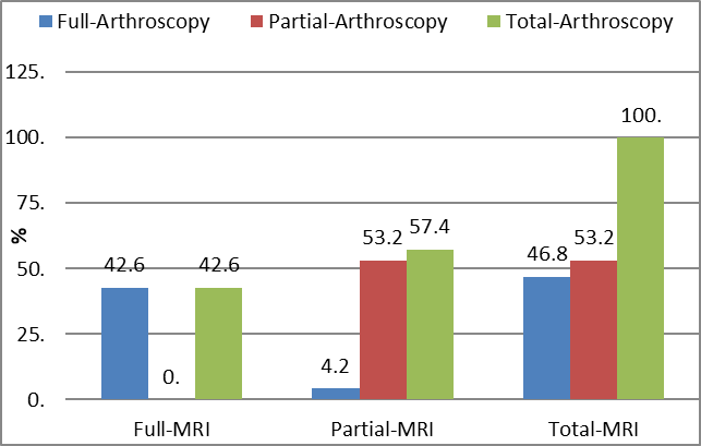
Fig. 10: Accuracy and efficacy of MRI finding in comparison to arthroscopy- supraspinatus tendon tear
Table-9 & Fig.11 shows the accuracy and efficacy of MRI shoulder in comparison to arthroscopy. Full thickness tear of Subscapularis tendon was correctly detected by MRI among 40% patients with sensitivity and specificity of 66.7% and 100% respectively.
Table-9: Accuracy and efficacy of MRI finding in comparison to arthroscopy- Subscapularis tendon tear
|
MRI findings |
Arthroscopy findings |
Total |
||||
|
Full |
Partial |
|||||
|
No. |
% |
No. |
% |
No. |
% |
|
|
Full |
6 |
40.0 |
0 |
0.0 |
6 |
40.0 |
|
Partial |
3 |
20.0 |
6 |
40.0 |
9 |
60.0 |
|
Total |
9 |
60.0 |
6 |
40.0 |
15 |
100.0 |
|
Predictive values |
|
|
|
|
|
|
|
Sensitivity |
66.7 |
|||||
|
Specificity |
100.0 |
|||||
|
PPV |
100.0 |
|||||
|
NPV |
66.7 |
|||||
|
Accuracy |
80.0 |
|||||
%age is from total no. of cases
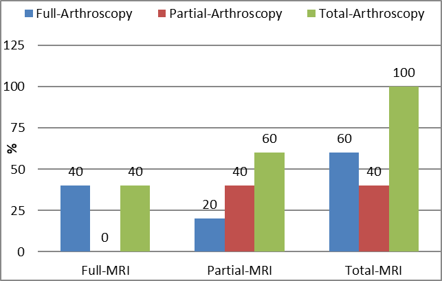
Fig. 11: Accuracy and efficacy of MRI finding in comparison to arthroscopy- Subscapularis tendon tear.
Table-10 & Fig.12 shows the accuracy and efficacy of MRI shoulder in comparison to arthroscopy. Full thickness tear of Infraspinatus tendon was correctly detected by MRI among 62.5% patients with sensitivity and specificity of 83.3% and 100% respectively.
Table-10: Accuracy and efficacy of MRI finding in comparison to arthroscopy- Infraspinatus tendon tear
|
MRI findings |
Arthroscopy findings |
Total |
||||
|
Full |
Partial |
|||||
|
No. |
% |
No. |
% |
No. |
% |
|
|
Full |
5 |
62.5 |
0 |
12.5 |
5 |
62.5 |
|
Partial |
1 |
12.5 |
2 |
25 |
3 |
37.5 |
|
Total |
6 |
75.0 |
2 |
25.0 |
8 |
100.0 |
|
Predictive values |
|
|
|
|
|
|
|
Sensitivity |
83.3 |
|||||
|
Specificity |
100.0 |
|||||
|
PPV |
100.0 |
|||||
|
NPV |
66.7 |
|||||
|
Accuracy |
87.5 |
|||||
%age is from total no. of cases
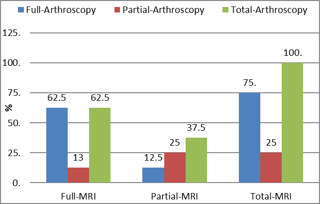
Fig. 12: Accuracy and efficacy of MRI finding in comparison to arthroscopy- Infraspinatus tendon tear
Table-11 & Fig.13 shows the overall accuracy and efficacy of MRI in comparison to arthroscopy. Full thickness was correctly detected by MRI among 44.3% patients with sensitivity and specificity of 83.8% and 100% respectively.
Table-11 Overall Accuracy and efficacy of MRI finding in comparison to arthroscopy
|
MRI findings |
Arthroscopy findings |
Total |
||||
|
Full |
Partial |
|||||
|
No. |
% |
No. |
% |
No. |
% |
|
|
Full |
31 |
44.3 |
0 |
0 |
31 |
44.3 |
|
Partial |
6 |
8.6 |
33 |
47.1 |
39 |
55.7 |
|
Total |
37 |
52.9 |
33 |
47.1 |
70 |
100.0 |
|
Predictive values |
|
|
|
|
|
|
|
Sensitivity |
83.8 |
|||||
|
Specificity |
100.0 |
|||||
|
PPV |
100.0 |
|||||
|
NPV |
84.6 |
|||||
|
Accuracy |
91.4 |
|||||
%age is from total no. of cases
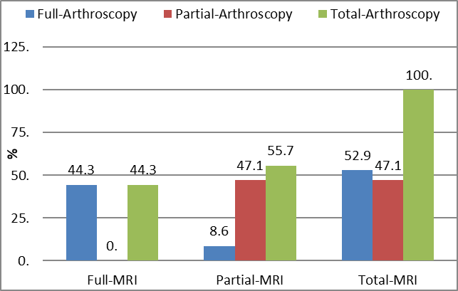
Fig.13: Overall accuracy and efficacy of MRI in comparison to arthroscopy.
CONCLUSION:
More than half of patients had pain & restriction (54.3%) followed by history of trauma (20%), restriction of movements/difficulty in abduction (17.1%) and pain in shoulder (8.6%). More than half of patients had right shoulder involvement (57.1%). Supraspinatus tear on MRI finding was among more than half of patients (67.1%) followed by Subscapularis (21.4%) and Infraspinatus (11.4%). Full thickness tear was in 42.6% of supraspinatus tendon on MRI finding. Full thickness tear was in 40% of Subscapularis tendon on MRI finding. On MRI findings of partial thickness tear of supraspinatus tendon, articular surface was among more than one third of patients (51.9%) followed by bursal (29.6%) and Inter-substance (18.5%). Full thickness tear was in 46.8% of supraspinatus tendon on arthroscopy finding. Full thickness tear was in 75% of Infraspinatus tendon on arthroscopy finding.
On Arthroscopy findings of partial thickness tear of supraspinatus tendon, articular surface was among more than one third of patients (48%) followed by Bursal (32%) and Inter-substance (20%). SASD bursal effusion was the most common ancillary MRI finding (28.6%). SC bursal effusion was the second most common ancillary MRI finding(25.7%). Hill sach's and bankarts & Labral tear were least common ancillary MRI finding each constituted 2.9%. Full thickness tear of supraspinatus tendon was correctly detected by MRI among 42.6% patients with sensitivity and specificity of 90.9% and 100% respectively. Full thickness tear of Subscapularis tendon was correctly detected by MRI among 40% patients with sensitivity and specificity of 66.7% and 100% respectively. Full thickness tear of Infraspinatus tendon was correctly detected by MRI among 62.5% patients with sensitivity and specificity of 83.3% and 100% respectively. Full thickness tear was correctly detected by MRI among 44.3% patients with sensitivity and specificity of 83.8% and 100% respectively.
The present study substantiates that MRI is effective in diagnosing rotator cuff tears. Arthroscopy still remains the gold standard in diagnosing shoulder lesions. MRI and arthroscopy have complimentary roles in diagnosing shoulder pathology.
This study has limitations of potential selection bias as all patients with rotator cuff injury severe enough to undergo arthroscopy were included in the study, missing out the people who could not undergo MRI in view of claustrophobia or metallic and pacemaker implants and patients who could not undergo arthroscopy in view of co morbidities.
REFERENCES:
1. Van der Windt DA, Koes BW, de Jong BA, Bouter LM. Shoulder disorders in general practice: incidence, patient characteristics, and management. Ann Rheum Dis. 1995;54:959–64.
2. Ostör AJ, Richards CA, Prevost AT, Speed CA, Hazleman BL. Diagnosis and relation to general health of shoulder disorders presenting to primary care. Rheumatology. 2005;44:800–5.
3. Badcock LJ, Lewis M, Hay EM, McCarney R, Croft PR. Chronic shoulder pain in the community: a syndrome of disability or distress? Ann Rheumatic Dis. 2002;61(2), 128–31.
4. Masters S, O’Doherty L, Mitchell GK, Yelland M. Acute shoulder pain in primary care-an observational study. Aust Fam Physician. 2007;36(6):473-6.
5. Kinare A. Musculoskeletal Ultrasound Symposium. Indian J Radiology and Imaging. 2007;17:194-200.
6. Teefey SA, Rubin DA, Middleton WD, Hildebolt CF, Leibold RA, Yamaguchi K. Detection and quantification of rotator cuff tears: Comparison of ultra sonographic, magnetic resonance imaging, and arthroscopic findings in 71 consecutive cases. J Bone Joint Surg Am. 2004;86:708– 16.
7. Jacobson JA, Lancaster S, Prasad A, van Holsbeeck MT, Craig JG, Kolowich P. Full-thickness and partial-thickness supraspinatus tendon tears: Value of US signs in diagnosis. Radiology. 2004;230:234–42.
8. Joseph O. de Jesus, Laurence Parker, Andrea J. Fangos, Levon N. Nazarian. Accuracy of MRI, MR Arthrography, and Ultrasound in the Diagnosis of Rotator Cuff tears: a meta-analysis. AJR. 2009;192:1701-1707.
9. Nam EK, Snyder SJ. The diagnosis and treatment of superior labrum, anterior and [2] posterior (SLAP) lesions. American Journal of Sports Medicine. 2003; 31(5):798- 810.