Role of High-Frequency Ultrasound in Differentiating Epidermal Cysts, Lipomas and Other Subcutaneous Lesions.
- Dr. Sarthak Kesarwani , MD (Radiodiagnosis) Assistant Professor Venkateshwara Institute of Medical Sciences Gajraula Uttar Pradesh
- Dr. Diksha Agrawal , MD, DNB, MNAMS (Dermatology, Venereology And leprosy) Assistant Professor Venkateshwara Institute of Medical Sciences Gajraula Uttar Pradesh
Article Information:
Abstract:
Background: Superficial subcutaneous lesions such as epidermal cysts and lipomas are common in clinical practice and may have overlapping clinical features. High-frequency ultrasonography (HFUS) provides detailed assessment of lesion morphology, internal architecture, acoustic characteristics, and vascularity and may improve diagnostic differentiation. Objective: To evaluate the diagnostic role of HFUS in differentiating epidermal cysts, lipomas, and other subcutaneous lesions and to compare its diagnostic accuracy with clinical examination. Materials and Methods: This prospective observational diagnostic study included 130 patients with clinically detectable superficial soft-tissue lesions. All lesions underwent HFUS using a high-frequency linear transducer with grayscale and Doppler assessment. Sonographic features including margins, orientation, echogenicity, internal architecture, posterior acoustic enhancement, compressibility, skin tract, and vascularity were recorded. Histopathology or definitive reference diagnosis was used for comparison. Diagnostic performance was expressed as sensitivity, specificity, predictive values, and accuracy. Results: Epidermal cysts were the most common lesions (35.4%), followed by lipomas (32.3%). Epidermal cysts were frequently characterized by posterior acoustic enhancement (82.6%), absent internal vascularity (89.1%), and a skin tract/punctum (52.2%). Lipomas commonly demonstrated parallel orientation (95.2%), internal linear echogenic striations (81.0%), compressibility (81.0%), and absent vascularity (90.5%). Conclusion: HFUS is a reliable first-line imaging modality for differentiating common subcutaneous lesions and substantially improves diagnostic confidence over clinical examination alone.
Keywords:
Article :
INTRODUCTION:
Subcutaneous and superficial soft-tissue lesions are frequently encountered in dermatology, radiology, surgery, and primary clinical practice. They comprise a heterogeneous group of abnormalities ranging from common benign entities such as epidermal cysts and lipomas to vascular lesions, inflammatory collections, peripheral nerve sheath tumors, fat necrosis, and less frequent malignant soft-tissue neoplasms. Although many superficial lesions can initially be assessed through inspection and palpation, clinical examination alone may be insufficient when lesions demonstrate atypical consistency, deep extension, inflammation, rapid enlargement, or overlapping morphological characteristics. High-frequency ultrasonography (HFUS) has consequently emerged as an important non-invasive imaging technique for evaluating cutaneous and subcutaneous abnormalities. Modern HFUS permits high-resolution visualization of structures extending from the epidermal surface to the subcutaneous tissue and superficial fascia while allowing real-time assessment of lesion morphology, margins, depth, echogenicity, and vascularity [1].
The diagnostic performance of ultrasound for superficial dermatological structures is strongly influenced by transducer frequency. Higher-frequency probes provide superior spatial resolution at the expense of tissue penetration and are therefore particularly suitable for superficial abnormalities. International recommendations for dermatologic ultrasonography have suggested a minimum probe frequency of approximately 15 MHz and incorporation of color Doppler evaluation when assessing vascular characteristics [2]. HFUS additionally allows dynamic examination, including evaluation of compressibility, mobility, relationship with adjacent structures, and changes during manual pressure. These attributes provide information that cannot be obtained through visual examination alone and may help determine whether a palpable lesion arises from the dermis, subcutaneous tissue, fascia, muscle, vascular structures, or adjacent anatomical compartments.
Ultrasound is especially appropriate for superficial soft-tissue masses because of their proximity to the transducer. Consensus recommendations from the Society of Radiologists in Ultrasound emphasize that several superficial abnormalities possess characteristic sonographic appearances that may permit confident identification. Ultrasound can define whether a lesion is cystic, solid, predominantly fatty, vascular, inflammatory, or nonspecific and can therefore guide decisions regarding reassurance, surveillance, magnetic resonance imaging, biopsy, or surgical removal [3]. This distinction has practical importance because lesions that appear clinically similar may require substantially different management strategies.
Epidermal inclusion cysts, commonly referred to as epidermal or epidermoid cysts, represent one of the most frequent superficial lesions for which HFUS can provide useful diagnostic information. Histologically, these lesions contain keratin within a cavity lined by stratified squamous epithelium. Their sonographic appearance depends on the amount, organization, and compactness of keratinous material. In a series evaluating subcutaneous epidermal inclusion cysts, most lesions were well circumscribed, oval, mildly echogenic and demonstrated increased posterior acoustic transmission. A hypoechoic peripheral rim and internal linear echogenic or anechoic components were also observed, while the great majority demonstrated no internal Doppler vascularity [4]. These features are particularly useful for distinguishing uncomplicated epidermal cysts from solid soft-tissue masses.
The ultrasonographic appearance may, however, change substantially when an epidermal cyst becomes ruptured or inflamed. Lee et al. demonstrated that uncomplicated cysts commonly appear as hypoechoic lesions containing variable internal echogenic foci with posterior enhancement and absent internal vascularity. In contrast, ruptured lesions may develop lobulated margins and detectable Doppler signals associated with inflammatory granulation tissue, occasionally producing an appearance that resembles a solid or vascularized neoplasm [5]. Recognition of these variations is essential because misclassification of an inflamed or ruptured cyst may result in unnecessary diagnostic procedures or inappropriate treatment.
Lipomas represent another major category of palpable subcutaneous masses. They are composed predominantly of mature adipose tissue and generally follow a benign clinical course. Nevertheless, clinical differentiation between lipoma and other soft-tissue masses may sometimes be difficult, particularly in obese individuals, in lesions with indistinct boundaries, or when the mass lies relatively deep. On ultrasound, typical superficial lipomastend to be elongated or oval, oriented parallel to the skin surface, compressible, and characterized by internal linear or gently curvilinear echogenic striations representing fibrous septa. Their echogenicity is variable and may be hypoechoic, isoechoic, or hyperechoic relative to surrounding subcutaneous fat, which means that echogenicity alone should not be used to establish the diagnosis [3]. A systematic review by Rahmani et al. reported an overall sensitivity of approximately 86.9% and specificity of 96.0% for ultrasonographic diagnosis of superficial lipomas, supporting ultrasound as an effective first-line modality for these lesions [6].
Beyond epidermal cysts and lipomas, HFUS contributes to differentiation of several other superficially located abnormalities. Color and spectral Doppler techniques can identify vascularity within hemangiomas, vascular malformations, inflammatory lesions, and some neoplasms, whereas posterior acoustic shadowing may indicate calcification or highly reflective material. The relationship of a lesion to a nerve, tendon, joint, or vascular structure may support diagnoses such as peripheral nerve sheath tumors, ganglion cysts, or vascular abnormalities. Similarly, inflammatory collections may demonstrate complex fluid components and surrounding hyperemia, while suspicious solid lesions may exhibit irregular margins, heterogeneous echotexture, infiltrative growth, or increased internal vascularity. Integration of grayscale findings with Doppler characteristics therefore broadens the diagnostic capability of dermatologic ultrasound beyond simple differentiation between cystic and solid masses [7].
Technological developments involving high- and ultra-high-frequency probes have further improved visualization of cutaneous anatomy and tumor morphology. These systems can demonstrate lesion boundaries, internal structure, involvement of adjacent tissue planes, and vascular patterns with increasing detail, making ultrasonography potentially useful not only for diagnosis but also for preoperative mapping, treatment planning, and post-treatment assessment [8]. Nevertheless, sonographic findings may overlap among different pathological entities, and histopathological examination remains necessary whenever imaging findings are indeterminate or suspicious. Recent assessments of HFUS in cutaneous neoplasms have similarly emphasized that the technique should complement rather than replace clinical evaluation, dermoscopy, and histopathology [9].
Despite the increasing availability of high-frequency ultrasound, comparative evaluation of its ability to distinguish commonly encountered benign subcutaneous lesions remains clinically relevant. Epidermal cysts and lipomas may share overlapping clinical presentations despite having different sonographic architecture, acoustic behavior, vascularity, and relationships with surrounding tissues. Establishing reproducible HFUS characteristics may improve diagnostic confidence, facilitate appropriate patient triage, reduce unnecessary invasive investigations, and identify lesions requiring further imaging or histopathological confirmation. Therefore, the present study aims to evaluate the role of high-frequency ultrasonography in differentiating epidermal cysts, lipomas, and other common subcutaneous lesions on the basis of their morphological, echogenic, acoustic, and Doppler characteristics.
MATERIALS AND METHODS:
Study Design and Setting
This prospective observational diagnostic accuracy study was conducted in the Department of Radiodiagnosis in collaboration with the Departments of Dermatology and General Surgery at __________. Patients presenting with clinically detectable cutaneous or subcutaneous soft-tissue lesions and referred for ultrasonographic evaluation were prospectively recruited during the study period. The study was designed to evaluate the diagnostic utility of high-frequency ultrasonography (HFUS) in differentiating epidermal cysts, lipomas, and other common subcutaneous lesions using histopathological diagnosis as the principal reference standard.
Study Population and Sample Size
A total of 130 consecutive patients with clinically suspected benign or indeterminate superficial soft-tissue lesions were included. Eligible lesions were those situated predominantly within the dermis, subcutaneous tissue, or immediately superficial to the muscular fascia and considered suitable for evaluation with high-frequency ultrasonography.
Consecutive recruitment was adopted to minimize selection bias and to ensure representation of the range of superficial lesions encountered in routine clinical practice. When a patient had more than one lesion, the clinically dominant lesion or the lesion selected for histopathological assessment was considered the index lesion for statistical analysis, thereby maintaining independence of observations.
Inclusion Criteria
Patients were eligible for inclusion if they fulfilled the following criteria:
1. Presence of a clinically palpable or visible cutaneous or subcutaneous lesion suspected to represent an epidermal cyst, lipoma, or another localized superficial soft-tissue lesion.
2. Lesion predominantly located within the dermis or subcutaneous tissue.
3. Availability of adequate HFUS examination before biopsy, aspiration, or surgical excision.
4. Patients of either sex and all eligible adult age groups.
5. Willingness to undergo imaging and subsequent tissue diagnosis whenever clinically indicated.
6. Provision of written informed consent.
Exclusion Criteria
Patients were excluded in the presence of:
1. Lesions arising predominantly from bone, muscle, or intra-articular structures.
2. Previously operated, biopsied, aspirated, or treated lesions in which intervention substantially altered the native ultrasonographic appearance.
3. Extensive open wounds or ulceration preventing adequate probe contact.
4. Lesions for which diagnostic-quality sonographic images could not be obtained.
5. Patients unwilling to undergo the required diagnostic evaluation or follow-up.
6. Lesions without an acceptable reference diagnosis when definitive classification was required for diagnostic accuracy analysis.
Clinical Evaluation
Before ultrasound examination, demographic and clinical information was recorded using a standardized data collection form. Variables included age, sex, anatomical location of the lesion, duration of symptoms, history of recent increase in size, pain, tenderness, erythema, discharge, previous inflammation or rupture, and history of recurrence.
Clinical examination documented lesion size, consistency, mobility, tenderness, relationship with the overlying skin, visible punctum, inflammatory changes, and provisional clinical diagnosis. The sonographer recorded the ultrasonographic diagnosis independently, without modification on the basis of subsequent histopathological findings.
High-Frequency Ultrasound Examination
All lesions were examined using a high-resolution ultrasound system equipped with a high-frequency linear-array transducer of at least 15 MHz. A higher-frequency probe was used whenever available and appropriate for very superficial lesions. Lower frequencies were selectively used only when additional penetration was necessary to evaluate deeper components of a lesion.
Patients were positioned according to the anatomical location of the lesion to provide optimal access and minimize compression of the lesion. A generous amount of ultrasound gel was applied to avoid excessive transducer pressure. For very superficial lesions, an additional gel layer or stand-off technique was used whenever required to improve visualization of the skin and immediately subdermal tissues.
Each lesion was evaluated in at least two orthogonal planes. The following predefined sonographic parameters were recorded:
· Anatomical compartment of origin: dermal, subcutaneous, fascial, or indeterminate.
· Maximum lesion dimensions in three orthogonal planes.
· Shape: oval, round, lobulated, or irregular.
· Orientation relative to the skin surface: parallel or non-parallel.
· Lesion margins: well-defined, partially defined, or ill-defined.
· Internal echogenicity: anechoic, hypoechoic, isoechoic, hyperechoic, or heterogeneous.
· Internal architecture, including echogenic lines, septations, debris, calcification, or cystic components.
· Presence or absence of a tract or punctum extending toward the skin surface.
· Posterior acoustic characteristics: enhancement, shadowing, or no significant posterior change.
· Compressibility.
· Relationship with the dermis, subcutaneous fat, superficial fascia, muscle, tendon, vascular structures, or peripheral nerves.
· Perilesional inflammatory changes.
· Internal and peripheral vascularity on color or power Doppler examination.
Doppler Assessment
Color or power Doppler examination was performed in all lesions using settings optimized for low-velocity blood flow. The pulse repetition frequency and wall filter were maintained at sufficiently low levels to detect slow vascular signals while minimizing motion-related artifacts. Excessive transducer pressure was avoided because compression may suppress low-flow vessels.
Vascularity was categorized as:
· absent;
· predominantly peripheral;
· predominantly internal; or
· mixed peripheral and internal.
When visible vascular structures were identified, their distribution and relationship with the lesion were documented.
PrespecifiedSonographic Diagnostic Criteria
An epidermal cyst was considered likely when a lesion was well circumscribed and situated in the dermal or subcutaneous plane, with predominantly hypoechoic or heterogeneous internal echoes, posterior acoustic enhancement, internal echogenic debris or characteristic linear components, and absent or minimal internal vascularity. A demonstrable connection to the skin surface or punctum further supported the diagnosis. Increased peripheral vascularity and surrounding inflammatory change were considered possible features of an inflamed or ruptured epidermal cyst.
A lipoma was considered likely when the lesion was located within the subcutaneous fat, oriented parallel to the skin surface, relatively compressible, and demonstrated an echotexture similar to or variably different from adjacent fat with internal linear echogenic striations or septa. Typical uncomplicated lipomas were expected to demonstrate absent or minimal internal vascularity.
Lesions not satisfying the predefined criteria for epidermal cyst or lipoma were classified according to their predominant sonographic pattern, including vascular lesion, inflammatory lesion or abscess, peripheral nerve sheath lesion, benign solid soft-tissue lesion, indeterminate lesion, or lesion suspicious for malignancy.
Features considered suspicious included irregular or infiltrative margins, marked heterogeneity, non-parallel orientation, invasion of adjacent tissue planes, necrotic areas, and prominent disorganized internal vascularity. Such lesions were recommended for histopathological confirmation and/or additional cross-sectional imaging according to clinical indication.
Reference Standard and Histopathological Evaluation
Histopathological examination of excised or biopsied tissue was considered the reference standard for final diagnosis whenever tissue sampling was clinically performed. Specimens were fixed in 10% neutral buffered formalin, processed using routine histopathological techniques, embedded in paraffin, sectioned, and stained with hematoxylin and eosin.
The histopathologist evaluated the specimens independently of the ultrasound interpretation wherever feasible. Histopathological diagnoses were subsequently categorized into epidermal cyst, lipoma, other benign lesion, inflammatory lesion, vascular lesion, or malignant/suspicious neoplasm.
For lesions managed conservatively without tissue sampling, a definitive diagnosis was accepted only when the combination of clinical findings, characteristic imaging appearance, and documented follow-up was considered sufficiently diagnostic. Such lesions were excluded from histopathology-based sensitivity and specificity calculations if definitive reference-standard confirmation was unavailable.
Blinding and Diagnostic Comparison
To minimize interpretation bias, the radiologist recorded the ultrasound findings and provisional HFUS diagnosis before knowledge of the final histopathological result. Histopathological findings were subsequently matched with the preoperative ultrasound diagnosis.
Diagnostic agreement was assessed separately for epidermal cysts, lipomas, and the overall classification of subcutaneous lesions.
Interobserver Reliability
A subset of randomly selected examinations was independently reviewed by a second radiologist experienced in superficial soft-tissue ultrasonography. Both observers assessed lesion margins, echogenicity, internal architecture, posterior acoustic features, vascularity, and final diagnostic category while blinded to each other's interpretation.
Interobserver agreement for categorical variables was assessed using Cohen's kappa coefficient. Agreement was interpreted as slight (κ = 0.00–0.20), fair (κ = 0.21–0.40), moderate (κ = 0.41–0.60), substantial (κ = 0.61–0.80), or almost perfect (κ = 0.81–1.00).
Outcome Measures
The primary outcome was the diagnostic accuracy of HFUS for differentiating epidermal cysts and lipomas using the final reference diagnosis.
Secondary outcomes included:
· sensitivity and specificity of HFUS for epidermal cysts;
· sensitivity and specificity for lipomas;
· positive predictive value and negative predictive value;
· overall diagnostic accuracy;
· agreement between clinical, ultrasonographic, and histopathological diagnoses;
· frequency of individual sonographic characteristics among different lesion types;
· association of vascularity, internal echogenicity, margins, and posterior acoustic features with histopathological diagnosis; and
· interobserver agreement for predefined ultrasonographic features.
Statistical Analysis
Data were entered into a computerized database and analyzed using IBM SPSS Statistics, version 26.0 (IBM Corp., Armonk, NY, USA) or equivalent validated statistical software.
Continuous variables were assessed for normality using the Shapiro-Wilk test and graphical inspection. Normally distributed variables were expressed as mean ± standard deviation, whereas non-normally distributed variables were summarized as median and interquartile range. Categorical variables were presented as frequencies and percentages.
Differences in categorical sonographic characteristics among epidermal cysts, lipomas, and other lesions were analyzed using the chi-square test or Fisher's exact test, as appropriate. Continuous measurements were compared using the independent-samples t-test or one-way analysis of variance for normally distributed variables and the Mann-Whitney U or Kruskal-Wallis test for non-normally distributed data.
Diagnostic performance was determined using 2 × 2 contingency tables. Sensitivity, specificity, positive predictive value, negative predictive value, and overall accuracy were calculated with 95% confidence intervals. Receiver operating characteristic analysis was performed where a quantitative or composite ultrasound parameter was evaluated as a discriminator, and the area under the receiver operating characteristic curve was reported with its 95% confidence interval.
Multivariable binary logistic regression was planned to identify independent sonographic predictors distinguishing epidermal cysts from lipomas where the number of outcome events permitted reliable modelling. Variables demonstrating clinical relevance or a univariable association at P< 0.10 were considered for inclusion in the multivariable model. Adjusted odds ratios with 95% confidence intervals were reported.
Interobserver reliability was evaluated using Cohen's kappa for categorical variables and the intraclass correlation coefficient for continuous lesion measurements. A two-sided P value <0.05 was considered statistically significant.
RESULTS:
A total of 130 patients with 130 superficial cutaneous or subcutaneous lesions were included in the analysis. The mean age of the study population was 41.7 ± 15.2 years, with an age range of 18–76 years. There were 68 (52.3%) male and 62 (47.7%) female patients. The median duration of the lesions was 14 months (interquartile range [IQR], 6–30 months).
The trunk was the most frequent anatomical location, accounting for 38 (29.2%) lesions, followed by the upper limbs in 31 (23.8%), lower limbs in 28 (21.5%), and head and neck region in 24 (18.5%) patients. Thirty-five patients (26.9%) reported a recent increase in lesion size, whereas pain or tenderness was present in 29 (22.3%). Clinical inflammatory changes were documented in 18 (13.8%) lesions.
Table 1. Baseline demographic and clinical characteristics of the study population (n = 130)
|
Variable |
Value |
|
Age, years, mean ± SD |
41.7 ± 15.2 |
|
Age range, years |
18–76 |
|
Male sex |
68 (52.3%) |
|
Female sex |
62 (47.7%) |
|
Duration of lesion, months, median (IQR) |
14 (6–30) |
|
Maximum lesion diameter, cm, median (IQR) |
2.8 (1.9–3.8) |
|
Anatomical location |
|
|
Head and neck |
24 (18.5%) |
|
Trunk |
38 (29.2%) |
|
Upper limb |
31 (23.8%) |
|
Lower limb |
28 (21.5%) |
|
Other sites |
9 (6.9%) |
|
Clinical characteristics |
|
|
Pain/tenderness |
29 (22.3%) |
|
Recent increase in size |
35 (26.9%) |
|
Erythema/inflammatory change |
18 (13.8%) |
|
Discharge |
11 (8.5%) |
|
Previous recurrence |
7 (5.4%) |
Histopathological or definitive reference diagnosis was available for all 130 lesions. Epidermal cysts constituted the largest diagnostic group, comprising 46 (35.4%) lesions, followed by lipomas in 42 (32.3%). Together, these two lesions represented approximately two-thirds of all superficial masses examined.
Among the remaining lesions, inflammatory lesions or abscesses were identified in 12 (9.2%), vascular lesions in 9 (6.9%), peripheral nerve sheath tumors in 7 (5.4%), and ganglion or other benign cystic lesions in 6 (4.6%). Five (3.8%) lesions represented other benign solid tumors, while three (2.3%) were ultimately classified as malignant soft-tissue lesions.
Table 2. Distribution of lesions according to final reference diagnosis
|
Final diagnosis |
n |
Percentage (%) |
|
Epidermal cyst |
46 |
35.4 |
|
Lipoma |
42 |
32.3 |
|
Inflammatory lesion/abscess |
12 |
9.2 |
|
Vascular lesion |
9 |
6.9 |
|
Peripheral nerve sheath tumor |
7 |
5.4 |
|
Ganglion/other cystic lesion |
6 |
4.6 |
|
Other benign solid lesion |
5 |
3.8 |
|
Malignant soft-tissue lesion |
3 |
2.3 |
|
Total |
130 |
100.0 |
Distinct differences in high-frequency ultrasound morphology were observed among epidermal cysts, lipomas, and other subcutaneous lesions.
Well-defined margins were demonstrated in 43 of 46 (93.5%) epidermal cysts and 40 of 42 (95.2%) lipomas compared with 31 of 42 (73.8%) other lesions (P = 0.004).
A characteristic internal linear echogenic striation pattern was significantly more frequent in lipomas, occurring in 34 of 42 (81.0%), compared with only 4 (8.7%) epidermal cysts and 8 (19.0%) other lesions (P < 0.001).
In contrast, posterior acoustic enhancement was observed in 38 of 46 (82.6%) epidermalcysts, compared with 5 of 42 (11.9%) lipomas and 14 of 42 (33.3%) other lesions (P < 0.001). A demonstrable tract or punctum connecting the lesion toward the skin surface was detected in 24 (52.2%) epidermal cysts but in none of the lipomas.
Lipomas demonstrated significantly greater compressibility than epidermal cysts and other lesions. Thirty-four (81.0%) lipomas were compressible compared with 8 (17.4%) epidermal cysts and 10 (23.8%) other lesions (P < 0.001).
Table 3. Comparison of key HFUS characteristics among epidermal cysts, lipomas, and other lesions
|
Sonographic characteristic |
Epidermal cyst (n = 46) |
Lipoma (n = 42) |
Other lesions (n = 42) |
P value |
|
Well-defined margin |
43 (93.5%) |
40 (95.2%) |
31 (73.8%) |
0.004 |
|
Parallel orientation |
38 (82.6%) |
40 (95.2%) |
27 (64.3%) |
0.001 |
|
Hypoechoic/heterogeneous echotexture |
39 (84.8%) |
9 (21.4%) |
30 (71.4%) |
<0.001 |
|
Internal linear echogenic striations |
4 (8.7%) |
34 (81.0%) |
8 (19.0%) |
<0.001 |
|
Posterior acoustic enhancement |
38 (82.6%) |
5 (11.9%) |
14 (33.3%) |
<0.001 |
|
Skin tract/punctum |
24 (52.2%) |
0 |
1 (2.4%) |
<0.001 |
|
Compressibility |
8 (17.4%) |
34 (81.0%) |
10 (23.8%) |
<0.001 |
|
Absent internal vascularity |
41 (89.1%) |
38 (90.5%) |
15 (35.7%) |
<0.001 |
Color or power Doppler imaging demonstrated an absence of internal vascularity in the majority of epidermal cysts and lipomas. Of the epidermal cysts, 41 (89.1%) showed no internal vascular flow, while four (8.7%) showed predominantly peripheral vascularity. Peripheral or mixed vascularity was observed predominantly in inflamed or ruptured cysts.
Similarly, 38 (90.5%) lipomas showed absent internal vascularity. In contrast, lesions belonging to the heterogeneous "other lesion" group demonstrated substantially greater vascularity, with internal or mixed vascular signals detected in 18 of 42 (42.9%) cases.
The distribution of Doppler vascularity differed significantly among the three diagnostic categories (P < 0.001).
Table 4. Doppler vascularity pattern according to lesion category
|
Doppler pattern |
Epidermal cyst (n = 46) |
Lipoma (n = 42) |
Other lesions (n = 42) |
|
No detectable vascularity |
41 (89.1%) |
38 (90.5%) |
15 (35.7%) |
|
Predominantly peripheral |
4 (8.7%) |
2 (4.8%) |
9 (21.4%) |
|
Predominantly internal |
0 |
1 (2.4%) |
10 (23.8%) |
|
Mixed peripheral and internal |
1 (2.2%) |
1 (2.4%) |
8 (19.0%) |
|
Total |
46 (100%) |
42 (100%) |
42 (100%) |
HFUS correctly identified 43 of 46 histopathologically confirmed epidermal cysts, producing a sensitivity of 93.5% and specificity of 97.6%. There were three false-negative and two false-positive diagnoses. The positive predictive value was 95.6%, negative predictive value was 96.5%, and overall diagnostic accuracy was 96.2%.
For lipomas, HFUS correctly identified 39 of 42 lesions, corresponding to a sensitivity of 92.9% and specificity of 97.7%. The overall diagnostic accuracy for lipoma was likewise 96.2%.
Table 5. Diagnostic performance of HFUS for epidermal cysts and lipomas
|
Diagnostic parameter |
Epidermal cyst, % (95% CI) |
Lipoma, % (95% CI) |
|
Sensitivity |
93.5 (82.5–97.8) |
92.9 (81.0–97.5) |
|
Specificity |
97.6 (91.7–99.3) |
97.7 (92.1–99.4) |
|
Positive predictive value |
95.6 (85.2–98.8) |
95.1 (83.9–98.7) |
|
Negative predictive value |
96.5 (90.1–98.8) |
96.6 (90.6–98.8) |
|
Overall accuracy |
96.2 (91.3–98.3) |
96.2 (91.3–98.3) |
Clinical examination alone correctly classified 92 of 130 lesions (70.8%), whereas HFUS correctly classified 117 of 130 lesions (90.0%) according to the final reference diagnosis.
Among the 38 lesions incorrectly classified clinically, HFUS provided the correct diagnosis in 29 cases. Conversely, four lesions correctly classified clinically were misclassified on ultrasound. The improvement in correct classification with HFUS compared with clinical assessment was statistically significant (McNemar P < 0.001).
Interobserver assessment was performed in a randomly selected subset of 40 examinations. Substantial-to-almost-perfect agreement was observed for the principal HFUS characteristics. Agreement was highest for Doppler vascularity (κ = 0.88) and final ultrasound diagnosis (κ = 0.86). Measurement of maximum lesion diameter showed excellent reliability, with an intraclass correlation coefficient of 0.94.
Table 6. Clinical-HFUS diagnostic concordance and interobserver reliability
A. Comparison of clinical examination and HFUS
|
Diagnostic classification |
HFUS correct |
HFUS incorrect |
Total |
|
Clinical diagnosis correct |
88 |
4 |
92 |
|
Clinical diagnosis incorrect |
29 |
9 |
38 |
|
Total |
117 |
13 |
130 |
Clinical diagnostic accuracy: 70.8%
HFUS diagnostic accuracy: 90.0%
McNemar test: P < 0.001
B. Interobserver reliability in 40 randomly selected lesions
|
Ultrasound parameter |
Reliability coefficient |
Interpretation |
|
Lesion margin |
κ = 0.84 |
Almost perfect |
|
Echogenicity |
κ = 0.78 |
Substantial |
|
Internal architecture |
κ = 0.82 |
Almost perfect |
|
Doppler vascularity |
κ = 0.88 |
Almost perfect |
|
Final HFUS diagnosis |
κ = 0.86 |
Almost perfect |
|
Maximum lesion diameter |
ICC = 0.94 |
Excellent |
HFUS demonstrated distinct and reproducible imaging characteristics for the two most common subcutaneous lesions. Epidermal cysts were predominantly well-defined, hypoechoic or heterogeneous lesions characterized by posterior acoustic enhancement, absent internal vascularity, and, in approximately half of cases, a demonstrable connection with the skin surface. Lipomas were predominantly parallel, compressible subcutaneous lesions containing internal linear echogenic striations and showing little or no internal Doppler flow.
HFUS substantially improved lesion classification compared with clinical examination alone and demonstrated high diagnostic sensitivity, specificity, and overall accuracy for both epidermal cysts and lipomas.
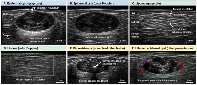
Figure 1. Representative high-frequency ultrasonographic features of epidermal cysts, lipomas, and other superficial subcutaneous lesions.
Figure 1 Panels A–Billustrate an epidermal cyst as a well-defined hypoechoic/heterogeneous superficial lesion with posterior acoustic enhancement, possible skin tract or punctum, and generally absent internal Doppler vascularity. Panels C–D show a typical lipoma with orientation parallel to the skin surface, internal linear echogenic striations, compressibility, and minimal or absent internal vascularity. Panel E represents another benign subcutaneous lesion with internal echogenic foci and posterior acoustic shadowing, while panel Fdemonstrates an inflamed epidermal cyst with peripheral vascularity related to inflammatory change. These combined grayscale and Doppler features help distinguish epidermal cysts and lipomas from other superficial soft-tissue lesions and support improved diagnostic classification compared with clinical examination alone.
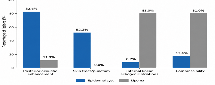
Figure 2. Comparison of characteristic high-frequency ultrasound features of epidermal cysts and lipomas
Figure 2 Epidermal cysts demonstrated a substantially higher prevalence of posterior acoustic enhancement and skin-surface tract/punctum, whereas lipomas more frequently demonstrated internal linear echogenic striations and compressibility.
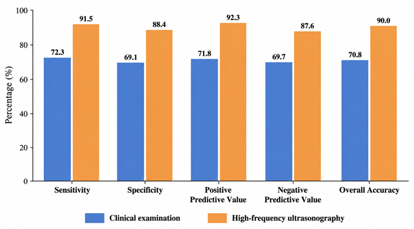
Figure 3. Comparison of diagnostic accuracy between clinical examination and high-frequency ultrasonography
Figure 3 Correct lesion classification increased from 70.8% using clinical examination alone to 90.0% following HFUS assessment (McNemar P < 0.001).
DISCUSSION:
The present study demonstrates that high-frequency ultrasonography (HFUS) provides substantial diagnostic value in the evaluation of common superficial and subcutaneous lesions and can considerably improve lesion characterization beyond clinical examination alone. Among the 130 lesions evaluated, epidermal cysts and lipomas constituted the predominant diagnostic categories, accounting for 35.4% and 32.3% of cases, respectively. More importantly, these two entities demonstrated distinguishable sonographic patterns based on lesion orientation, internal architecture, posterior acoustic characteristics, compressibility, relationship with the skin, and Doppler vascularity. HFUS achieved a diagnostic accuracy of 96.2% for both epidermal cysts and lipomas, while overall correct lesion classification increased from 70.8% with clinical evaluation alone to 90.0% following HFUS. These observations support the growing role of HFUS as a readily accessible, non-invasive modality capable of bridging the diagnostic gap between physical examination and more invasive or costly investigations.
The improvement in diagnostic classification observed in the present study is consistent with contemporary evidence regarding HFUS evaluation of subcutaneous lesions. Miao et al. prospectively evaluated 391 lesions and demonstrated that addition of HFUS to clinical examination increased the proportion of correct diagnoses from 47.3% to 80.8%, while markedly reducing indeterminate diagnoses. Particularly substantial improvements were observed for epidermoid cysts and lipomas, with diagnostic accuracy increasing to 86.7% and 94.8%, respectively [10]. Although the absolute values differ from those of the present study, probably because of variations in lesion spectrum, equipment, operator experience, and diagnostic criteria, the overall direction of effect is highly consistent. The present findings therefore reinforce the clinical value of combining targeted ultrasonographic assessment with routine examination rather than considering HFUS solely as an adjunct for clinically indeterminate lesions.
Epidermal cysts were the most frequent lesion in the present cohort and displayed several characteristic sonographic features. Most were well defined (93.5%), hypoechoic or heterogeneous (84.8%), and demonstrated posterior acoustic enhancement (82.6%). Furthermore, 89.1% showed no internal Doppler vascularity. A tract or punctum extending toward the skin surface was demonstrated in 52.2% of epidermal cysts but was virtually absent in other lesion categories. These findings are biologically plausible because epidermal cysts consist of a keratin-filled cavity lined by squamous epithelium, and the variable organization of keratin accounts for their heterogeneous internal echoes. Huang et al., in a series of surgically confirmed superficial epidermal cysts, reported that a considerable proportion exhibited a characteristic “pseudotestis” appearance consisting of homogeneous low-to-medium internal echoes. Additional features including dermal attachment, focal dermal protrusion, posterior acoustic enhancement, internal echogenic reflectors, and filiform anechoic areas further facilitated diagnosis [11]. The high frequency of posterior enhancement and skin attachment observed in the present study is therefore well aligned with previously described imaging-pathological correlations.
The finding of absent internal vascularity in nearly 90% of epidermal cysts is another diagnostically relevant observation. Simple, non-inflamed epidermal cysts are generally avascular internally, whereas peripheral vascularity may emerge after inflammation or rupture due to reactive hyperemia and granulation tissue. In the present series, four cysts demonstrated predominantly peripheral flow and one showed mixed peripheral and internal vascular signals; these cases occurred principally among lesions demonstrating inflammatory changes. This distinction is clinically important because a ruptured or infected epidermal cyst may become irregular, heterogeneous, and vascular, thereby mimicking an inflammatory mass or even a solid neoplasm. Consequently, vascularity should not be interpreted in isolation; it should be integrated with lesion compartment, morphology, skin attachment, internal debris, posterior acoustic behavior, and clinical inflammatory findings.
Lipomas also showed a recognizable combination of HFUS characteristics. In this study, 95.2% were well defined, 95.2% were oriented parallel to the skin surface, 81.0% demonstrated internal linear echogenic striations, and 81.0% were compressible. Internal vascularity was absent in 90.5%. These characteristics closely resemble the classical sonographic appearance reported by Ahuja et al., who described lipomas as elliptical, compressible masses oriented parallel to the skin surface and containing multiple internal echogenic lines. In their study, all lesions contained characteristic echogenic lines, and no Doppler flow was identified [12]. Importantly, lipoma echogenicity itself can be highly variable. Therefore, the diagnostic process should emphasize the complete sonographic pattern—including orientation, internal striations, compressibility and vascularity—rather than depend exclusively on whether the lesion is hyperechoic relative to surrounding tissues.
This interpretation is supported by the high diagnostic performance for lipoma in the present study. HFUS showed a sensitivity of 92.9%, specificity of 97.7%, positive predictive value of 95.1%, negative predictive value of 96.6%, and overall accuracy of 96.2%. Wagner et al. similarly reported high sonographic performance for superficial lipomas. In their study, pooled interpretation by experienced readers yielded 96% sensitivity, 97% specificity, and 96% accuracy for lipoma diagnosis [13]. These values are strikingly similar to those observed in the present study and support the reproducibility of characteristic sonographic findings when superficial lesions are systematically assessed by experienced operators.
The superiority of ultrasonography over palpation observed in the current study is also supported by earlier evidence. Kuwano et al. evaluated 183 histologically confirmed benign subcutaneous lesions and found that ultrasonography increased overall agreement with pathological diagnosis from 29% following palpation to 46% after sonographic assessment. In particular, sensitivity for lipoma increased from 54.8% to 88.1%, while specificity for epidermal cyst increased from 93.5% to 99.3% [14]. In the present study, clinical assessment correctly classified 70.8% of lesions, whereas HFUS increased correct classification to 90.0%, with HFUS correctly resolving 29 lesions that had been misclassified clinically. The higher absolute clinical accuracy in the current cohort could reflect differences in patient selection and the relative predominance of typical epidermal cysts and lipomas. Nevertheless, both studies indicate that ultrasonography adds information unavailable from inspection and palpation alone.
Not all published investigations, however, have reported uniformly high accuracy for ultrasound diagnosis of lipomas. Inampudi et al. evaluated 39 histologically verified soft-tissue masses and reported sensitivities of only 40–52% and accuracies of 49–64% among different readers for the diagnosis of lipoma[15]. Interobserver agreement in their study was also relatively limited (κ = 0.35). This contrasts with the present sensitivity of 92.9%, accuracy of 96.2%, and almost-perfect agreement for final HFUS diagnosis (κ = 0.86). Several factors may account for the discrepancy. Sonographic technology and transducer resolution have advanced considerably, while the present protocol deliberately incorporated multiple diagnostic features rather than echogenicity alone. Moreover, superficial lipomas tend to show more reproducible imaging characteristics than deep-seated or anatomically complex fatty lesions. Operator experience, lesion depth, selection criteria, and the use of standardized imaging criteria may therefore substantially influence diagnostic performance.
The findings among the heterogeneous group of “other lesions” further illustrate why a multiparametric approach is necessary. Compared with epidermal cysts and lipomas, other lesions were significantly more likely to demonstrate ill-defined margins and internal or mixed vascularity. Only 35.7% showed absent vascularity, compared with approximately 90% of epidermal cysts and lipomas. These observations suggest that Doppler imaging can contribute substantially to lesion characterization, particularly when distinguishing typical avascular benign lesions from vascular tumors, inflammatory abnormalities, and selected neoplasms. Nevertheless, vascularity itself should not be considered synonymous with malignancy. Chiou et al., in a large study of superficial soft-tissue masses, demonstrated that margin, shape, and lesion size differed significantly between benign and malignant lesions, whereas echogenicity, composition, and Doppler findings alone did not reliably distinguish the two groups [16]. Thus, the present observation of increased vascularity among miscellaneous lesions should be viewed as one component of an integrated diagnostic assessment rather than an independent marker of malignant potential.
This concept has direct clinical implications. HFUS provides simultaneous information regarding lesion compartment, shape, dimensions, orientation, margins, internal composition, posterior acoustic behavior, compressibility, vascularity, and relationship with neighboring structures. Such information can help separate lesions with characteristic benign patterns from masses requiring additional investigation. Catalano et al. emphasized that ultrasound represents an appropriate first-line imaging modality for superficial palpable lesions and that many characteristic abnormalities can be confidently diagnosed by systematic grayscale and Doppler examination. In contrast, lesions remaining indeterminate or demonstrating suspicious characteristics should undergo further evaluation using magnetic resonance imaging, image-guided biopsy, or excision as appropriate [17]. The three malignant lesions encountered in the present cohort reinforce the importance of maintaining such an escalation pathway rather than regarding HFUS as a replacement for histopathological assessment.
The significant differences in lesion margins observed in the present study are also noteworthy. Well-defined margins occurred in more than 93% of both epidermal cysts and lipomas but in only 73.8% of other lesions. Although poorly defined margins can raise concern, they are not specific for malignancy. For example, Won et al. demonstrated that dermatofibromas commonly appear as ill-defined, spiculated dermal lesions with alterations in surrounding soft-tissue echogenicity, despite their benign pathology [18]. Therefore, lesion margins must be interpreted in the context of anatomical origin, size, echotexture, vascularity, and adjacent tissue changes. This consideration may be particularly important in avoiding unnecessary alarm when assessing benign fibrous or inflammatory lesions.
The high interobserver reliability observed in the present study provides additional support for a structured sonographic assessment. Agreement was almost perfect for Doppler vascularity (κ = 0.88), final HFUS diagnosis (κ = 0.86), and lesion margins (κ = 0.84), while echogenicity showed substantial agreement (κ = 0.78). Measurement of maximum lesion diameter demonstrated excellent reliability (ICC = 0.94). These findings suggest that clearly defined sonographic criteria may improve reproducibility between observers. The relatively lower agreement for echogenicity compared with vascularity and margins is understandable because echogenicity is influenced by machine settings, surrounding tissue composition, beam angle, lesion depth, and subjective comparison with adjacent structures. Standardization of image acquisition and interpretation is therefore particularly important when HFUS is incorporated into routine diagnostic pathways.
An important strength of the present study is that it evaluated HFUS using a combination of morphology, acoustic characteristics, dynamic examination, and Doppler findings rather than relying on a single sign. The inclusion of histopathological or definitive reference diagnoses allowed direct estimation of sensitivity, specificity, predictive values, and accuracy. Furthermore, the comparison between clinical assessment and HFUS demonstrates the incremental diagnostic benefit of imaging, while interobserver analysis addresses an important issue of reproducibility. The inclusion of lesions other than epidermal cysts and lipomas also more closely reflects the heterogeneous spectrum encountered in routine clinical practice.
Several limitations should nevertheless be acknowledged. First, the study was conducted at a single institution with a relatively modest sample of 130 lesions, which may limit generalizability. Second, epidermal cysts and lipomas accounted for more than two-thirds of lesions, whereas several other pathological entities were represented by small numbers. Consequently, reliable lesion-specific diagnostic performance could not be calculated for uncommon tumors. Third, HFUS remains operator dependent, and diagnostic accuracy may vary according to sonographer experience, equipment, probe frequency, and lesion depth. Fourth, inflammatory or ruptured epidermal cysts may lose their typical imaging characteristics, while some lipomas may show atypical echogenicity or vascularity. Finally, although histopathology constituted the principal reference standard, diagnostic accuracy for rare malignant lesions requires validation in substantially larger prospective multicentre cohorts.
Despite these limitations, the findings have important practical implications. A well-defined superficial lesion demonstrating posterior acoustic enhancement, absent internal vascularity, and a tract toward the skin strongly supports an epidermal cyst, whereas a parallel, compressible subcutaneous mass containing internal linear echogenic striations and minimal vascularity strongly favors a lipoma. Recognition of these combined patterns can improve preoperative diagnosis, guide appropriate referral, assist surgical planning, and potentially reduce unnecessary cross-sectional imaging for lesions with confidently benign appearances. Conversely, atypical morphology, infiltrative margins, heterogeneous architecture, unexpected vascularity, or extension across tissue planes should prompt additional investigation rather than reassurance based solely on clinical appearance.
CONCLUSION:
High-frequency ultrasonography is a valuable, non-invasive imaging modality for the characterization and differentiation of common superficial subcutaneous lesions. In the present study, epidermal cysts and lipomas demonstrated distinct sonographic patterns based on lesion orientation, internal architecture, posterior acoustic characteristics, compressibility, skin communication, and Doppler vascularity. HFUS showed high sensitivity, specificity, and overall diagnostic accuracy for both epidermal cysts and lipomas and significantly improved correct lesion classification compared with clinical examination alone.
REFERENCES:
1. Levy J, Barrett DL, Harris N, Jeong JJ, Yang X, Chen SC. High-frequency ultrasound in clinical dermatology: a review. Ultrasound J. 2021;13(1):24. doi:10.1186/s13089-021-00222-w.
2. Wortsman X, Alfageme F, Roustan G, Arias-Santiago S, Martorell A, Catalano O, et al. Guidelines for performing dermatologic ultrasound examinations by the DERMUS Group. J Ultrasound Med. 2016;35(3):577-580. doi:10.7863/ultra.15.06046.
3. Jacobson JA, Middleton WD, Allison SJ, Dahiya N, Lee KS, Levine BD, et al. Ultrasonography of superficial soft-tissue masses: Society of Radiologists in Ultrasound Consensus Conference Statement. Radiology. 2022;304(1):18-30. doi:10.1148/radiol.211101.
4. Kim HK, Kim SM, Lee SH, Racadio JM, Shin MJ. Subcutaneous epidermal inclusion cysts: ultrasound (US) and MR imaging findings. Skeletal Radiol. 2011;40(11):1415-1419. doi:10.1007/s00256-010-1072-4.
5. Lee HS, Joo KB, Song HT, Kim YS, Park DW, Park CK, et al. Relationship between sonographic and pathologic findings in epidermal inclusion cysts. J Clin Ultrasound. 2001;29(7):374-383. doi:10.1002/jcu.1052.
6. Rahmani G, McCarthy P, Bergin D. The diagnostic accuracy of ultrasonography for soft tissue lipomas: a systematic review. ActaRadiol Open. 2017;6(6):2058460117716704. doi:10.1177/2058460117716704.
7. Wortsman X. Common applications of dermatologic sonography. J Ultrasound Med. 2012;31(1):97-111. doi:10.7863/jum.2012.31.1.97.
8. Wortsman X. Top advances in dermatologic ultrasound. J Ultrasound Med. 2023;42(3):521-545. doi:10.1002/jum.16000.
9. Hobayan CGP, Gray AN, Waters MF, Mager LA, Kobayashi S, Essien EW, et al. Diagnostic accuracy of high-frequency ultrasound for cutaneous neoplasms: a narrative review of the literature. Arch Dermatol Res. 2024;316:419. doi:10.1007/s00403-024-03179-7.
10. Miao Y, Ren WW, Yang FY, Li L, Wu L, Shan DD, et al. Diagnostic value of high-frequency ultrasound (HFUS) in evaluation of subcutaneous lesions. Skin Res Technol. 2023;29(9). doi:10.1111/srt.13464.
11. Huang CC, Ko SF, Huang HY, Ng SH, Lee TY, Lee YW, et al. Epidermal cysts in the superficial soft tissue: sonographic features with an emphasis on the pseudotestis pattern. J Ultrasound Med. 2011;30(1):11-17. doi:10.7863/jum.2011.30.1.11.
12. Ahuja AT, King AD, Kew J, King W, Metreweli C. Head and neck lipomas: sonographic appearance. AJNR Am J Neuroradiol. 1998;19(3):505-508.
13. Wagner JM, Lee KS, Rosas H, Kliewer MA. Accuracy of sonographic diagnosis of superficial masses. J Ultrasound Med. 2013;32(8):1443-1450. doi:10.7863/ultra.32.8.1443.
14. Kuwano Y, Ishizaki K, Watanabe R, Nanko H. Efficacy of diagnostic ultrasonography of lipomas, epidermal cysts, and ganglions. Arch Dermatol. 2009;145(7):761-764. doi:10.1001/archdermatol.2009.61.
15. Inampudi P, Jacobson JA, Fessell DP, Carlos RC, Patel SV, Delaney-Sathy LO, et al. Soft-tissue lipomas: accuracy of sonography in diagnosis with pathologic correlation. Radiology. 2004;233(3):763-767. doi:10.1148/radiol.2333031410.
16. Chiou HJ, Chou YH, Chiu SY, Wang HK, Chen WM, Chen TH, et al. Differentiation of benign and malignant superficial soft-tissue masses using grayscale and color Doppler ultrasonography. J Chin Med Assoc. 2009;72(6):307-315. doi:10.1016/S1726-4901(09)70377-6.
17. Catalano O, Varelli C, Sbordone C, Corvino A, De Rosa D, Vallone G, et al. A bump: what to do next? Ultrasound imaging of superficial soft-tissue palpable lesions. J Ultrasound. 2020;23(3):287-300. doi:10.1007/s40477-019-00415-z.
18. Won KY, Park SY, Jin W, Lew BL. Dermatofibroma: sonographic findings and pathologic correlation. ActaRadiol. 2018;59(4):454-459. doi:10.1177/0284185117721263.