Clinical Spectrum, Etiologic Patterns, and Serum Biomarker Profiles in Uveitis: A Prospective Observational Study.
- Swathi Pola , Assistant Professor, Department of Ophthalmology, ESIC Medical College and Hospital, Sanathnagar, Hyderabad, Telangana, India.
- Dharani Padam , Assistant Professor, Department of Ophthalmology, ESIC Medical College and Hospital, Sanathnagar, Hyderabad, Telangana, India.
- Beulah Sabatina Gunnala , Assistant Professor, Department of Ophthalmology, ESIC Medical College and Hospital, Sanathnagar, Hyderabad, Telangana, India.
- Tejaswini Medishetty , Senior Resident, Department of Ophthalmology, ESIC Medical College and Hospital, Sanathnagar, Hyderabad, Telangana, India.
Article Information:
Abstract:
Background: Uveitis is a heterogeneous intraocular inflammatory disorder with overlapping clinical, infectious, and immune features. Serum biomarkers may provide supportive information, but their role in routine etiologic assessment remains uncertain. The study is designed to describe the clinical and etiologic spectrum of uveitis and explore serum biomarker positivity across anatomic and disease categories. Methods: This prospective observational study included 60 patients (78 eyes) with uveitis presenting to ESIC Medical College & Hospital, Hyderabad, from July 2022 to July 2023. Uveitis was classified using Standardization of Uveitis Nomenclature principles. Clinical evaluation was supplemented by routine and targeted systemic investigations. Serum immunoglobulins, alpha-1-acid glycoprotein, IFN-gamma, TNF-alpha, transforming growth factor-beta, cytokines, homocysteine, and C-peptide were assessed by enzyme-linked immunosorbent assay. Results: Mean age was 44.5 +/- 13 years; 80% were aged 18-60 years. Females constituted 51.6%, and 68.3% had unilateral disease. Anterior uveitis was most frequent (53.3%), followed by panuveitis (23.3%), posterior uveitis (21.6%), and intermediate uveitis (1.7%). Etiology remained idiopathic in 65%; 20% were infectious and 15% immune-related. Tuberculosis was the leading identified infectious cause. Cytokine positivity was most frequent (24/60, 40%), followed by immunoglobulin positivity (10/60, 16.7%), IFN-gamma (6/60, 10%), alpha-1-acid glycoprotein (5/60, 8.3%), TNF-alpha (4/60, 6.7%), and C-peptide (1/60, 1.7%); homocysteine was not elevated. Disease-linked patterns included IFN-gamma positivity in tubercular uveitis and cytokine positivity in herpetic and sarcoid uveitis. Conclusion: Anterior and idiopathic uveitis predominated in this tertiary-care cohort, while tuberculosis remained the main recognized infectious association. Serum biomarkers showed potentially useful disease-linked patterns, but the findings are exploratory and do not establish diagnostic sensitivity or specificity. Larger controlled studies with quantitative assays and paired ocular-fluid measurements are required.
Keywords:
Article :
INTRODUCTION:
Uveitis encompasses inflammatory disorders affecting the uveal tract and, in many patients, adjacent structures such as the vitreous, retina, and optic nerve. Its clinical expression ranges from a short episode of anterior chamber inflammation to persistent posterior or pan-uveal disease with macular edema, glaucoma, cataract, retinal injury, and permanent visual loss. Because the same anatomic pattern may result from infection, systemic immune disease, or an apparently isolated ocular process, etiologic diagnosis remains one of the central challenges in uveitis practice [1,2].
The distribution of uveitis differs across geography, age, referral setting, and local disease prevalence. Population and tertiary-center studies have consistently shown that anterior uveitis is common, but the relative contribution of infectious and immune-mediated causes varies considerably [3-10]. In India, endemic infections, especially tuberculosis, remain important in the diagnostic work-up, while HLA-B27-associated disease, sarcoidosis, spondyloarthropathy, and other immune disorders contribute to the non-infectious spectrum [3,5-9]. These differences make a locally derived clinical profile useful for both diagnostic planning and resource allocation.
Inflammatory mediators provide an additional layer of biologic information. Cytokines and chemokines regulate leukocyte trafficking, T-cell differentiation, macrophage activation, and disruption of ocular immune privilege. Studies of aqueous humor, tears, and peripheral blood have identified changes in IL-1, IL-2, IL-6, IL-8, IL-17, IFN-gamma, TNF-alpha, and other mediators across several uveitic entities [11-14]. However, marker concentrations can overlap between diseases, and systemic levels may not directly reflect the intraocular microenvironment [21-29]. Biomarkers are therefore best considered adjuncts to clinical and etiologic assessment rather than stand-alone tests.
The present study was undertaken to characterize the clinical and etiologic profile of patients with uveitis attending a tertiary-care ophthalmology service in Hyderabad and to explore the pattern of selected serum biomarkers across anatomic and disease categories. The principal objective was descriptive: to determine whether recognizable biomarker patterns accompanied specific clinical diagnoses and could support a more focused diagnostic approach.
MATERIALS AND METHODS:
Study design and setting
A prospective observational study was conducted in the Department of Ophthalmology, ESIC Medical College & Hospital, Sanathnagar, Hyderabad, Telangana, India, from July 2022 to July 2023. All eligible patients presenting with clinical features of uveitis during the study period were considered for inclusion. Sixty patients, involving 78 eyes, were evaluated.
Eligibility criteria
Patients of any age with signs and symptoms consistent with uveitis were eligible. Patients were excluded when ocular inflammation occurred in the early postoperative period, followed ocular trauma, represented endophthalmitis, or when the patient did not consent to participation. The study used the term idiopathic when the inflammatory pattern could not be assigned to a recognized uveitic entity or linked to a specific systemic or infectious cause after the planned evaluation.
Clinical assessment
Each participant underwent a detailed ocular and systemic history, including onset, duration, recurrence, laterality, previous treatment, systemic symptoms, and relevant comorbidity. Ophthalmic examination included Snellen visual acuity, slit-lamp biomicroscopy, applanation tonometry, dilated fundus examination with a 90-diopter lens and indirect ophthalmoscopy. Gonioscopy and visual-field testing were performed when clinically indicated. B-scan ultrasonography was used when media opacity prevented adequate posterior-segment visualization. Optical coherence tomography and fundus fluorescein angiography were obtained in selected posterior-segment cases.
The predominant anatomic site of inflammation was categorized as anterior, intermediate, posterior, or panuveitis. Anterior chamber cells and flare and vitreous inflammation were assessed clinically in keeping with Standardization of Uveitis Nomenclature principles [1]. The final anatomic and etiologic diagnosis integrated ocular findings, laboratory results, imaging, and systemic specialist assessment when required.
Laboratory and systemic evaluation
Baseline investigations included complete blood count, erythrocyte sedimentation rate, C-reactive protein, tuberculin skin testing, venereal disease research laboratory testing, serum angiotensin-converting enzyme, rheumatoid factor, viral serology, chest radiography, TORCH profile, and HLA-B27 testing. Additional investigations were selected according to clinical suspicion. These included high-resolution computed tomography of the chest for suspected sarcoidosis, QuantiFERON testing for tuberculosis, antinuclear antibody and C-ANCA testing, and radiography of the lumbosacral spine and sacroiliac joints in suspected spondyloarthropathy.
Serum biomarker assessment
The biomarker panel comprised immunoglobulins (IgG, IgA, and IgM), alpha-1-acid glycoprotein, IFN-gamma, TNF-alpha, transforming growth factor-beta, a cytokine panel, serum homocysteine, and C-peptide. The dissertation records that these markers were analyzed by enzyme-linked immunosorbent assay. Individual cytokines documented in the study records included IL-1, IL-2, IL-6, IL-8, and IL-17. Biomarker findings were recorded as positive or raised.
Treatment and multidisciplinary care
Treatment was based on the clinical diagnosis and severity. Topical anti-inflammatory therapy was used when appropriate, with systemic anti-inflammatory treatment and other supportive measures added according to disease extent and systemic involvement. Patients with relevant systemic findings were referred to the appropriate medical specialty, and subsequent evaluation and treatment were coordinated with that service.
Statistical analysis
Data were compiled and analyzed using SPSS version 20 (IBM Corp., Armonk, NY, USA), as documented in the dissertation. Continuous age data are presented as mean +/- standard deviation, while categorical variables are summarized as frequencies and percentages. Biomarker counts are interpreted as positive test findings; an individual patient could contribute more than one positive marker.
Ethical considerations
The study received approval from the Institutional Ethics Committee of ESIC Medical College & Hospital and ESIC Super Speciality Hospital, Hyderabad (Ref. No. 799/U/IEC/ESICMC/S0165/07/2022; approval letter dated 26 July 2022). Written informed consent was obtained from participants as applicable to the approved protocol. The study was conducted in accordance with institutional ethical requirements.
RESULTS:
Participant characteristics
Sixty patients with uveitis were included, representing 78 affected eyes. The mean age at presentation was 44.5 +/- 13 years. Forty-eight patients (80%) were 18-60 years old, nine (15%) were older than 60 years, and three (5%) were younger than 18 years (Table 1; Figure 1). There was a slight female predominance, with 31 women (51.6%) and 29 men (48.3%). Unilateral disease was reported in 41 patients (68.3%), while 19 (31.7%) had bilateral involvement.
Table 1. Demographic and clinical profile of the study population (n=60)
|
Characteristic |
Category |
n |
% |
|
Age group |
<18 years |
3 |
5.0 |
|
18-60 years |
48 |
80.0 |
|
|
>60 years |
9 |
15.0 |
|
|
Sex |
Male |
29 |
48.3 |
|
Female |
31 |
51.6 |
|
|
Laterality |
Unilateral |
41 |
68.3 |
|
Bilateral |
19 |
31.7 |
|
|
Anatomic location |
Anterior uveitis |
32 |
53.3 |
|
Intermediate uveitis |
1 |
1.7 |
|
|
Posterior uveitis |
13 |
21.7 |
|
|
Panuveitis |
14 |
23.3 |
Mean age: 44.5 +/- 13 years. Percentages are based on the 60-patient cohort.
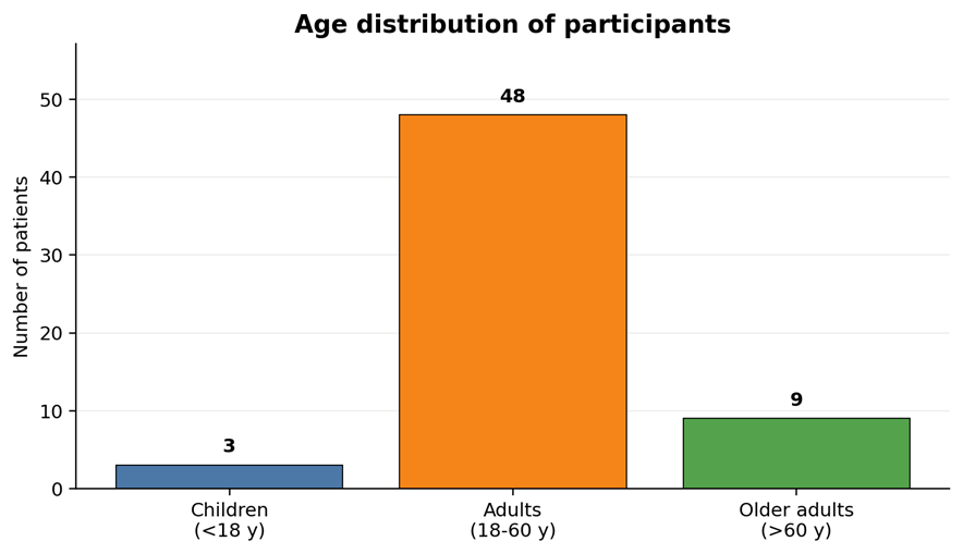
Figure 1: Age distribution of patients with uveitis. Adults aged 18-60 years formed the largest group
Anatomic pattern of uveitis
Anterior uveitis was the most frequent anatomic diagnosis, accounting for 32 patients (53.3%). Panuveitis was present in 14 (23.3%), posterior uveitis in 13 (21.6%), and intermediate uveitis in one patient (1.7%) (Figure 2). The age-stratified distribution is shown in Table 2. In children, posterior and pan-uveal involvement predominated, whereas anterior uveitis was the leading pattern among adults and older patients.
Table 2. Age-wise distribution of anatomic uveitis patterns
|
Age group |
Anterior |
Intermediate |
Posterior |
Panuveitis |
Total |
|
<18 years |
0 |
0 |
2 |
1 |
3 |
|
18-60 years |
27 |
1 |
8 |
12 |
48 |
|
>60 years |
5 |
0 |
3 |
1 |
9 |
|
Total |
32 |
1 |
13 |
14 |
60 |
Values are patient counts. The anatomic category reflects the predominant site of inflammation.
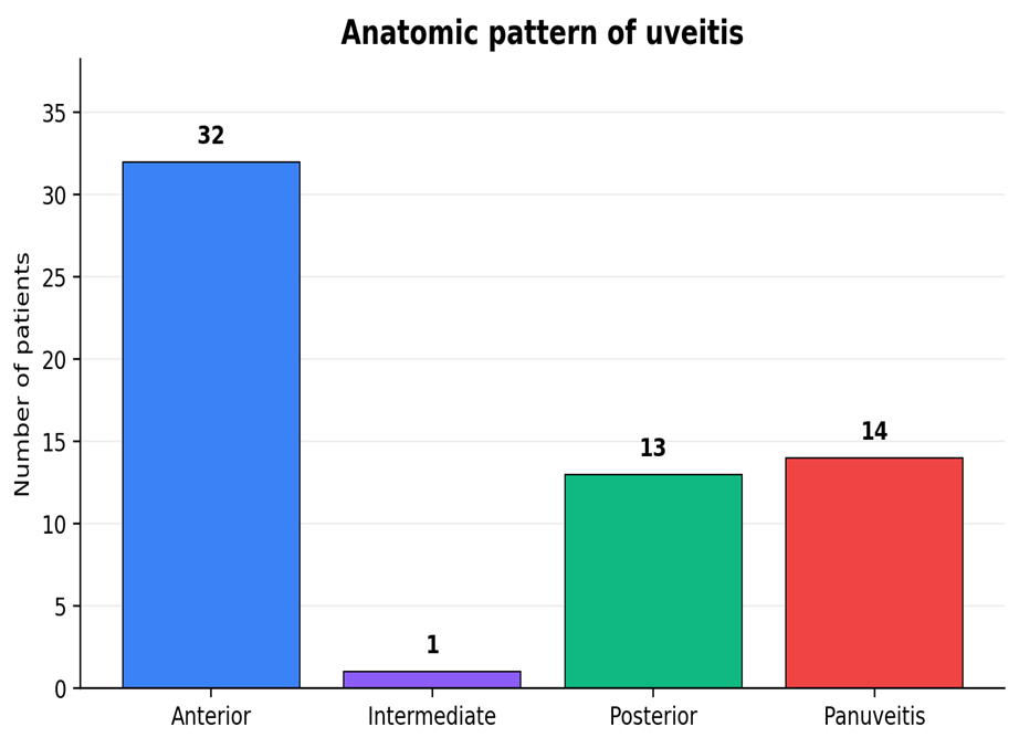
Figure 2: Distribution of uveitis according to anatomic classification.
Etiologic spectrum
The etiology remained idiopathic in 39 patients (65%). Twelve cases (20%) were classified as infectious and nine (15%) as immune-related (Figure 3). Idiopathic disease was the most frequent category in anterior, intermediate, and posterior uveitis. In contrast, infectious causes contributed most strongly to the panuveitis subgroup, accounting for six of 14 cases (Table 3).
Table 3. Etiologic classification according to anatomic location
|
Etiology |
Anterior |
Intermediate |
Posterior |
Panuveitis |
Total |
|
Idiopathic |
25 |
1 |
9 |
4 |
39 |
|
Immune-related |
4 |
0 |
1 |
4 |
9 |
|
Infectious |
3 |
0 |
3 |
6 |
12 |
|
Total |
32 |
1 |
13 |
14 |
60 |
Etiologic assignment was based on clinical features, laboratory investigations, imaging, and systemic evaluation
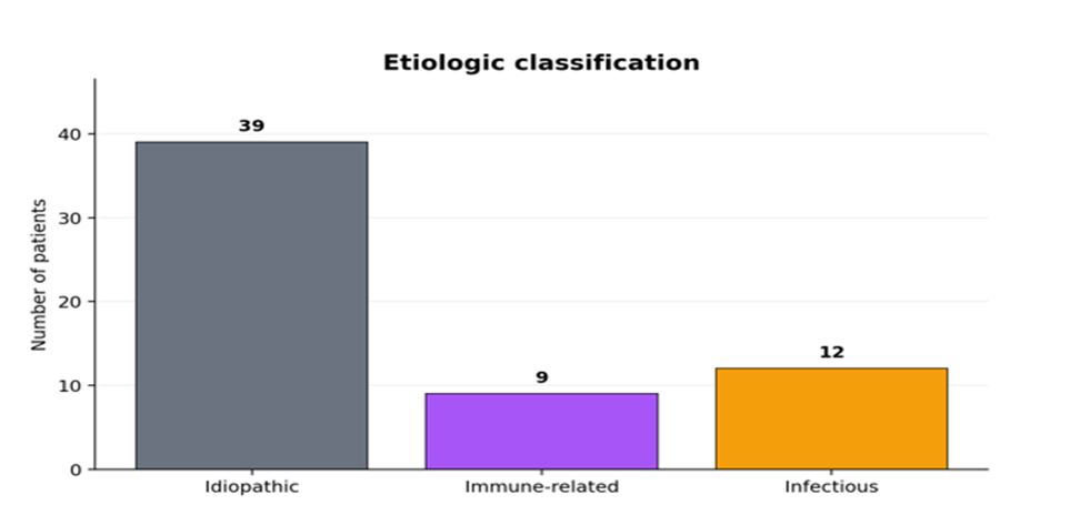
Figure 3: Etiologic distribution of the 60 uveitis cases
Infectious and immune-associated causes
Tuberculosis was the most frequent identified infectious association, with eight cases distributed across anterior, posterior, and pan-uveal disease. Herpetic uveitis accounted for two cases, and toxoplasmosis for two posterior uveitis cases (Table 4). No fungal uveitis was identified in the reported cohort. Immune-associated diagnoses recorded in the study included HLA-B27-related disease, rheumatoid factor positivity, antinuclear antibody-associated presentations, sarcoidosis, psoriasis, and spondyloarthropathy. Because several immune labels were reported across different aggregate tables, the immune subgroup is best interpreted as a clinically defined category rather than a single laboratory-positive group.
Table 4. Distribution of identified infectious agents by anatomic pattern
|
Infectious agent |
Anterior |
Intermediate |
Posterior |
Panuveitis |
Total |
|
Tuberculosis |
2 |
0 |
1 |
5 |
8 |
|
Herpes virus |
1 |
0 |
0 |
1 |
2 |
|
Toxoplasma gondii |
0 |
0 |
2 |
0 |
2 |
|
Total |
3 |
0 |
3 |
6 |
12 |
Counts reproduce the aggregate infectious-disease table in the dissertation. Tuberculosis was the dominant identified infectious association
Serum biomarker profile
Cytokine positivity was the most frequent serum biomarker finding, recorded in 24 patients (40%). Immunoglobulin positivity was reported in 10 (16.7%), IFN-gamma in six (10%), alpha-1-acid glycoprotein in five (8.3%), TNF-alpha in four (6.7%), and C-peptide in one (1.7%). No patient was reported to have raised serum homocysteine. Most cytokine-positive findings occurred in anterior uveitis, whereas IFN-gamma positivity was distributed across anterior, posterior, and pan-uveal disease (Table 5; Figure 4). Because individual patients could have more than one positive biomarker, these categories are not mutually exclusive and should not be summed to estimate the proportion of patients with any positive test.
Table 5. Positive serum biomarker findings by anatomic location
|
Biomarker |
Anterior |
Intermediate |
Posterior |
Panuveitis |
Overall n (%) |
|
IgG/IgA/IgM |
5 |
0 |
2 |
3 |
10 (16.7) |
|
Alpha-1-acid glycoprotein |
5 |
0 |
0 |
0 |
5 (8.3) |
|
IFN-gamma |
2 |
0 |
1 |
3 |
6 (10.0) |
|
Cytokines |
19 |
0 |
3 |
2 |
24 (40.0) |
|
Homocysteine |
0 |
0 |
0 |
0 |
0 (0) |
|
TNF-alpha |
2 |
0 |
2 |
0 |
4 (6.7) |
|
C-peptide |
0 |
0 |
0 |
1 |
1 (1.7) |
Biomarker categories overlap because more than one marker could be positive in the same patient. "Cytokines" represents the composite cytokine category.
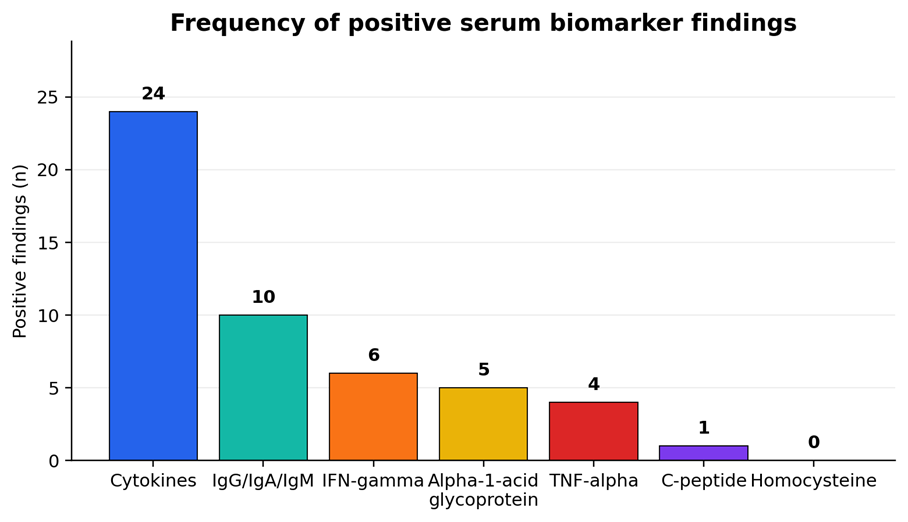
Figure 4: Overall frequency of positive serum biomarker findings
Disease-linked biomarker observations
Exploratory subgroup patterns were also observed. IFN-gamma was the predominant raised marker in tubercular uveitis, with six positive findings reported. Herpetic and sarcoid uveitis cases showed cytokine positivity, while toxoplasmosis cases were associated with positive immunoglobulin findings. Within HLA-B27-associated disease, TNF-alpha and cytokine positivity were recorded. Idiopathic cases accounted for much of the overall cytokine signal (Table 6).
Table 6: Disease-linked serum biomarker patterns reported in the study
|
Clinical/etiologic category |
Reported positive serum marker pattern |
|
HLA-B27-associated uveitis |
Cytokines (2 findings), TNF-alpha (3 findings), C-peptide (1 finding) |
|
Tubercular uveitis |
IFN-gamma (6 findings), immunoglobulins (1 finding) |
|
Herpetic uveitis |
Cytokines (2 findings) |
|
Sarcoid uveitis |
Cytokines (3 findings) |
|
Toxoplasmosis |
Immunoglobulins (2 findings) |
|
Idiopathic uveitis |
Immunoglobulins (7), alpha-1-acid glycoprotein (5), cytokines (17), TNF-alpha (1) |
DISCUSSION:
This prospective tertiary-care study demonstrates the heterogeneity of uveitis in routine clinical practice while also showing a clear dominance of anterior and idiopathic presentations. Four observations are especially relevant. First, the disease burden was concentrated in working-age adults. Second, anterior uveitis represented more than half of the cohort. Third, although most cases remained idiopathic after evaluation, infectious uveitis still accounted for one fifth of the series, with tuberculosis as the leading identified infection. Fourth, serum inflammatory markers showed recognizable disease-linked patterns, but the overlap between markers and the small disease subgroups limit their use as independent diagnostic tests.
Age, sex, and laterality
The mean age at presentation was 44.5 years, and 80% of participants were between 18 and 60 years. This pattern is consistent with the well-recognized concentration of uveitis in economically active age groups, although elderly-onset uveitis is increasingly recognized as populations age [16,17,20]. The modest female predominance in the present cohort differs from several Indian referral-center series in which men were more frequent, but population-level studies have not consistently shown a large sex difference [4,17,18]. Such variation probably reflects the mix of infectious, systemic immune, and referral patterns at individual centers rather than a uniform sex-specific susceptibility.
Unilateral disease was more common than bilateral disease. This is clinically plausible in a cohort dominated by anterior uveitis, where unilateral acute presentations are frequent, particularly in HLA-B27-associated disease [5,15,35,36]. Bilaterality becomes more common in several posterior and pan-uveal syndromes, including sarcoidosis and other systemic inflammatory disorders [6-9].
Anatomic distribution
Anterior uveitis accounted for 53.3% of cases, followed by panuveitis and posterior uveitis. This ordering is broadly compatible with earlier Indian and international experience, where anterior uveitis is often the largest anatomic category [4,15,18,19,30]. Differences in the relative frequency of posterior and pan-uveal disease across studies are expected because tertiary referral services tend to accumulate more severe or diagnostically complex cases. The very small intermediate uveitis subgroup in this series should not be interpreted as a population estimate.
Etiology and the importance of tuberculosis
A specific cause could not be assigned in 65% of participants. A high idiopathic proportion is not unusual in uveitis, particularly in anterior disease, because the phenotype may remain isolated even after a focused systemic evaluation [1,2,15]. At the same time, the 20% infectious fraction underscores the continuing importance of infection-directed assessment in India. Tuberculosis was the leading identified infectious association and was represented across anterior, posterior, and pan-uveal patterns. This agrees with the established clinical diversity of intraocular tuberculosis and with Indian referral-center experience [3,4,11,28,31].
Tuberculosis illustrates why etiologic interpretation in uveitis cannot rely on a single laboratory result. Ocular disease may arise in the setting of active infection, latent infection with immunologic responses, or a compatible ocular phenotype without microbiologic confirmation. The diagnostic value of IFN-gamma-related testing therefore depends on pretest probability, ocular findings, systemic evaluation, and exclusion of competing diagnoses [3,11,28,31]. In this cohort, IFN-gamma was the dominant serum marker among tubercular cases, a finding consistent with the central role of type 1 cellular immunity in tuberculosis-associated inflammation.
Serum cytokines and other biomarkers
Cytokine positivity was the most common biomarker finding and was particularly frequent in anterior and idiopathic uveitis. This is biologically plausible because uveitis involves coordinated T-cell, macrophage, and chemokine signaling rather than a single disease-specific mediator. Elevated IL-17A has been described in peripheral blood from uveitis patients, while multiplex aqueous-humor studies have shown distinct but overlapping inflammatory profiles across clinical phenotypes [21,23-29]. The present findings therefore support the presence of systemic inflammatory activation but do not indicate that a composite serum cytokine result can identify a unique etiology.
The disease-linked observations are nevertheless informative. Herpetic cases were cytokine-positive, which accords with prior work showing inflammatory cytokine signatures in viral uveitis [29]. Sarcoid cases also showed cytokine positivity, consistent with the immune activation described in ocular sarcoidosis and rheumatic inflammatory disease [6-9,22]. HLA-B27-associated cases showed TNF-alpha and cytokine positivity, fitting the known inflammatory biology of spondyloarthropathy-associated anterior uveitis [5,22,36]. Toxoplasmosis cases were immunoglobulin-positive, but serology must be interpreted carefully because IgG commonly reflects prior exposure and does not by itself confirm active ocular toxoplasmosis [10,32].
The broader literature suggests that intraocular fluids may provide stronger disease discrimination than serum because aqueous humor more directly reflects local ocular inflammation [11,13,24-29]. At the same time, aqueous or vitreous sampling is invasive and is not justified for every patient. A clinically useful future strategy may therefore combine a parsimonious serum panel with ocular phenotype, imaging, and targeted intraocular testing only when the diagnosis remains uncertain.
Clinical implications
The practical message from this study is that biomarkers should refine, not replace, the conventional uveitis work-up. A careful history, anatomic classification, recognition of infectious or systemic clues, and tailored testing remain the foundation of diagnosis. Serum markers may be most useful when they increase or decrease the plausibility of a diagnosis already suggested by the clinical picture. This approach also reduces the risk of broad, non-specific laboratory screening that can produce incidental positive results and unnecessary treatment.
Strengths and limitations
The study prospectively characterized a real-world tertiary-care cohort and incorporated a relatively broad serum biomarker panel alongside clinical and systemic evaluation. It also provides local data from a region where both infectious and immune-mediated uveitis are relevant. Several limitations, however, are important. The sample was small and drawn from a single tertiary hospital, which limits generalizability and subgroup analysis. Biomarker results were recorded mainly as positive or negative, without uniform quantitative concentrations, assay-specific reference ranges, or diagnostic cut-offs. There was no healthy or disease-control group, no paired aqueous or vitreous biomarker analysis, and no serial sampling to assess change with treatment. Some patients had more than one positive marker, so the biomarker counts are not mutually exclusive. Finally, the study was not designed to estimate sensitivity, specificity, predictive values, or causal relationships. These limitations make the biomarker findings hypothesis-generating rather than confirmatory.
CONCLUSION:
In this tertiary-care cohort, uveitis most often affected adults and was usually unilateral. Anterior uveitis was the leading anatomic form, and most cases remained idiopathic after clinical and laboratory evaluation. Infectious disease accounted for one fifth of cases, with tuberculosis as the most frequent identified infection. Serum cytokines were the most commonly positive biomarker category, while IFN-gamma showed a notable association with tubercular uveitis and cytokine positivity was observed in herpetic and sarcoid disease. These patterns may support targeted evaluation, but they are not sufficient to establish biomarker-based diagnosis. Larger controlled studies using quantitative assays, prespecified thresholds, serial measurements, and paired serum-ocular fluid analysis are needed before these markers can be incorporated into routine diagnostic algorithms.
REFERENCES:
1. Nussenblatt RB, Whitcup SM. Uveitis: Fundamentals and Clinical Practice. 4th ed. Philadelphia: Mosby/Elsevier; 2010.
2. Foster CS, Vitale AT. Diagnosis and Treatment of Uveitis. Philadelphia: WB Saunders; 2002.
3. Gupta V, Gupta A, Rao NA. Intraocular tuberculosis - an update. Surv Ophthalmol. 2007;52(6):561-587.
4. Biswas J, Narain S, Das D, Ganesh SK. Pattern of uveitis in a referral uveitis clinic in India. Int Ophthalmol. 1996-1997;20(4):223-228.
5. Chang JH, McCluskey PJ, Wakefield D. Acute anterior uveitis and HLA-B27. Surv Ophthalmol. 2005;50(4):364-388.
6. Herbort CP, Rao NA, Mochizuki M; Scientific Committee of the First International Workshop on Ocular Sarcoidosis. International criteria for the diagnosis of ocular sarcoidosis: results of the First International Workshop on Ocular Sarcoidosis. Ocul Immunol Inflamm. 2009;17(3):160-169.
7. Jones N, Mochizuki M. Sarcoidosis: epidemiology and clinical features. Ocul Immunol Inflamm. 2010;18(2):72-79.
8. Agrawal R, Gonzalez-Lopez JJ, Meier F, Gupta B, Pavesio CE. Ocular and systemic features of sarcoidosis and correlation with the International Workshop for Ocular Sarcoidosis diagnostic criteria. Sarcoidosis Vasc Diffuse Lung Dis. 2015;32(3):237-245. PMID: 26422569.
9. Kawaguchi T, Hanada A, Horie S, Sugamoto Y, Sugita S, Mochizuki M. Evaluation of characteristic ocular signs and systemic investigations in ocular sarcoidosis patients. Jpn J Ophthalmol. 2007;51(2):121-126.
10. Gaddi PJ, Yap GS. Cytokine regulation of immunopathology in toxoplasmosis. Immunol Cell Biol. 2007;85:155-159.
11. Ang M, Cheung G, Vania M, Chen J, Yang H, Li J, et al. Aqueous cytokine and chemokine analysis in uveitis associated with tuberculosis. Mol Vis. 2012;18:565-573.
12. Aggarwal S, Ghilardi N, Xie MH, de Sauvage FJ, Gurney AL. Interleukin-23 promotes a distinct CD4 T cell activation state characterized by the production of interleukin-17. J Biol Chem. 2003;278:1910-1914.
13. Xu J, Qin Y, Chang R, Tan H, Wang Q, Su G, et al. Aqueous cytokine levels in four common uveitis entities. Int Immunopharmacol. 2020;78:106021.
14. Agrawal R, Iyer J, Connolly J, Iwata D, Teoh S. Cytokines and biologics in non-infectious autoimmune uveitis: bench to bedside. Indian J Ophthalmol. 2014;62:74-81.
15. Mathur G, Biswas J. Systemic associations of anterior uveitis in a tertiary care ophthalmic centre in South India. Int Ophthalmol. 2012;32(5):417-421.
16. Abdulaal MR, Abiad BH, Hamam RN. Uveitis in the aging eye: incidence, patterns, and differential diagnosis. J Ophthalmol. 2015;2015:509456.
17. Acharya NR, Tham VM, Esterberg E, Borkar DS, Parker JV, Vinoya AC, et al. Incidence and prevalence of uveitis: results from the Pacific Ocular Inflammation Study. JAMA Ophthalmol. 2013;131(11):1405-1412.
18. Rathinam SR, Krishnadas R, Ramakrishnan R, Thulasiraj RD, Tielsch JM, Katz J, et al. Population-based prevalence of uveitis in Southern India. Br J Ophthalmol. 2011;95(4):463-467.
19. Dogra M, Singh R, Agarwal A, Sharma A, Singh SR, Gautam N, et al. Epidemiology of uveitis in a tertiary-care referral institute in North India. Ocul Immunol Inflamm. 2017;25(Suppl 1):S46-S53.
20. BenEzra D, Cohen E, Maftzir G. Uveitis in children and adolescents. Br J Ophthalmol. 2005;89:444-448.
21. Balamurugan S, Das D, Hasanreisoglu M, Toy BC, Akhter M, Anuradha VK, et al. Interleukins and cytokine biomarkers in uveitis. Indian J Ophthalmol. 2020;68(9):1750-1763. doi:10.4103/ijo.IJO_564_20.
22. Hysa E, Cutolo CA, Gotelli E, Pacini G, Schenone C, Kreps EO, et al. Immunopathophysiology and clinical impact of uveitis in inflammatory rheumatic diseases: an update. Eur J Clin Invest. 2021;51(8):e13572. doi:10.1111/eci.13572.
23. Jawad S, Liu B, Agron E, Nussenblatt RB, Sen HN. Elevated serum levels of interleukin-17A in uveitis patients. Ocul Immunol Inflamm. 2013;21:434-439.
24. Curnow SJ, Falciani F, Durrani OM, Cheung CM, Ross EJ, Wloka K, et al. Multiplex bead immunoassay analysis of aqueous humor reveals distinct cytokine profiles in uveitis. Invest Ophthalmol Vis Sci. 2005;46:4251-4259.
25. Ooi KGJ, Galatowicz G, Calder VL, Lightman SL. Cytokines and chemokines in uveitis - is there a correlation with clinical phenotype? Clin Med Res. 2006;4:294-309. doi:10.3121/cmr.4.4.294.
26. Abu El-Asrar AM, Al-Obeidan SS, Kangave D, Geboes K, Opdenakker G, Van Damme J, et al. CXC chemokine expression profiles in aqueous humor of patients with different clinical entities of endogenous uveitis. Immunobiology. 2011;216:1004-1009. doi:10.1016/j.imbio.2011.03.007.
27. Kuiper JJW, Mutis T, de Jager W, de Groot-Mijnes JDF, Rothova A. Intraocular interleukin-17 and proinflammatory cytokines in HLA-A29-associated birdshot chorioretinopathy. Am J Ophthalmol. 2011;152:177-182.e1. doi:10.1016/j.ajo.2011.01.031.
28. Abu El-Asrar AM, Struyf S, Kangave D, Al-Obeidan SA, Opdenakker G, Geboes K, et al. Cytokine and CXC chemokine expression patterns in aqueous humor of patients with presumed tuberculous uveitis. Cytokine. 2012;59:377-381. doi:10.1016/j.cyto.2012.04.030.
29. Lahmar I, Abou-Bacar A, Abdelrahman T, Guinard M, Babba H, Ben Yahia S, et al. Cytokine profiles in toxoplasmic and viral uveitis. J Infect Dis. 2009;199:1239-1249. doi:10.1086/597478.
30. Das D, Bhattacharjee H, Bhattacharyya PK, Jain L, Panicker MJ, Das K, et al. Pattern of uveitis in North East India: a tertiary eye care center study. Indian J Ophthalmol. 2009;57:144-146.
31. Agarwal A, Deokar A, Sharma R, Singh N, Aggarwal K, Sharma S, et al. Longitudinal analysis of serum cytokine profile among patients with tubercular multifocal serpiginoid choroiditis: a pilot study. Eye (Lond). 2019;33(1):129-135. doi:10.1038/s41433-018-0157-5.
32. Park SW, Kim SH, Kwon HJ, Lee SM, Byon IS, Lee JE. Diagnostic value of positive findings of Toxoplasma gondii-specific immunoglobulin M serum antibody in uveitis patients to confirm ocular toxoplasmosis. Ocul Immunol Inflamm. 2019;27(4):583-590. doi:10.1080/09273948.2018.1433303.
33. Ferguson TA, Fletcher S, Herndon J, Griffith TS. Neuropeptides modulate immune deviation induced via the anterior chamber of the eye. J Immunol. 1995;155:1746-1756.
34. Jang MH, Seoh JY, Miyasaka M. Cytokines, chemokines, and their receptors: targets for immunomodulation. J Leukoc Biol. 2006;80(2):217-219. doi:10.1189/jlb.1105671.
35. Brewerton DA, Hart FD, Nicholls A, Caffrey M, James DC, Sturrock RD. Ankylosing spondylitis and HLA-B27. Lancet. 1973;1(7809):904-907.
36. Mishra MN, Bharucha KM. HLA-B27 association with uveitis in an Asian Indian population. Iran J Immunol. 2011;8(2):85-89.