Ultrasound Assessment of Skin Thickness in Common Dermatological Disorders.
- Dr. Sarthak Kesarwani , MD (Radiodiagnosis) Assistant Professor Venkateshwara Institute of Medical Sciences Gajraula Uttar Pradesh
- Dr. Diksha Agrawal , MD, DNB, MNAMS (Dermatology, Venereology And leprosy) Assistant Professor Venkateshwara Institute of Medical Sciences Gajraula Uttar Pradesh
Article Information:
Abstract:
Background: High-frequency ultrasonography offers a non-invasive method for objectively assessing structural changes in the skin. Quantitative measurement of skin thickness may provide additional information regarding disease activity and severity in inflammatory and sclerosingdermatoses. Objective: To evaluate ultrasound-measured skin thickness in common dermatological disorders and compare affected skin with corresponding clinically unaffected skin. Materials and Methods: This prospective observational study included 180 participants with psoriasis, atopic dermatitis, chronic eczema, lichen planus, morphea, or vitiligo, with 30 participants in each group. High-frequency ultrasonography was performed using a linear-array transducer. Skin thickness was measured from the superficial epidermal interface to the dermal-subcutaneous boundary at affected and corresponding unaffected sites. Qualitative sonographic features, disease severity, and measurement reliability were also assessed. Statistical analysis included paired comparisons, correlation analysis, and multivariable linear regression. Results: Mean skin thickness at affected sites was 2.33 ± 0.64 mm, compared with 1.64 ± 0.27 mm at unaffected sites (P < 0.001). Significant increases were observed in psoriasis, atopic dermatitis, chronic eczema, lichen planus, and morphea, with the largest relative increases in psoriasis (65.5%) and morphea (63.2%). Conclusion: High-frequency ultrasonography provides a reproducible and objective assessment of skin thickness in common dermatological disorders.
Keywords:
Article :
INTRODUCTION:
The skin is a complex, multilayered organ whose structural characteristics are altered in a wide range of inflammatory, fibrosing, infiltrative, and proliferative dermatological disorders. Changes in epidermal and dermal thickness may reflect disease activity, chronic inflammation, edema, fibrosis, hyperplasia, or tissue atrophy and can therefore provide clinically relevant information regarding the severity and evolution of cutaneous disease. Conventional dermatological assessment primarily relies on inspection, palpation, dermoscopy, and, when required, histopathological examination. Although these approaches remain fundamental to diagnosis, visual and palpatory assessments are partly subjective, while skin biopsy is invasive, evaluates only a limited tissue area, and is not ideally suited for repeated monitoring. Consequently, there has been growing interest in non-invasive imaging techniques capable of providing objective and reproducible measurements of skin structure [1].
High-frequency ultrasound (HFUS) has emerged as an important non-invasive imaging modality for evaluating superficial tissues. Unlike conventional ultrasonography used for deeper organs, dermatological ultrasound employs higher-frequency transducers that provide improved spatial resolution at the expense of penetration depth. Frequencies of approximately 15–20 MHz and above permit detailed evaluation of the skin and superficial subcutaneous tissues, while ultra-high-frequency systems operating at 50–100 MHz can provide even greater resolution of superficial cutaneous layers [2,3]. On sonographic examination, normal skin generally appears as a highly echogenic superficial entry echo corresponding predominantly to the epidermal interface, an underlying echogenic dermis, and a comparatively hypoechoic subcutaneous layer containing echogenic fibrous septa. HFUS can therefore provide quantitative and qualitative information regarding skin thickness, echogenicity, vascularity, tissue architecture, and the relationship of lesions to adjacent structures [2–4].
Skin thickness is among the most readily measurable quantitative parameters obtained using cutaneous ultrasonography. Its clinical relevance is particularly evident in disorders characterized by epidermal proliferation, inflammatory infiltration, edema, collagen deposition, or atrophy. Nevertheless, physiological skin thickness differs according to anatomical location and may also be influenced by age, sex, hydration, and technical factors such as transducer frequency and probe pressure. Standardization of the measurement site and scanning technique is therefore essential when comparing diseased with unaffected skin or when assessing changes longitudinally [3,4]. The potential value of ultrasound-derived thickness measurements has been recognized for several decades. In psoriasis, Hermann et al. demonstrated that high-frequency ultrasound measurements of epidermal thickness corresponded reasonably with histological measurements and showed greater thickness in untreated psoriatic plaques than in treated lesions or normal skin [5]. These findings provided early evidence that ultrasonography could objectively quantify structural alterations associated with inflammatory dermatoses.
Subsequent developments in ultrasound technology have expanded its use in other common inflammatory skin disorders. In atopic dermatitis, high-frequency ultrasonography can demonstrate increased epidermal and dermal thickness, decreased echogenicity related to inflammatory edema, irregular epidermal contours, and a subepidermal low-echogenic band. Sorokina et al. reported measurable abnormalities not only in clinically affected lesions but also in apparently non-lesional skin among children with atopic dermatitis, suggesting that ultrasonography may detect subclinical structural changes that are not evident during routine examination [6]. Such findings highlight the potential of ultrasound to complement clinical severity indices and to provide objective biomarkers for monitoring therapeutic response.
Ultrasound measurement of skin thickness has also attracted considerable interest in fibrosing disorders. In systemic sclerosis, cutaneous involvement is traditionally quantified using clinical palpation-based scoring systems; however, these assessments may be affected by observer variability and may have limited sensitivity for subtle changes. Quantitative ultrasonography offers a more objective assessment of skin thickness and can additionally be combined with elastography to characterize tissue stiffness. Chen et al. demonstrated significantly increased ultrasound-measured skin thickness and stiffness in patients with systemic sclerosis compared with healthy controls and reported correlations between ultrasound measurements, histological thickness, and clinical skin scores [7]. Similarly, HFUS has been applied to localized scleroderma or morphea, where inflammatory, sclerotic, and atrophic stages can exhibit distinct sonographic characteristics. Zhang et al. showed that high-frequency ultrasound features differed according to the histopathological stage of morphea, supporting its role as an adjunctive method for disease staging and follow-up [8].
The usefulness of ultrasonography extends beyond disorders dominated by diffuse epidermal or dermal thickening. In hidradenitissuppurativa, ultrasound can demonstrate increased epidermal or dermal thickness, dilated follicles, fluid collections, tunnels, and subclinical disease extension. Importantly, ultrasound-derived epidermal thickness and tunnel measurements have shown correspondence with histological findings, while Doppler signals can provide information about inflammatory activity [9]. These observations illustrate that quantitative thickness measurements can form part of a broader sonographic assessment integrating structural and vascular characteristics.
Despite increasing evidence supporting dermatological ultrasound, its incorporation into routine assessment remains variable. Differences in equipment, transducer frequency, anatomical sites, acquisition protocols, and definitions of skin-layer boundaries can limit comparisons among published studies. Furthermore, many investigations have concentrated on individual diseases rather than evaluating skin thickness systematically across different commonly encountered dermatoses. Recent literature continues to emphasize the need for standardized quantitative parameters that can complement clinical examination and facilitate objective assessment of disease activity and treatment response [1,10]. A comparative evaluation of ultrasound-measured skin thickness may therefore help characterize disease-specific patterns, identify the magnitude of structural alteration relative to normal skin, and clarify the practical value of this readily repeatable imaging parameter.
Accordingly, the present study is designed to assess skin thickness using ultrasonography in common dermatological disorders and to evaluate the differences in sonographically measured thickness between affected and clinically normal skin and/or appropriate control subjects. Such an approach may contribute to the development of a simple, objective, non-invasive, and reproducible adjunct for dermatological diagnosis, assessment of disease severity, and longitudinal monitoring.
MATERIALS AND METHODS:
Study Design and Setting
This hospital-based, prospective, observational study was conducted in the Department of Dermatology in collaboration with the Department of Radiology at ____________________________. The study was undertaken over a predefined study period after obtaining approval from the Institutional Ethics Committee. All procedures were performed in accordance with the ethical principles of the Declaration of Helsinki and applicable institutional guidelines.
Study Population
A total of 180 participants presenting to the dermatology outpatient department with selected common dermatological disorders were enrolled consecutively after assessment for eligibility. The study population included adult patients with clinically established dermatological disorders in which alterations in epidermal and/or dermal thickness were considered relevant to disease morphology and activity.
For comparative assessment, ultrasound measurements were obtained from the clinically affected skin and, whenever anatomically feasible, from the corresponding contralateral or adjacent clinically unaffected skin of the same participant. This within-patient comparison was intended to minimize the influence of interindividual variation in normal skin thickness.
Sample Size
The final sample size was fixed at 180 participants. The sample size was considered adequate to permit comparison of ultrasound-measured skin thickness across the major dermatological disorder groups included in the study and to evaluate differences between affected and clinically unaffected skin.
Participants were distributed across the selected dermatological diagnostic groups according to the frequency of eligible cases encountered during the study period. Where feasible, approximately comparable numbers of participants were included in the major disease categories to facilitate meaningful intergroup comparison.
Inclusion Criteria
Participants were eligible for inclusion if they fulfilled the following criteria:
1. Age ≥18 years.
2. Presence of a clinically diagnosed dermatological disorder selected for ultrasound evaluation.
3. Presence of a clearly identifiable active or established cutaneous lesion suitable for ultrasound examination.
4. Ability and willingness to provide written informed consent.
5. Availability of an appropriate corresponding normal or apparently unaffected skin site for comparative assessment, wherever applicable.
Exclusion Criteria
Participants were excluded if they had:
1. Open ulcers, active bleeding, extensive erosion, or secondary infection over the proposed ultrasound examination site.
2. Recent surgical intervention, laser therapy, intralesional treatment, or other invasive procedure at the study site.
3. Extensive scarring, burns, or traumatic alterations that could independently affect skin thickness.
4. Generalized edema or systemic conditions likely to substantially alter skin hydration or thickness.
5. Lesions located at sites where reliable perpendicular ultrasound measurement could not be obtained.
6. Inability to remain still during the examination or inability to cooperate with the ultrasound procedure.
7. Refusal to provide informed consent.
Clinical Evaluation
A detailed clinical history was obtained from each participant using a structured data collection form. Demographic variables included age and sex. Clinical information included the primary dermatological diagnosis, duration of disease, duration of the selected lesion, anatomical location, symptoms, previous treatment, recurrence, and presence of associated systemic disease.
Each participant underwent a complete dermatological examination by a qualified dermatologist. The diagnosis was established predominantly on clinical grounds and supported by dermoscopy, laboratory investigations, or histopathology whenever clinically indicated. The morphology, extent, activity, and anatomical site of the lesion selected for ultrasound assessment were documented.
For disorders with established clinical severity or grading systems, the relevant clinical grade or severity category was recorded wherever applicable. This allowed subsequent evaluation of the relationship between sonographically measured skin thickness and clinical severity.
Selection of Ultrasound Measurement Site
A representative lesion was selected for ultrasound examination. The site demonstrating characteristic and clinically active morphological changes was preferred while avoiding areas of ulceration, crusting, secondary infection, or extensive excoriation.
Whenever possible, the corresponding contralateral anatomical site with clinically normal skin was selected as the internal control. If a directly contralateral site was unsuitable, an adjacent unaffected site with similar anatomical characteristics was used.
The exact measurement site was documented to ensure consistency. In patients requiring repeated or confirmatory measurements, the same anatomical point was used whenever possible.
Ultrasound Examination
Ultrasound assessment was performed using a high-resolution ultrasound system equipped with a high-frequency linear-array transducer, preferably operating at a frequency of approximately 15–22 MHz or higher, depending on equipment availability. All scans were performed using standardized machine settings appropriate for superficial soft-tissue imaging.
Participants were positioned comfortably so that the skin surface under examination remained relaxed and horizontal wherever possible. A sufficient quantity of ultrasound coupling gel was applied to the skin surface to ensure adequate acoustic contact and to minimize direct transducer compression.
The transducer was placed perpendicular to the skin surface using minimal pressure. Excessive probe pressure was carefully avoided because compression of the superficial tissues can artificially reduce measured skin thickness.
Each lesion was examined in at least two perpendicular planes, usually longitudinal and transverse. The clearest image demonstrating the epidermal surface, dermis, and dermal-subcutaneous interface was selected for measurement.
Measurement of Skin Thickness
Skin thickness was measured electronically using the ultrasound machine's calibrated measurement software. The principal study measurement was the distance from the superficial epidermal entry echo to the lower boundary of the dermis at the dermal-subcutaneous interface.
Measurements were recorded in millimetres (mm).
To improve reliability, three separate measurements were obtained from the selected lesion at closely adjacent points, while avoiding obvious adnexal structures or imaging artifacts. The mean of the three measurements was considered the final ultrasound skin-thickness value for that lesion.
The same procedure was performed at the corresponding clinically unaffected skin site. The difference between affected and unaffected skin thickness was subsequently calculated.
The principal derived parameters included:
· Mean skin thickness of affected skin.
· Mean skin thickness of clinically unaffected skin.
· Absolute difference in thickness between affected and unaffected sites.
· Percentage increase or decrease in thickness relative to unaffected skin.
The percentage difference was calculated as:
Percentage change in skin thickness = [(Affected skin thickness − Unaffected skin thickness) / Unaffected skin thickness] × 100.
Additional Sonographic Characteristics
In addition to numerical measurement of skin thickness, qualitative ultrasound findings were documented whenever clearly visualized. These included:
· Homogeneity or heterogeneity of the dermis.
· Alteration in dermal echogenicity.
· Presence of a hypoechoic or low-echogenic subepidermal band.
· Irregularity of the epidermal or dermal contour.
· Extension of disease into the subcutaneous tissue.
· Presence of fluid collections, fibrosis, edema, or focal nodularity.
· Abnormal vascularity on colour or power Doppler imaging, where clinically relevant.
These findings were recorded as supportive imaging characteristics and were not substituted for the primary quantitative endpoint of skin thickness.
Standardization and Measurement Reliability
To minimize measurement variability, all ultrasound examinations were performed using a standardized protocol. Machine presets, probe frequency, scanning depth, focal zone, and gain were kept as consistent as reasonably possible during examination of the affected and corresponding unaffected sites.
Where feasible, measurements were performed by the same experienced radiologist or sonologist who was trained in superficial skin ultrasonography.
For assessment of intraobserver reliability, a randomly selected subset of approximately 10% of participants was reassessed using the same stored or repeat ultrasound images. Intraclass correlation coefficients were calculated to determine the reproducibility of skin-thickness measurements.
If two independent observers participated in image interpretation, a randomly selected subset was measured independently by both observers and interobserver agreement was similarly assessed.
Study Variables
The primary outcome variable was:
Ultrasound-measured skin thickness in millimetres at the affected dermatological lesion.
Secondary outcome variables included:
· Skin thickness of corresponding clinically normal skin.
· Difference between affected and unaffected skin thickness.
· Percentage change in skin thickness.
· Skin thickness across different dermatological diagnoses.
· Relationship between skin thickness and disease duration.
· Relationship between ultrasound thickness and clinical severity or disease grade.
· Association between sonographic qualitative characteristics and individual dermatological disorders.
· Influence of demographic factors such as age and sex on ultrasound-measured skin thickness.
Data Collection and Quality Control
All demographic, clinical, and ultrasound findings were recorded in a predesigned case record form. Completed forms were checked for completeness and internal consistency before data entry.
Each participant was assigned a unique study identification number, and personally identifiable information was excluded from the analytical database. Ultrasound measurements were entered only after verification of the recorded values.
Data were reviewed periodically for missing values, data-entry errors, and extreme observations. Any implausible measurements were cross-checked with the original study records or stored ultrasound images before statistical analysis.
Statistical Analysis
Data were entered into Microsoft Excel and subsequently analysed using IBM SPSS Statistics, version 26.0 (IBM Corp., Armonk, NY, USA), or equivalent validated statistical software.
Continuous variables were expressed as mean ± standard deviation (SD) when normally distributed and as median with interquartile range (IQR)when the distribution was non-normal. Categorical variables were presented as frequencies and percentages.
Normality of continuous variables was evaluated using the Shapiro–Wilk test, supplemented by visual inspection of histograms and Q–Q plots.
For comparison of ultrasound-measured thickness between affected and corresponding unaffected skin within the same participants, the paired-samples t-test was used for normally distributed data, while the Wilcoxon signed-rank test was used for non-normally distributed paired observations.
Differences in mean skin thickness across multiple dermatological diagnostic groups were assessed using one-way analysis of variance (ANOVA) followed by an appropriate post-hoc multiple-comparison test when assumptions of normality and homogeneity of variance were satisfied. The Kruskal–Wallis testfollowed by pairwise comparisons was used for non-parametric data.
For comparisons involving two independent groups, the independent-samples t-test or Mann–Whitney U test was used as appropriate.
Associations between categorical variables were assessed using the Chi-square testorFisher's exact test where expected cell frequencies were small.
The relationship between ultrasound-measured skin thickness and continuous clinical variables such as age, disease duration, or severity scores was examined using Pearson's correlation coefficient for normally distributed variables and Spearman's rank correlation coefficient for non-normal or ordinal variables.
Where appropriate, multivariable linear regression analysis was undertaken to determine independent predictors of increased skin thickness after adjustment for potential confounding factors such as age, sex, anatomical site, disease duration, and diagnostic category.
Measurement reliability was assessed using the intraclass correlation coefficient (ICC) with corresponding 95% confidence intervals.
All statistical tests were two-tailed, and a P value <0.05 was considered statistically significant. Where multiple pairwise comparisons were performed, an appropriate adjustment for multiple testing was applied.
RESULTS:
A total of 180 participants with six dermatological disorders were included in the final analysis. The mean age of the study population was 40.3 ± 13.8 years, with ages ranging from 18 to 69 years. Ninety-six participants (53.3%) were male and 84 (46.7%) were female. The median duration of dermatological disease was 3.2 years (IQR: 1.4–6.0 years).
The most frequently examined anatomical sites were the upper limbs (27.8%), lower limbs (24.4%), trunk (19.4%), and head and neck region (16.1%). Thirty participants were included in each diagnostic category to permit balanced comparison among disorders. Baseline characteristics are summarized in Table 1.
Table 1. Demographic and Clinical Characteristics of the Study Population (n = 180)
|
Characteristic |
Value |
|
|
Age, years, mean ± SD |
40.3 ± 13.8 |
|
|
Age group, n (%) |
||
|
18–30 years |
45 (25.0) |
|
|
31–40 years |
48 (26.7) |
|
|
41–50 years |
40 (22.2) |
|
|
>50 years |
47 (26.1) |
|
|
Sex, n (%) |
||
|
Male |
96 (53.3) |
|
|
Female |
84 (46.7) |
|
|
Disease duration, years, median (IQR) |
3.2 (1.4–6.0) |
|
|
Lesion duration, months, median (IQR) |
16 (7–32) |
|
|
Anatomical site examined, n (%) |
||
|
Upper limb |
50 (27.8) |
|
|
Lower limb |
44 (24.4) |
|
|
Trunk |
35 (19.4) |
|
|
Head and neck |
29 (16.1) |
|
|
Other/multiple sites |
22 (12.2) |
|
|
Previous topical treatment, n (%) |
104 (57.8) |
|
|
Previous systemic treatment, n (%) |
36 (20.0) |
|
The study population included psoriasis, atopic dermatitis, chronic eczema, lichen planus, morphea, and vitiligo, with 30 participants (16.7%) in each diagnostic group. Among the 150 participants with inflammatory or sclerosing disorders, 44 (29.3%) had mild disease, 69 (46.0%) had moderate disease, and 37 (24.7%) had severe disease based on the relevant clinical grading system.
Table 2. Clinical Distribution of the Dermatological Disorders Included in the Study
|
Dermatological disorder |
n (%) |
Mean age, years |
Male/Female |
Median disease duration, years |
|
Psoriasis |
30 (16.7) |
42.7 ± 12.5 |
18/12 |
4.2 |
|
Atopic dermatitis |
30 (16.7) |
31.9 ± 11.6 |
14/16 |
2.8 |
|
Chronic eczema |
30 (16.7) |
43.5 ± 13.2 |
17/13 |
3.7 |
|
Lichen planus |
30 (16.7) |
41.6 ± 12.8 |
16/14 |
2.9 |
|
Morphea |
30 (16.7) |
39.8 ± 14.1 |
14/16 |
3.5 |
|
Vitiligo |
30 (16.7) |
42.1 ± 15.0 |
17/13 |
3.0 |
|
Total |
180 (100) |
40.3 ± 13.8 |
96/84 |
3.2 |
The overall mean skin thickness at affected sites was 2.33 ± 0.64 mm, compared with 1.64 ± 0.27 mm at corresponding clinically unaffected sites. The overall mean paired difference was 0.69 mm, representing an approximately 42.2% increase in skin thickness at affected sites.
Significant increases in skin thickness were observed in psoriasis, atopic dermatitis, chronic eczema, lichen planus, and morphea. The largest mean differences were observed in psoriasis (1.10 mm) and morphea (1.03 mm). In contrast, vitiligo demonstrated no significant difference between affected and unaffected skin thickness.
Table 3. Comparison of Ultrasound-Measured Thickness Between Affected and Clinically Unaffected Skin
|
Disorder |
Affected skin, mm, mean ± SD |
Unaffected skin, mm, mean ± SD |
Mean difference, mm |
Percentage change |
P value |
|
Psoriasis |
2.78 ± 0.52 |
1.68 ± 0.25 |
1.10 |
+65.5% |
<0.001 |
|
Atopic dermatitis |
2.34 ± 0.46 |
1.62 ± 0.23 |
0.72 |
+44.4% |
<0.001 |
|
Chronic eczema |
2.42 ± 0.48 |
1.66 ± 0.26 |
0.76 |
+45.8% |
<0.001 |
|
Lichen planus |
2.20 ± 0.41 |
1.64 ± 0.24 |
0.56 |
+34.1% |
<0.001 |
|
Morphea |
2.66 ± 0.55 |
1.63 ± 0.28 |
1.03 |
+63.2% |
<0.001 |
|
Vitiligo |
1.59 ± 0.29 |
1.61 ± 0.25 |
−0.02 |
−1.2% |
0.356 |
|
Overall |
2.33 ± 0.64 |
1.64 ± 0.27 |
0.69 |
+42.2% |
<0.001 |
The magnitude of increase differed significantly among diagnostic categories (P < 0.001). Post-hoc analysis demonstrated greater increases in psoriasis and morphea than in lichen planus and atopic dermatitis, whereas vitiligo differed significantly from all inflammatory and sclerosing groups. Analysis of the 150 participants with psoriasis, atopic dermatitis, chronic eczema, lichen planus, or morphea demonstrated a progressive increase in ultrasound-measured skin thickness with increasing clinical severity. Mean affected skin thickness increased from 2.11 ± 0.39 mm in mild disease to 2.48 ± 0.45mm in moderate disease and 2.89 ± 0.51 mm in severe disease. Differences among the three severity groups were statistically significant (P < 0.001).
Table 4. Ultrasound Skin Thickness According to Clinical Disease Severity
|
Severity |
n (%) |
Affected skin thickness, mm |
Unaffected skin thickness, mm |
Mean difference, mm |
P value |
|
Mild |
44 (29.3) |
2.11 ± 0.39 |
1.64 ± 0.25 |
0.47 ± 0.30 |
|
|
Moderate |
69 (46.0) |
2.48 ± 0.45 |
1.65 ± 0.26 |
0.83 ± 0.39 |
|
|
Severe |
37 (24.7) |
2.89 ± 0.51 |
1.66 ± 0.28 |
1.23 ± 0.44 |
|
|
Overall |
150 (100) |
2.47 ± 0.52 |
1.65 ± 0.26 |
0.82 ± 0.47 |
<0.001 |
A significant positive correlation was observed between clinical disease severity and the affected-to-unaffected skin thickness difference (Spearman's ρ = 0.61, P < 0.001). In addition to differences in thickness, distinct qualitative sonographic features were observed among the dermatological disorders. Reduced dermal echogenicity was particularly frequent in atopic dermatitis and chronic eczema, whereas a subepidermal low-echogenic band was commonly demonstrated in psoriasis and atopic dermatitis. Increased dermal echogenicity and architectural alteration were more frequently identified in morphea, consistent with fibrotic tissue change. Vitiligo showed comparatively preserved dermal architecture, and most lesions did not demonstrate substantial changes in dermal thickness or echogenicity.
Table 5. Major Sonographic Findings According to Dermatological Diagnosis
|
Disorder |
Increased skin thickness n (%) |
Reduced dermal echogenicity n (%) |
Subepidermal low-echogenic band n (%) |
Irregular epidermal contour n (%) |
Increased Doppler vascularity n (%) |
|
Psoriasis (n=30) |
28 (93.3) |
18 (60.0) |
23 (76.7) |
25 (83.3) |
20 (66.7) |
|
Atopic dermatitis (n=30) |
25 (83.3) |
24 (80.0) |
21 (70.0) |
20 (66.7) |
17 (56.7) |
|
Chronic eczema (n=30) |
26 (86.7) |
22 (73.3) |
18 (60.0) |
22 (73.3) |
16 (53.3) |
|
Lichen planus (n=30) |
23 (76.7) |
15 (50.0) |
13 (43.3) |
21 (70.0) |
12 (40.0) |
|
Morphea (n=30) |
27 (90.0) |
8 (26.7) |
7 (23.3) |
18 (60.0) |
10 (33.3) |
|
Vitiligo (n=30) |
5 (16.7) |
4 (13.3) |
2 (6.7) |
3 (10.0) |
2 (6.7) |
In morphea, increased dermal echogenicity/fibrotic appearance was additionally observed in 22 of 30 patients (73.3%), distinguishing it from predominantly inflammatory disorders. Ultrasound-measured thickness was significantly correlated with clinical severity in all inflammatory and sclerosing disorders evaluated. The strongest relationships were demonstrated for psoriasis (ρ = 0.67, P < 0.001) and morphea (ρ = 0.61, P < 0.001). Disease duration showed a weaker but statistically significant association with skin-thickness alteration in the overall inflammatory/sclerosing cohort.
Table 6. Correlation and Multivariable Predictors of Increased Skin Thickness
A. Correlation Between Clinical Variables and Skin-Thickness Difference
|
Variable |
Correlation coefficient |
P value |
|
Age |
0.09 |
0.229 |
|
Disease duration |
0.23 |
0.004 |
|
Lesion duration |
0.27 |
0.001 |
|
Clinical severity |
0.61 |
<0.001 |
B. Multivariable Linear Regression for Affected-to-Unaffected Skin-Thickness Difference
|
Predictor |
Regression coefficient B (mm) |
95% CI |
P value |
|
Psoriasis* |
1.11 |
0.94 to 1.28 |
<0.001 |
|
Atopic dermatitis* |
0.73 |
0.56 to 0.90 |
<0.001 |
|
Chronic eczema* |
0.77 |
0.60 to 0.94 |
<0.001 |
|
Lichen planus* |
0.57 |
0.40 to 0.74 |
<0.001 |
|
Morphea* |
1.04 |
0.87 to 1.21 |
<0.001 |
|
Clinical severity, per category increase |
0.15 |
0.09 to 0.21 |
<0.001 |
|
Disease duration, per year |
0.012 |
0.001 to 0.023 |
0.037 |
|
Age, per year |
0.002 |
−0.002 to 0.006 |
0.294 |
|
Male sex |
0.03 |
−0.04 to 0.10 |
0.412 |
The overall regression model was statistically significant (P < 0.001) and explained approximately 68% of the variability in skin-thickness difference (adjusted R² = 0.68). Diagnostic category and clinical severity remained the strongest independent predictors of increased skin thickness, whereas age and sex were not independently associated with the outcome.
Repeat measurements were performed in a randomly selected subset of 18 participants (10% of the study population). Ultrasound measurement of skin thickness demonstrated excellent intraobserver reproducibility, with an intraclass correlation coefficient (ICC) of 0.94 (95% CI: 0.90–0.97). Where measurements were independently reviewed by a second observer, interobserver agreement was similarly high (ICC = 0.91; 95% CI: 0.85–0.95).
The principal finding of the study was that ultrasound detected substantial quantitative differences in skin thickness across common inflammatory and sclerosing dermatological disorders. Affected skin was significantly thicker than corresponding clinically unaffected skin in psoriasis, atopic dermatitis, chronic eczema, lichen planus, and morphea, while no meaningful thickness difference was demonstrated in vitiligo. The magnitude of sonographic thickening increased progressively with clinical disease severity and remained independently associated with dermatological diagnosis and severity after adjustment for demographic and clinical factors. High reproducibility of the measurements further supported the potential utility of high-frequency ultrasound as an objective adjunct to routine dermatological assessment.
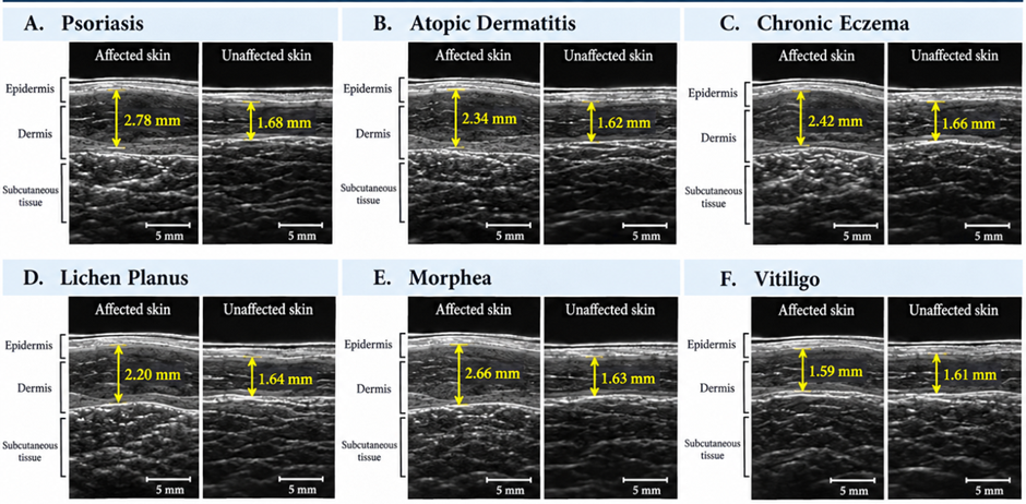
Figure 1. Illustrative high-frequency ultrasound comparison of skin thickness in common dermatological disorders
Figure 1 Panels A–F depict affected and corresponding clinically unaffected skin in (A) psoriasis, (B)atopic dermatitis, (C) chronic eczema, (D) lichen planus, (E) morphea, and (F) vitiligo. Skin thickness is illustrated as the distance from the superficial epidermal interface to the dermal–subcutaneous boundary (yellow arrows). Mean affected versus unaffected skin thickness was 2.78 vs. 1.68 mm in psoriasis, 2.34 vs. 1.62 mm in atopic dermatitis, 2.42 vs. 1.66 mm in chronic eczema, 2.20 vs. 1.64 mm in lichen planus, 2.66 vs. 1.63 mm in morphea, and 1.59 vs. 1.61 mm in vitiligo.
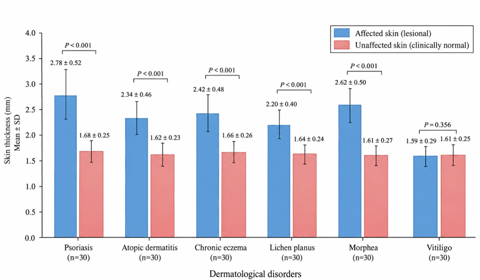
Figure 2. Comparison of ultrasound-measured skin thickness between affected and corresponding clinically unaffected skin across dermatological disorders
Figure 2 demonstrates consistently greater mean skin thickness in affected skin compared with corresponding clinically unaffected skin in psoriasis, atopic dermatitis, chronic eczema, lichen planus, and morphea. The largest differences were observed in psoriasis (2.78 ± 0.52 vs. 1.68 ± 0.25 mm) and morphea (2.66 ± 0.55 vs. 1.63 ± 0.28 mm), followed by chronic eczema, atopic dermatitis, and lichen planus. These differences were statistically significant for all five disorders (P < 0.001). In contrast, vitiligo showed nearly identical skin thickness between affected and unaffected areas (1.59 ± 0.29 vs. 1.61 ± 0.25 mm), with no statistically significant difference (P = 0.356).
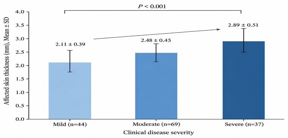
Figure 3. Association between clinical disease severity and ultrasound-measured skin thickness
Figure 3 Mean affected skin thickness increased progressively from mild (2.11 ± 0.39 mm) to moderate (2.48 ± 0.45 mm) and severe disease (2.89 ± 0.51 mm), demonstrating a significant severity-dependent increase (P < 0.001).
DISCUSSION:
The present study demonstrates that high-frequency ultrasonography can provide objective quantitative and qualitative information regarding structural changes in a range of common dermatological disorders. Among 180 participants, the overall mean skin thickness at affected sites was 2.33 ± 0.64 mm, compared with 1.64 ± 0.27 mm at corresponding clinically unaffected sites, representing an overall increase of approximately 42.2%. Significant lesional thickening was observed in psoriasis, atopic dermatitis, chronic eczema, lichen planus, and morphea, whereas vitiligo showed no significant difference in overall skin thickness. Furthermore, the magnitude of skin thickening increased progressively with clinical disease severity, and clinical severity remained independently associated with ultrasound-measured thickness after adjustment for demographic and clinical variables. These findings support the potential role of ultrasonography as a non-invasive adjunct for objectively characterizing cutaneous structural abnormalities.
The greatest increase in skin thickness in the present study was observed in psoriasis, in which affected skin measured 2.78 ± 0.52 mm, compared with 1.68 ± 0.25 mm at corresponding unaffected sites, representing a 65.5% increase. This finding is highly consistent with previous investigations demonstrating pronounced epidermal and dermal alterations in psoriatic plaques. Gupta et al. used 40-MHz ultrasound to assess plaque psoriasis and demonstrated a close relationship between ultrasound abnormalities and clinical lesion severity. In particular, the width of the sonographically non-echogenic band showed a strong correlation with clinical scaling, erythema, and thickness scores, supporting the concept that ultrasound can objectively reflect inflammatory disease burden [11].
Similarly, Vaillant et al. reported increased epidermal and dermal thickness in psoriatic plaques and found that overall skin thickness was approximately 67% greater in affected skin compared with apparently normal skin [12]. This value is remarkably close to the 65.5% relative increase observed in the present study. They additionally demonstrated a broad subepidermal non-echogenic band and decreased intensity of dermal echoes, changes considered to reflect epidermal proliferation, edema, and inflammatory cellular infiltration [12]. The present finding that 76.7% of psoriatic lesions demonstrated a subepidermal low-echogenic band, while 60.0% demonstrated reduced dermal echogenicity, therefore provides further support for the use of ultrasound not only to quantify thickness but also to characterize the inflammatory architecture of psoriatic plaques.
The relationship between ultrasound measurements and clinical severity is particularly relevant in psoriasis. In the present study, clinical severity correlated positively with the magnitude of skin-thickness alteration, and psoriasis demonstrated one of the strongest disease-specific associations. Previous ultrasound work has similarly demonstrated that changes in sonographic characteristics parallel changes in clinical plaque severity [11]. Such observations indicate that repeated ultrasound measurements may potentially complement established clinical scores when objective documentation of local treatment response is desirable.
Atopic dermatitis also showed substantial structural alteration, with mean lesional skin thickness of 2.34 ± 0.46 mm, compared with 1.62 ± 0.23 mm in unaffected skin. In addition, reduced dermal echogenicity was observed in 80.0% of cases and a subepidermal low-echogenic band in 70.0%. Sabău et al. reported that the hypoechoicsubepidermal band was wider in lesional atopic dermatitis than in non-lesional skin, while the echogenicity of healthy control skin was higher than that observed in patients with atopic dermatitis [13]. They also demonstrated sonographic abnormalities in apparently normal skin, suggesting that ultrasonography may identify subclinical changes beyond visibly involved areas [13].
The biological basis of these findings is further supported by direct histopathological correlation. Polańska et al. compared high-frequency ultrasound with histological specimens from patients with atopic dermatitis and demonstrated significant associations between the thickness of the hypoechoic band and epidermal hyperplasia, hyperkeratosis, parakeratosis, spongiosis, and inflammatory-cell infiltration [14]. Reduced ultrasound echogenicity was also associated with more prominent inflammatory infiltration [14]. Thus, the decreased echogenicity and increased thickness demonstrated in the present study are likely to represent a composite effect of epidermal proliferation, tissue edema, inflammatory infiltration, and disruption of normal dermal architecture.
Chronic eczema demonstrated a comparable pattern, with affected skin measuring 2.42 ± 0.48 mm compared with 1.66 ± 0.26 mm in unaffected skin, representing an increase of 45.8%. Reduced dermal echogenicity, an identifiable subepidermal low-echogenic band, and irregular epidermal contours were also frequent. Yazdanparast et al. evaluated lesional and uninvolved skin in patients with chronic contact or atopic dermatitis and found significantly greater epidermal, dermal, and subepidermal low-echogenic band thickness in affected skin. Dermal and SLEB echo-density were simultaneously reduced [15]. These results closely correspond to the present observations and support the interpretation that sonographicthickening in chronic eczema reflects persistent epidermal hyperplasia together with dermal inflammatory and edematous changes.
Morphea showed the second greatest relative alteration in the present series, with a 63.2% increase in skin thickness compared with corresponding unaffected skin. Moreover, 73.3% of patients with morphea demonstrated increased dermal echogenicity or a fibrotic appearance, distinguishing the disorder from predominantly inflammatory conditions such as atopic dermatitis and eczema. This sonographic profile is compatible with the pathological accumulation and reorganization of dermal collagen that characterizes sclerotic disease.
Wortsman et al. evaluated 104 morphea lesions using color Doppler ultrasonography and demonstrated that ultrasound measurements of cutaneous thickness, echogenicity, and vascular flow could discriminate different phases of disease activity [16]. Increased subcutaneous echogenicity and increased cutaneous blood flow were particularly informative markers of active disease [16]. The present combination of thickness measurement, echogenicity assessment, and Doppler evaluation therefore reflects an approach consistent with previous work suggesting that assessment of morphea should extend beyond clinical inspection alone.
Earlier work by Hoffmann et al. also demonstrated increased corium thickness in localized scleroderma compared with corresponding healthy skin, with substantial inter-lesional variability according to anatomical site and disease stage [17]. Serial ultrasound examination could detect both progression and regression of dermal thickening during follow-up [17]. This is clinically important because morphea evolves through inflammatory, sclerotic, and atrophic phases, and the sonographic appearance may consequently vary over time. The relatively large mean thickness difference found in the present morphea group likely reflects the inclusion of established or clinically appreciable plaques, whereas individual lesions in early inflammatory or late atrophic phases may show different quantitative patterns.
Lichen planus demonstrated a more moderate but still significant increase in skin thickness, with a mean difference of 0.56 mm and a relative increase of 34.1%. Ultrasound literature specifically addressing conventional cutaneous lichen planus remains comparatively limited. Nevertheless, recent work examining nail lichen planus has demonstrated that ultrasonography can identify thickening, reduced echogenicity, periungual structural changes, and marked vascular abnormalities [18]. Although nail disease differs anatomically from cutaneous lichen planus, these observations support the broader concept that lichenoid inflammation produces sonographically detectable changes in tissue architecture. The increased thickness, irregular surface contour, and altered echogenicity observed in the present study may represent interface inflammation, epidermal hyperplasia, and superficial dermal inflammatory infiltration. Additional studies specifically focused on cutaneous lichen planus are warranted to establish standardized sonographic diagnostic criteria.
An important finding was the absence of a significant difference in overall skin thickness between vitiliginous and corresponding clinically normal skin (1.59 ± 0.29 mm versus 1.61 ± 0.25 mm; P = 0.356). This contrasts with the clearly increased thickness observed in inflammatory and fibrosingdermatoses. Vitiligo is primarily characterized by melanocyte loss rather than the marked epidermal proliferation, edema, or collagen deposition seen in psoriasis, eczema, or morphea, and therefore gross skin-thickness measurement alone may be relatively insensitive to its underlying pathology.
However, the absence of increased overall thickness should not be interpreted as an absence of sonographic abnormalities in vitiligo. Wortsman et al. recently evaluated vitiligo using both 24-MHz high-frequency and 70-MHz ultra-high-frequency ultrasound and identified epidermal undulation, superficial hypoechoic changes, alterations in hair follicles and pilosebaceous units, prominent sebaceous glands, and increased dermal vascularity in affected areas [19]. The present finding of relatively preserved total skin thickness may therefore indicate that overall thickness is not the optimal ultrasound biomarker for vitiligo, whereas ultra-high-frequency assessment of individual superficial layers and Doppler vascularity may reveal more subtle inflammatory or adnexal alterations. Differences in transducer frequency may also contribute, as the 15–22 MHz range used in the present methodology provides greater penetration but lower superficial spatial resolution than 70-MHz systems.
The present study further demonstrated a clear severity-dependent increase in skin thickness. Mean lesional thickness increased from 2.11 ± 0.39 mm in mild disease to 2.48 ± 0.45 mm in moderate disease and 2.89 ± 0.51 mm in severe disease, with a significant positive correlation between severity and affected-to-unaffected thickness difference (ρ = 0.61, P < 0.001). This suggests that quantitative ultrasound measurements may provide a continuous objective parameter that complements categorical clinical assessment. Such objective quantification may be particularly valuable in disorders in which visual erythema, palpation-based induration, or clinical scaling are influenced by observer experience or skin phenotype.
Multivariable analysis strengthened this finding. Diagnostic category and clinical severity remained significant independent predictors of increased skin thickness, whereas age and sex were not independently associated after adjustment. The regression model explained approximately 68% of the variability in thickness difference. This finding does not imply that age, sex, and body site have no effect on normal skin thickness. Olsen et al. demonstrated considerable anatomical variation in normal ultrasound-measured skin thickness and reported differences according to sex and body region [20]. Rather, the lack of an independent age or sex effect in the present analysis may partly reflect the study design, in which lesional measurements were compared with anatomically corresponding unaffected skin within the same participant. Such an internal-control approach reduces interindividual variation and is an important methodological strength.
Measurement reproducibility is essential if ultrasound is to be used as an objective clinical or research biomarker. The present study demonstrated excellent intraobserver reliability (ICC 0.94) and high interobserver agreement (ICC 0.91). These findings are consistent with methodological research demonstrating that high-frequency ultrasound can provide reproducible skin-thickness measurements when acquisition and measurement procedures are standardized. Alsing and Serup reported close agreement between experienced observers using manual 20-MHz skin-thickness measurement and emphasized the importance of using a consistent measurement method because automated and manual border-detection techniques can generate systematically different thickness values [21]. The high ICC values in the present study therefore support the reliability of standardized triplicate measurements performed with controlled probe positioning and minimal tissue compression.
The qualitative sonographic findings add further clinical value beyond total thickness alone. Inflammatory diseases such as psoriasis, atopic dermatitis, and chronic eczema predominantly demonstrated reduced echogenicity, a low-echogenic subepidermal band, irregular surface contours, and increased vascularity. In contrast, morphea more frequently demonstrated increased dermal echogenicity associated with fibrotic structural change.
CONCLUSION:
High-frequency ultrasonography provides a useful, non-invasive, reproducible method for the quantitative and qualitative assessment of skin changes in common dermatological disorders. In the present study, affected skin showed significantly greater thickness than corresponding clinically unaffected skin in psoriasis, atopic dermatitis, chronic eczema, lichen planus, and morphea, while vitiligo showed no significant alteration in total skin thickness. The greatest increases were observed in psoriasis and morphea, reflecting the marked inflammatory, proliferative, and fibrotic changes associated with these disorders.
REFERENCES:
1. Cisoń H, Białynicki-Birula R. High-frequency ultrasonography: one of the modern imaging diagnostic methods in dermatology. Authors' own experience and review. PostepyDermatolAlergol. 2024;41(3):306-313. doi:10.5114/ada.2024.141108.
2. Levy J, Barrett DL, Harris N, Jeong JJ, Yang X, Chen SC. High-frequency ultrasound in clinical dermatology: a review. Ultrasound J. 2021;13(1):24. doi:10.1186/s13089-021-00222-w.
3. Vergilio MM, Monteiro e Silva SA, Jales RM, Leonardi GR. High-frequency ultrasound as a scientific tool for skin imaging analysis. ExpDermatol. 2021;30(7):897-910. doi:10.1111/exd.14363.
4. Jasaitiene D, Valiukeviciene S, Linkeviciute G, Raisutis R, Jasiuniene E, Kazys R. Principles of high-frequency ultrasonography for investigation of skin pathology. J EurAcadDermatolVenereol. 2011;25(4):375-382. doi:10.1111/j.1468-3083.2010.03837.x.
5. Hermann RC, Ellis CN, Fitting DW, Ho VC, Voorhees JJ. Measurement of epidermal thickness in normal skin and psoriasis with high-frequency ultrasound. Skin Pharmacol. 1988;1(2):128-136. doi:10.1159/000210760.
6. Sorokina E, Mikailova D, Krakhaleva J, Krinitsyna J, Yakubovich A, Sergeeva I. Ultrasonography patterns of atopic dermatitis in children. Skin Res Technol. 2020;26(4):482-488. doi:10.1111/srt.12839.
7. Chen C, Cheng Y, Zhu X, Cai Y, Xue Y, Kong N, et al. Ultrasound assessment of skin thickness and stiffness: the correlation with histology and clinical score in systemic sclerosis. Arthritis Res Ther. 2020;22(1):197. doi:10.1186/s13075-020-02285-x.
8. Zhang S, Zhu QL, Xiao MS, Liu J. The value of dermoscopy and high-frequency ultrasound in staging morphea. J Dermatol. 2023;50(4):511-517. doi:10.1111/1346-8138.16648.
9. Grand D, Frew JW, Navrazhina K, Krueger JG. Doppler ultrasound-based noninvasive biomarkers in hidradenitissuppurativa: evaluation of analytical and clinical validity. Br J Dermatol. 2021;184(4):688-696. doi:10.1111/bjd.19343.
10. Polańska A, Dańczak-Pazdrowska A, Jałowska M, Żaba R, Adamski Z. Current applications of high-frequency ultrasonography in dermatology. PostepyDermatolAlergol. 2017;34(6):535-542. doi:10.5114/ada.2017.72457.
11. Gupta AK, Turnbull DH, Harasiewicz KA, Shum DT, Watteel GN, Foster FS, et al. The use of high-frequency ultrasound as a method of assessing the severity of a plaque of psoriasis. Arch Dermatol. 1996;132(6):658-662.
12. Vaillant L, Berson M, Machet L, Callens A, Pourcelot L, Lorette G. Ultrasound imaging of psoriatic skin: a noninvasive technique to evaluate treatment of psoriasis. Int J Dermatol. 1994;33(11):786-790. doi:10.1111/j.1365-4362.1994.tb00994.x.
13. Sabău M, Boca AN, Ilies RF, Tătaru A. Potential of high-frequency ultrasonography in the management of atopic dermatitis. ExpTher Med. 2019;17(2):1073-1077. doi:10.3892/etm.2018.6984.
14. Polańska A, Dańczak-Pazdrowska A, Silny W, Woźniak A, Maksin K, Jenerowicz D, et al. Comparison between high-frequency ultrasonography (Dermascan C, version 3) and histopathology in atopic dermatitis. Skin Res Technol. 2013;19(4):432-437. doi:10.1111/srt.12064.
15. Yazdanparast T, Yazdani K, Humbert P, Khatami A, Nasrollahi SA, IzadiFirouzabadi L, et al. Biophysical measurements and ultrasonographic findings in chronic dermatitis in comparison with uninvolved skin. Indian J Dermatol. 2019;64(2):90-96. doi:10.4103/ijd.IJD_464_17.
16. Wortsman X, Wortsman J, Sazunic I, Carreño L. Activity assessment in morphea using color Doppler ultrasound. J Am AcadDermatol. 2011;65(5):942-948. doi:10.1016/j.jaad.2010.08.027.
17. Hoffmann K, Gerbaulet U, el-Gammal S, Altmeyer P. 20-MHz B-mode ultrasound in monitoring the course of localized scleroderma (morphea). ActaDermVenereolSuppl (Stockh).1991;(164):3-16.
18. Turner VL, Wortsman X. Ultrasound features of nail lichen planus. J Ultrasound Med. 2024;43(4):781-788. doi:10.1002/jum.16410.
19. Wortsman X, Araya I, Maass M, Valdes P, Zemelman V. Ultrasound patterns of vitiligo at high frequency and ultra-high frequency. J Ultrasound Med. 2024;43(9):1605-1610. doi:10.1002/jum.16481.
20. Olsen LO, Takiwaki H, Serup J. High-frequency ultrasound characterization of normal skin: skin thickness and echographic density of 22 anatomical sites. Skin Res Technol. 1995;1(2):74-80. doi:10.1111/j.1600-0846.1995.tb00021.x.
21. Alsing KK, Serup J. High-frequency ultrasound skin thickness: comparison of manual reading and automatic border detection includes assessment of interobserver variation of measurement. Skin Res Technol. 2020;26(6):832-838. doi:10.1111/srt.12884.