Auricular Anthropometric Norms for the Adult Indian Female - A Non-Interventional, Cross-Sectional Observational Study with Implications for Aesthetic and Reconstructive Surgery.
- Dr. Sushil Bhojraj Machhale , Senior Resident, Department of Plastic and Reconstructive Surgery, Government Medical College and Hospital, Nagpur, Maharashtra, India.
- Dr. Surendra Kumar Patil , Professor & HOD, Department of Plastic and Reconstructive Surgery, Government Medical College and Hospital, Nagpur, Maharashtra, India.
Article Information:
Abstract:
Background: Population-specific auricular anthropometry is relevant to aesthetic otoplasty, reconstruction of congenital or acquired auricular defects, and restoration of bilateral facial balance. Indian female-specific reference values remain limited. Objectives: To establish bilateral linear and proportional auricular reference values in adult Indian females and to quantify right-left differences in ear and lobular dimensions. Methods: This non-interventional, cross-sectional observational study analysed 500 healthy Indian females. Although the prespecified eligibility range was 18-45 years, the enrolled cohort was aged 18-36 years; no participant aged 37-45 years was represented. Accordingly, the anthropometric estimates apply to the observed 18-36-year cohort. Bilateral ear length, ear width, lobule length, and lobule width were measured with a digital Vernier caliper. Auricular and lobular indices were derived from width-to-length ratios. Descriptive statistics, observed central 95% limits (P2.5-P97.5), and paired right-left comparisons were calculated. Results: Mean age was 26.73 ± 5.21 years. Mean right and left ear lengths were 56.36 ± 1.91 mm and 54.90 ± 1.88 mm, and mean ear widths were 28.20 ± 1.58 mm and 27.63 ± 1.55 mm, respectively. Right and left lobule lengths measured 21.01 ± 1.43 mm and 20.11 ± 1.43 mm, while lobule widths measured 23.73 ± 1.35 mm and 22.87 ± 1.35 mm. All four right-sided linear dimensions were significantly greater than their left-sided counterparts (p<0.001). Conversely, left auricular index (50.30 ± 1.10) and left lobular index (113.84 ± 1.41) were modestly higher than the corresponding right-sided indices (49.99 ± 1.12 and 113.01 ± 1.29; both p<0.001). Conclusion: mong the Indian women aged 18-36 years represented in this cohort, small but consistent side-specific differences were observed. Absolute auricular and lobular dimensions were larger on the right, whereas width-to-length proportional indices were higher on the left. These cohort-derived values may support dimensional planning in aesthetic and reconstructive surgery, particularly when a satisfactory contralateral ear is unavailable, but they should not be extrapolated to older adult women or all regional Indian populations without external validation.
Keywords:
Article :
INTRODUCTION:
The external ear occupies a relatively small part of the craniofacial complex, yet minor deviations in its size, contour, projection, or bilateral relationship can be visually conspicuous. For plastic and reconstructive surgeons, therefore, a quantitative description of normal auricular morphology has practical value. It assists preoperative assessment, reconstruction of congenital or acquired defects, correction of disproportionate ears, and evaluation of postoperative symmetry. Farkas emphasized that population norms are particularly useful when the amount and position of tissue required for reconstruction must be estimated rather than judged visually.[1]
Auricular morphology is not constant across the life course or between sexes. Age-related elongation, especially of the lobule, and sex-related differences in total pinna dimensions have been repeatedly demonstrated.[2] Three-dimensional investigations have also shown that bilateral similarity does not necessarily imply exact equality, and that ear dimensions and proportional indices vary with age.[3] These findings make it difficult to justify a single universal set of auricular dimensions for surgical planning.
Population background introduces another layer of variation. In central Indian men, Purkait and Singh documented distinct dimensions and bilateral differences and concluded that metric standards derived from other ethnic groups cannot simply be transferred to Indian patients.[4] A large Maharashtrian investigation similarly demonstrated age- and sex-associated variation in external-ear dimensions and highlighted the clinical relevance of population-specific anthropometry.[5]
This issue becomes especially important in auricular reconstruction. In unilateral deformity, the unaffected contralateral ear usually provides the most individualized template. That option is limited in bilateral microtia, bilateral trauma, previous contralateral surgery, or marked pre-existing asymmetry. Population-based reference measurements can then help define a plausible framework height, width, and lobular proportion. Contemporary anthropometric work has explicitly advocated such baseline data for reconstruction when a normal opposite ear cannot serve as a reliable guide.[6]
Indian data specifically focused on adult women remain less extensive than mixed-sex or male datasets. The present study was therefore undertaken to establish bilateral auricular and lobular measurements and proportional indices in a large cohort of adult Indian females. A secondary objective was to quantify right-left differences rather than presuming mirror symmetry, because even small side-specific differences may matter when millimetric precision is sought in aesthetic or reconstructive surgery.
MATERIALS AND METHODS:
Study Design and Setting
A non-interventional, cross-sectional observational anthropometric study was conducted on OPD and IPD patients of Plastic Surgery Department at Government Medical College and Hospital (GMCH) Nagpur from February 2024 to February 2026. State/city identifiers, the number of recruitment sites, and urban-rural residence were not available for stratified analysis; consequently, the findings are not presented as region-specific estimates.
Study Population
The study included 500 healthy Indian females. Prespecified eligibility was 18-45 years; however, the enrolled sample ranged from 18 to 36 years and contained no participant aged 37-45 years. Recruitment therefore did not cover the full eligible age band, and all descriptive reference estimates in this manuscript are restricted to the observed 18-36-year cohort.
Sample Size and Precision
A total of 500 female participants of central India were included. For this descriptive study, precision of the estimated means was assessed from the final sample. The standard errors of the principal linear measurements ranged from approximately 0.06 to 0.09 mm, indicating narrow uncertainty around the cohort means.
Inclusion Criteria
Healthy Indian females within the eligible age range with normally developed bilateral external ears were included.
Exclusion Criteria
Women with congenital auricular deformity, previous traumatic injury involving the ear, previous otoplasty or other ear surgery, active local infection, or dermatological disease affecting the pinna were excluded.
Anthropometric Measurements
Measurements were recorded in millimetres using a digital Vernier caliper (Mitutoyo; stated accuracy 0.01 mm). A non-stretchable flexible measuring tape was available for contour confirmation. Ear length was defined as the maximum vertical dimension from the superior aspect of the helix to the inferior portion of the lobule. Ear width was the maximum horizontal width perpendicular to the long axis. Lobule length represented the vertical dimension of the lobule, and lobule width was its maximum horizontal width. Each parameter was recorded separately for the right and left ears.
Measurement Reproducibility
Formal interobserver and intraobserver reliability coefficients were not available for analysis. Consequently, the repeatability of the caliper measurements cannot be quantified from this cohort and should be considered when interpreting small right-left differences.
Derived Indices
Auricular index was calculated as ear width/ear length × 100. Lobular index was calculated as lobule width/lobule length × 100. The indices were calculated separately for the right and left sides.
Statistical Analysis
Continuous variables were summarized as mean, standard deviation, minimum, maximum, and empirical 2.5th and 97.5th percentiles, reported as the observed central 95% limits (P2.5-P97.5). These limits describe the central 95% distribution of the observed cohort and are not presented as universally validated clinical reference intervals for all Indian women. Right-left measurements were treated as paired observations and compared using paired-samples t tests. Mean paired differences and 95% confidence intervals were calculated. All tests were two-sided, and p<0.05 was considered statistically significant.
Ethical Considerations
Institutional ethical clearance was obtained before commencement of the study. No therapeutic allocation, drug administration, device implantation, or alteration of routine clinical care was involved.
RESULTS:
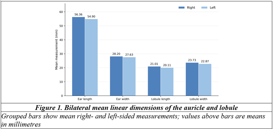
All 500 participants were included in the analysis. Mean age was 26.73 ± 5.21 years (range, 18-36 years); no participant aged 37-45 years was represented. The results below therefore describe the enrolled 18-36-year cohort. The overall distribution of measured dimensions is summarized in Table 1. Right ear length averaged 56.36 ± 1.91 mm and left ear length 54.90 ± 1.88 mm. Mean right and left ear widths were 28.20 ± 1.58 mm and 27.63 ± 1.55 mm, respectively. The corresponding lobule measurements were 21.01 ± 1.43 mm versus 20.11 ± 1.43 mm for length and 23.73 ± 1.35 mm versus 22.87 ± 1.35 mm for width.
Table 1. Descriptive anthropometric measurements in the observed cohort of Indian women aged 18-36 years (N=500)
|
Parameter |
Mean ± SD |
Range |
Observed central 95% limits (P2.5-P97.5) |
|
Age (years) |
26.73 ± 5.21 |
18-36 |
18-36 |
|
Right ear length (mm) |
56.36 ± 1.91 |
53.10-59.80 |
53.25-59.60 |
|
Left ear length (mm) |
54.90 ± 1.88 |
51.70-58.40 |
51.85-58.20 |
|
Right ear width (mm) |
28.20 ± 1.58 |
25.60-31.20 |
25.70-31.00 |
|
Left ear width (mm) |
27.63 ± 1.55 |
25.10-30.60 |
25.20-30.40 |
|
Right lobule length (mm) |
21.01 ± 1.43 |
18.60-23.80 |
18.70-23.60 |
|
Left lobule length (mm) |
20.11 ± 1.43 |
17.70-22.90 |
17.80-22.70 |
|
Right lobule width (mm) |
23.73 ± 1.35 |
21.30-26.30 |
21.40-26.10 |
|
Left lobule width (mm) |
22.87 ± 1.35 |
20.50-25.50 |
20.60-25.30 |
Absolute dimensions were greater on the right for each of the four linear parameters. The mean right-left difference was 1.47 mm for total ear length, 0.56 mm for ear width, 0.90 mm for lobule length, and 0.85 mm for lobule width. The bilateral mean values are displayed in Figure 1.
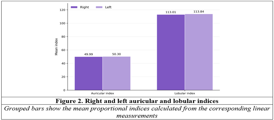
The proportional indices are shown in Table 2. Although the right ear was larger in absolute dimensions, its auricular index was slightly lower than the left, indicating that the relative increase in right-sided length exceeded the proportional increase in width. A similar pattern was observed for the lobule. Bilateral index values are illustrated in Figure 2.
Table 2. Auricular and lobular indices in the observed cohort (N=500)
|
Index |
Mean ± SD |
Range |
Observed central 95% limits (P2.5-P97.5) |
|
Right auricular index |
49.99 ± 1.12 |
48.21-52.17 |
48.31-52.01 |
|
Left auricular index |
50.30 ± 1.10 |
48.55-52.40 |
48.65-52.23 |
|
Right lobular index |
113.01 ± 1.29 |
110.50-114.66 |
110.59-114.58 |
|
Left lobular index |
113.84 ± 1.41 |
111.06-115.93 |
111.45-115.85 |
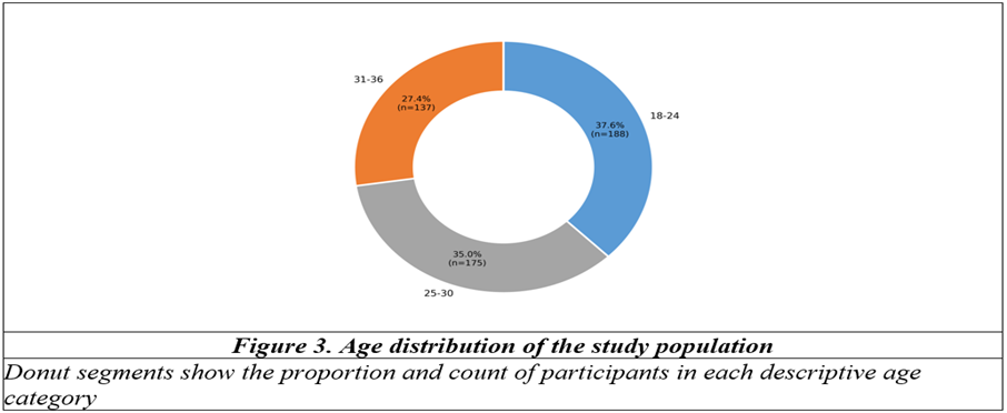
For descriptive presentation, 188 participants (37.6%) were aged 18-24 years, 175 (35.0%) were aged 25-30 years, and 137 (27.4%) were aged 31-36 years (Figure 3).
Paired right-left comparisons are presented in Table 3. All linear dimensions were significantly greater on the right (p<0.001), whereas the right auricular and lobular indices were significantly lower than their left-sided counterparts (p<0.001).
Table 3. Paired comparison of right and left auricular measurements
|
Parameter |
Right, mean ± SD |
Left, mean ± SD |
Mean difference (R-L) |
95% CI of difference |
p-value |
|
Ear length |
56.36 ± 1.91 |
54.90 ± 1.88 |
1.467 |
1.463 to 1.471 |
<0.001 |
|
Ear width |
28.20 ± 1.58 |
27.63 ± 1.55 |
0.564 |
0.559 to 0.568 |
<0.001 |
|
Lobule length |
21.01 ± 1.43 |
20.11 ± 1.43 |
0.899 |
0.898 to 0.901 |
<0.001 |
|
Lobule width |
23.73 ± 1.35 |
22.87 ± 1.35 |
0.852 |
0.847 to 0.857 |
<0.001 |
|
Auricular index |
49.99 ± 1.12 |
50.30 ± 1.10 |
-0.311 |
-0.316 to -0.305 |
<0.001 |
|
Lobular index |
113.01 ± 1.29 |
113.84 ± 1.41 |
-0.827 |
-0.854 to -0.801 |
<0.001 |
Linear measurements are in millimetres. R-L: right minus left; CI: confidence interval. Paired-difference estimates and CIs are displayed to three decimal places to preserve the narrow statistical intervals; this does not imply measurement resolution beyond the stated caliper specification.
The observed central 95% limits (P2.5-P97.5) for the major linear variables are depicted in Figure 4. These limits summarize the central distribution of this cohort and should not be interpreted as validated population-wide clinical reference intervals.
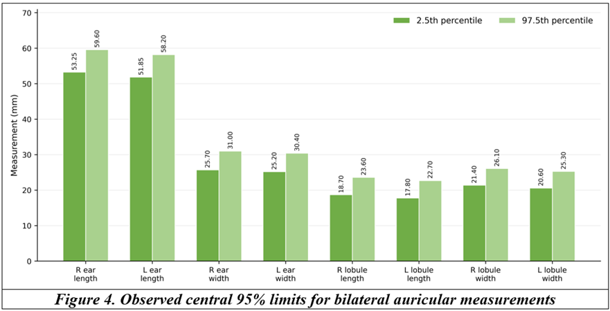
Bars represent P2.5 and P97.5 for each linear measurement in the observed cohort.
DISCUSSION:
This study provides a female-specific set of bilateral external-ear measurements in a comparatively large Indian adult cohort. The principal finding is not simply the absolute size of the ear, but the coexistence of two different asymmetry patterns. Right-sided ear and lobule dimensions were consistently larger in millimetres, whereas the proportional auricular and lobular indices were modestly higher on the left. This distinction is clinically relevant because matching overall height alone does not necessarily reproduce the shape or proportional balance of the opposite ear.
Indian auricular data are heterogeneous, even before age and sex are considered. Singh et al. studied 130 North Indian adults aged 18-25 years and reported mean auricular heights of 62.8 mm on the right and 62.3 mm on the left in the combined sample; their sex-stratified analysis also showed larger auricular dimensions in males.[7] These height values exceed the means observed in the present female cohort. Direct numerical comparison remains limited by the mixed-sex composition of their overall estimates and by potential differences in measurement protocol, but the contrast reinforces a practical point: pooled or regionally derived measurements should not automatically be treated as universal Indian standards.
Rani et al. also demonstrated substantial variability in external-ear anthropometry in an Indian population, with effects related to sex and side.[8] The current results extend that observation by showing that absolute and proportional asymmetry may move in opposite directions. A right ear can be longer and wider yet still possess a lower width-to-length index. For reconstruction, therefore, the relevant question is not only 'how large should the ear be?' but also 'how should its dimensions relate to one another?'.
Comparisons with non-Indian populations further illustrate the importance of population specificity. Bozkir et al. measured young Turkish adults with a digital caliper and reported female total ear heights of approximately 59.5-59.7 mm and ear widths near 31 mm.[9] These values are larger than the corresponding means in the present cohort. Such differences may reflect population biology, but measurement landmarks, caliper orientation, participant age, and sampling strategy can also materially influence the final estimate. Normative values should therefore be interpreted as method-dependent as well as population-dependent.
Ethnic variation has been demonstrated directly. Alexander et al., in a multiethnic sample, found systematic differences in ear morphology according to ethnicity, sex, and age, with Indian-subcontinent participants tending toward larger auricular dimensions than some comparator groups.[10] Thai anthropometric data likewise showed sex- and age-related differences and were explicitly developed to improve reconstructive planning within that population.[11] Taken together, these studies argue against an aesthetically universal ear and support population-aware planning while preserving individual facial harmony.
The age distribution deserves particular attention. Although the prespecified eligibility range extended to 45 years, recruitment did not include any woman aged 37-45 years. The study therefore does not provide empirical coverage of that segment of the intended adult age band. This is important because auricular morphology does not remain static after developmental maturity. Experimental and population studies indicate that ear length matures later than width during growth,[12-14] while Indian longitudinal work shows that different auricular subunits reach mature dimensions at different ages.[13] Adult aging, particularly lobular elongation, may then continue. The present estimates should therefore be interpreted as reference values for the observed 18-36-year cohort rather than for the full 18-45-year eligibility range or the entire adult female lifespan.
From an aesthetic-surgery perspective, proportional indices may be useful when evaluating whether a planned correction preserves the overall relationship between ear width and height. The mean auricular index in this cohort was close to 50 bilaterally, but the side difference was statistically consistent. Aesthetic judgement cannot be reduced to a numerical index, yet anthropometry supplies an objective frame against which individual anatomy and patient preference can be considered.
The reconstructive application is more direct. A structurally normal contralateral ear remains the preferred template for unilateral reconstruction because it is patient-specific. Population-based measurements become more valuable when the opposite ear is absent, malformed, previously operated on, or markedly asymmetric. Prasetyo and Putri similarly emphasized that anthropometric baseline data can guide framework dimensions in bilateral cases where no satisfactory contralateral model exists.[6] In such circumstances, the observed central 95% limits (P2.5-P97.5) reported here may be more useful than a single mean value, because they express a central range of naturally occurring dimensions rather than implying one ideal ear size.
Several limitations should be recognized. Only women were studied, which is appropriate for a female-specific reference but prevents internal assessment of sexual dimorphism. The sample did not include women aged 37-45 years despite their inclusion in the prespecified eligibility range, and the findings therefore cannot be extrapolated beyond 36 years from these data alone. Although recruitment occurred through screening camps across diverse regions of India, state- or city-level distribution, the number of recruitment sites, and urban-rural composition were not available for stratified analysis. The cohort therefore cannot be assumed to represent India's substantial regional diversity in proportion to the national population. Formal interobserver and intraobserver reliability coefficients were also unavailable, so the reproducibility of the caliper measurements cannot be quantified. The study used linear measurements and did not include auricular projection, auriculocephalic angle, conchal dimensions, cartilage thickness, surface area, or three-dimensional contour. Ear-piercing and ornament-use history was not captured. Because lobule length and width were primary outcomes, this omission is particularly relevant in Indian women, in whom prolonged piercing or ornament load may modify lobular morphology; an earlier northwest Indian lobule study excluded women specifically because of this concern.[15] Piercing-related variation therefore represents a potentially important residual source of heterogeneity. Collectively, these constraints mean that the present values should be treated as cohort-specific anthropometric estimates, not comprehensive national norms for all adult Indian women. A further methodological point concerns interpretation of statistical significance. The large sample made very small side differences highly statistically detectable. The absolute differences were generally below 1.5 mm, so clinical relevance should not be inferred from p-values alone. For operative planning, the magnitude of the difference, facial proportionality, and the quality of the contralateral ear remain more important than statistical significance in isolation.
Despite these constraints, the study offers a substantial side-specific database for adult Indian females. Its main practical contribution is the simultaneous description of linear measurements, proportional indices, and empirical central reference limits. These parameters can support, rather than replace, individualized clinical judgement in aesthetic and reconstructive surgery.
CONCLUSION:
In this cohort of 500 Indian women aged 18-36 years, mean ear length was 56.36 mm on the right and 54.90 mm on the left, while mean ear width was 28.20 mm and 27.63 mm, respectively. Lobular measurements showed the same direction of side difference. Absolute dimensions were consistently larger on the right, whereas auricular and lobular width-to-length indices were modestly higher on the left. These findings support the use of side-specific, population-aware measurements rather than assuming exact bilateral symmetry. The values may aid aesthetic assessment and auricular reconstruction when an intact contralateral ear is unavailable or unsuitable as a template. Broader multicentric studies should deliberately sample the full adult age spectrum, document regional and urban-rural background, record piercing and ornament-use history, assess measurement reliability, and incorporate auricular projection, conchal measurements, and three-dimensional morphology before national normative standards are proposed.
REFERENCES:
1. Farkas LG. Anthropometry of the normal and defective ear. Clin Plast Surg 1990;17(2):213-21.
2. Brucker MJ, Patel J, Sullivan PK. A morphometric study of the external ear: age- and sex-related differences. Plast Reconstr Surg 2003;112(2):647-52.
3. Sforza C, Grandi G, Binelli M, Tommasi DG, Rosati R, Ferrario VF. Age- and sex-related changes in the normal human ear. Forensic Sci Int 2009;187(1-3):110.e1-7.
4. Purkait R, Singh P. Anthropometry of the normal human auricle: a study of adult Indian men. Aesthetic Plast Surg 2007;31(4):372-9.
5. Japatti SR, Engineer PJ, Reddy BM, Tiwari AU, Siddegowda CY, Hammannavar RB. Anthropometric assessment of the normal adult human ear. Ann Maxillofac Surg 2018;8(1):42-50.
6. Prasetyo AT, Putri IL. Anthropometric study of human ear: a baseline data for ear reconstruction. J Craniofac Surg 2022;33(4):1245-9.
7. Singh AB, Gupta P, Singh P. Anthropometric assessment of human auricle in North Indian population. Natl J Maxillofac Surg 2022;13(2):234-7.
8. Rani D, Krishan K, Sahani R, Baryah N, Kanchan T. Variability in human external ear anthropometry: anthropological and forensic applications. Clin Ter 2021;172(6):531-41.
9. Bozkir MG, Karakas P, Yavuz M, Dere F. Morphometry of the external ear in our adult population. Aesthetic Plast Surg 2006;30(1):81-5.
10. Alexander KS, Stott DJ, Sivakumar B, Kang N. A morphometric study of the human ear. J Plast Reconstr Aesthet Surg 2011;64(1):41-7.
11. Khobkhun P, Pungrasmi P, Suwajo P, Nilprapha K, Meevassana J, Promniyom P, et al. An anthropometric study of the external ear in the Thai population. J Plast Reconstr Aesthet Surg 2022;75(4):1417-23.
12. Zhao S, Li D, Liu Z, Wang Y, Liu L, Jiang D, et al. Anthropometric growth study of the ear in a Chinese population. J Plast Reconstr Aesthet Surg 2018;71(4):518-23.
13. Purkait R. Progression of growth in the external ear from birth to maturity: a 2-year follow-up study in India. Aesthetic Plast Surg 2013;37(3):605-16.
14. Farkas LG, Posnick JC, Hreczko TM. Anthropometric growth study of the ear. Cleft Palate Craniofac J 1992;29(4):324-9.
15. Sharma A, Sidhu NK, Sharma MK, Kapoor K, Singh B. Morphometric study of ear lobule in northwest Indian male subjects. Anat Sci Int 2007;82(2):98-104.