Visual Recovery after Traumatic Hyphema in Closed-Globe Injury: Prognostic Factors and Clinical Outcomes in a Prospective Study.
- Bhavya Annadevula , Minto Eye Hospital, Regional Institiute of Ophthalmology, BMCRI, Bangaluru
- Nandini H , Minto Eye Hospital, Regional Institute of Ophthalmology, BMCRI, Bangaluru
- Uzma Nausheen , KBN Medical College and Hospital, Kalaburgi
Article Information:
Abstract:
Background: Traumatic hyphema is a common consequence of blunt ocular trauma and may be associated with significant visual impairment, elevated intraocular pressure, and concomitant anterior or posterior segment injury. Identifying clinical factors associated with visual recovery is important for prognosis and timely management. Methods: This hospital-based prospective observational study included 60 patients with traumatic hyphema following closed-globe injury who presented to a tertiary ophthalmic care centre between February 2021 and August 2022. Patients underwent detailed ophthalmic evaluation including visual acuity assessment, hyphema grading, intraocular pressure measurement, anterior segment examination, and posterior segment evaluation. Follow-up assessments were performed up to 12 weeks. Final visual outcome was analysed in relation to demographic characteristics, time to presentation, initial visual acuity, grade of hyphema, mode of injury, and associated ocular findings. Results: Most patients were male and ≤30 years of age. At presentation, 40% had visual acuity of hand movements or worse, while only 30% had vision between 6/6 and 6/18. By the final follow-up, 76.7% achieved good visual acuity of 6/6–6/18, representing a significant improvement from baseline (p<0.001). Grade 1 hyphema was the most common presentation, and 91.7% of hyphemas resolved within one week. Intraocular pressure was <21 mmHg in 90% of patients at presentation and in all patients by final follow-up (p=0.008). Conclusion: Visual recovery following traumatic hyphema in closed-globe injury was generally favourable. Early presentation and better presenting visual acuity were the most important prognostic indicators of final vision.
Keywords:
Article :
INTRODUCTION:
Ocular trauma remains an important cause of preventable visual morbidity, particularly among young and economically active individuals. The clinical spectrum of mechanical ocular injury ranges from relatively minor anterior segment damage to severe disruption of intraocular structures with permanent loss of vision. According to the Birmingham Eye Trauma Terminology system, a closed-globe injury is characterized by the absence of a full-thickness wound of the cornea or sclera and commonly results from blunt impact, producing contusion-related damage to multiple ocular tissues [1]. Among the most recognizable anterior segment manifestations of closed-globe trauma is traumatic hyphema, defined as the accumulation of blood within the anterior chamber. Although an isolated hyphema may resolve without permanent visual impairment, its presence can indicate substantial transfer of mechanical energy to the globe and may coexist with injuries that significantly influence ultimate visual recovery.
Traumatic hyphema most frequently follows blunt ocular trauma, in which sudden anteroposterior compression of the globe accompanied by equatorial expansion places considerable stress on the iris, ciliary body, anterior chamber angle, and their vascular structures. Rupture of vessels at the iris root or anterior ciliary body permits blood to enter the anterior chamber [2]. The clinical appearance can vary from circulating erythrocytes detectable only on slit-lamp examination to a layered hyphema or complete filling of the anterior chamber. The volume of anterior chamber blood is clinically relevant because larger hyphemas may be associated with delayed clearance, increased intraocular pressure (IOP), corneal blood staining, and a greater burden of associated ocular injury [3].
Visual impairment during the acute phase may result directly from obstruction of the visual axis by blood; however, persistent reduction in vision is often determined by associated structural injury or subsequent complications. Important complications include secondary hemorrhage, ocular hypertension, peripheral anterior synechiae, corneal blood staining, optic nerve damage, and traumatic glaucoma [3,4]. Rebleeding is of particular concern during the early post-traumatic period because disruption of the initially formed clot may result in a larger secondary hemorrhage and increased risk of complications. Nevertheless, evidence regarding the independent effect of rebleeding on final visual outcome has not been uniform, emphasizing that the severity of the initial ocular injury may be more important than hyphema alone in determining long-term vision [4].
Elevation of IOP represents another important determinant of clinical course. Blood cells, inflammatory debris, fibrin, and clot material can interfere with aqueous outflow through the trabecular meshwork, while angle recession, trabecular injury, lens displacement, inflammation, or other trauma-related mechanisms may further compromise aqueous dynamics [5]. Prolonged or marked ocular hypertension can threaten the optic nerve and may increase the likelihood of permanent visual impairment. Furthermore, angle recession following blunt trauma can predispose patients to glaucoma months or even years after the acute injury, demonstrating that apparently successful short-term recovery does not necessarily exclude late sequelae [5]. Careful monitoring of IOP and evaluation of the anterior chamber angle are therefore important components of both acute and subsequent follow-up.
Previous investigations have identified several clinical variables associated with visual prognosis following traumatic hyphema. In a large series of 425 patients, Ng et al. demonstrated that the initial size of the hyphema and the presence of retinal damage were significant predictors of poorer final visual outcome [6]. Cho et al. similarly reported that poor presenting visual acuity, glaucoma, vitreous hemorrhage, traumatic cataract, lens subluxation, corneal blood staining, and posterior segment injuries were associated with unfavorable final vision [7]. Importantly, posterior segment abnormalities—including retinal hemorrhage, macular injury, choroidal rupture, and related retinal pathology—may have a greater effect on long-term vision than the presence of anterior chamber hemorrhage itself [7]. These observations reinforce the need to regard traumatic hyphema as a potential marker of broader ocular injury rather than merely an isolated anterior segment condition.
Initial visual acuity and pupillary findings also provide valuable prognostic information following blunt ocular trauma. Clinical studies have demonstrated that a relative afferent pupillary defect or abnormal pupillary response may indicate severe ocular damage and an increased likelihood of poor visual outcome [8]. More recent analyses of blunt ocular trauma continue to emphasize the contribution of posterior segment abnormalities, particularly macular involvement, to persistent visual impairment, whereas many anterior segment abnormalities—including uncomplicated hyphema—may demonstrate substantial recovery with appropriate management [9]. Thus, evaluation of traumatic hyphema should extend beyond quantification of anterior chamber blood and incorporate presenting visual acuity, IOP, pupillary response, lens status, vitreous and retinal findings, and other associated injuries.
Contemporary reviews also emphasize that management must be individualized according to hyphema severity, IOP, associated ocular findings, systemic risk factors, and the likelihood of complications [10]. Conservative measures and pharmacological therapy are commonly sufficient in uncomplicated cases, while surgical evacuation may be required in selected patients with uncontrolled IOP, persistent large hyphema, or impending corneal blood staining. Despite improvements in diagnostic evaluation and management, accurate prediction of the magnitude and time course of visual recovery remains clinically challenging because patients with apparently similar grades of hyphema may experience markedly different outcomes.
Much of the available prognostic evidence specific to traumatic hyphema has been derived from retrospective clinical series, while more recent studies frequently evaluate blunt ocular trauma as a broader diagnostic category. Prospective characterization of visual recovery in patients specifically presenting with traumatic hyphema following closed-globe injury may therefore provide more precise information regarding the independent and combined effects of presenting visual acuity, hyphema grade, IOP, rebleeding, pupillary findings, and associated anterior and posterior segment injuries. Accordingly, the present prospective study was undertaken to evaluate the pattern of visual recovery and clinical outcomes following traumatic hyphema in closed-globe injury and to identify factors associated with favorable and unfavorable final visual outcomes. Identification of such prognostic indicators may facilitate early risk stratification, optimize follow-up intensity, improve patient counselling, and help clinicians recognize individuals who require closer surveillance or early intervention.
MATERIALS AND METHODS:
Study Design and Setting
This hospital-based, prospective observational study was conducted at the Minto Ophthalmic Hospital, Regional Institute of Ophthalmology, Bangalore Medical College and Research Institute, Bengaluru, Karnataka, India, over an 18-month period from February 2021 to August 2022. Patients presenting to the outpatient department or admitted to the inpatient services with traumatic hyphema following closed-globe ocular injury were prospectively evaluated and followed according to a predefined clinical protocol.
The study included 60 eyes of 60 consecutive eligible patients with traumatic hyphema secondary to blunt ocular trauma. Patients of all age groups were considered for enrolment. Each patient was followed from the initial presentation through the final assessment at 12 weeks to evaluate the course of visual recovery and identify clinical factors associated with the final visual outcome.
Study Population
Patients diagnosed clinically with traumatic hyphema occurring in association with a closed-globe injury were screened for eligibility. Closed-globe injury was considered an ocular injury without a full-thickness wound of the cornea or sclera, consistent with the Birmingham Eye Trauma Terminology framework used in the dissertation. The analysis was performed on one affected eye per patient.
Inclusion Criteria
Patients were included when they fulfilled all of the following criteria:
1. Patients of any age group presenting with traumatic hyphema following blunt, closed-globe ocular injury.
2. Patients willing to participate in the study and provide written informed consent.
3. Patients clinically diagnosed with hyphema secondary to blunt ocular trauma.
4. Patients willing and able to comply with the scheduled follow-up examinations.
For participants aged 12–18 years, assent was obtained in addition to consent from the parent or legally authorized guardian, in accordance with the protocol described in the dissertation.
Exclusion Criteria
Patients were excluded if they had an open-globe injury, a non-traumatic cause of hyphema, were unwilling to provide informed consent, or failed to complete the required follow-up. Hyphema arising from spontaneous, neovascular, iatrogenic, inflammatory, neoplastic, or other non-traumatic causes was therefore not included in the study cohort.
SAMPLE SIZE ESTIMATION
A total of 60 eyes of 60 patients were included in the study. The sample size was estimated based on a previous study in which 90% of patients achieved a good visual outcome (≥6/24) at the final follow-up.
Proportion of patients with good visual outcome at final follow-up (p) = 90% = 0.90
q = 1 − p = 1 − 0.90 = 0.10
Relative precision (d) = 8% = 0.08
Z(1 − alpha/2) at 5% level of significance = 1.96
The sample size was calculated using the formula:
n = [Z(1 − alpha/2)² × p × q] / d²
n = [(1.96)² × 0.90 × 0.10] / (0.08)²
n = 54
After accounting for an anticipated dropout rate of 10% of the calculated sample size, the final sample size was rounded to 60. Therefore, 60 eyes of 60 patients were included in the study.
Baseline Clinical Assessment
At enrolment, demographic and clinical information was recorded using a structured case-record form. Data collected included age, sex, affected eye, presenting complaints, mechanism and circumstances of injury, time elapsed between injury and presentation to the hospital, history of previous ocular trauma, history of ocular surgery, and relevant systemic illnesses.
The time from ocular injury to hospital presentation was recorded as an important potential prognostic variable. For descriptive analyses, presentation was categorized as within 6 hours, >6–24 hours, >24 hours to 1 week, or more than 1 week after injury. For assessment of the influence of delayed presentation on final visual outcome, patients could additionally be compared as those presenting within 24 hours and those presenting after 24 hours.
Visual Acuity Assessment
Visual acuity was assessed at presentation using a Snellen visual acuity chart wherever possible. Best-corrected visual acuity (BCVA) was documented following appropriate refractive correction. In patients with severe visual impairment who were unable to read the Snellen chart, vision was recorded using standard clinical categories including counting fingers, hand movements, perception of light, as clinically applicable.
Visual acuity was reassessed during each follow-up visit to determine the trajectory of visual recovery.
For analysis of the principal clinical outcome, final visual acuity was categorized according to the classification employed in the dissertation:
· Good visual outcome: BCVA from 6/6 to 6/18
· Poor visual outcome: BCVA of 6/24 or worse
The primary visual endpoint was the best-corrected visual acuity at 12 weeks.
Anterior Segment Examination and Hyphema Grading
A comprehensive anterior segment examination was performed using slit-lamp biomicroscopy. Particular attention was given to the extent of anterior chamber hemorrhage and associated traumatic findings involving the cornea, iris, anterior chamber, and lens.
Hyphema was graded according to the proportion of the anterior chamber occupied by blood:
· Grade 0 (microhyphema): circulating red blood cells without macroscopically visible layering;
· Grade 1: layered blood occupying less than one-third of the anterior chamber;
· Grade 2: blood occupying one-third to one-half of the anterior chamber;
· Grade 3: blood occupying more than one-half but less than the entire anterior chamber;
· Grade 4: total filling of the anterior chamber with blood.
Associated anterior segment abnormalities were documented systematically, including traumatic anterior uveitis, traumatic cataract, iridodialysis, lens subluxation, secondary glaucoma and corneal blood staining when present.
Intraocular Pressure Assessment
Intraocular pressure (IOP) was measured using a Goldmann applanation tonometer whenever clinically feasible. In circumstances where Goldmann applanation was unsuitable, particularly in patients with corneal epithelial abnormalities, an iCare rebound tonometer was used.
IOP measurements were documented at initial presentation and during follow-up. The presence of ocular hypertension or secondary glaucoma following trauma was recorded as a potential determinant of visual outcome.
Posterior Segment Evaluation
Following appropriate pupillary dilatation when clinically permissible, posterior segment examination was performed using indirect ophthalmoscopy with a 20-dioptre lens. The vitreous, retina, macula, choroid, and optic nerve were assessed for trauma-related abnormalities.
Particular attention was given to clinically important posterior segment injuries, including vitreous hemorrhage, retinal detachment, choroidal rupture, macular injury and other retinal abnormalities.
When the posterior segment could not be adequately visualized because of dense hyphema, media opacity, cataract, or vitreous hemorrhage, B-scan ultrasonography was performed to detect or exclude underlying posterior segment pathology.
Treatment and Clinical Management
All patients were managed according to the standard clinical protocol used at the study institution. Treatment was individualized according to hyphema severity, IOP, associated ocular injuries, and the clinical course.
Conservative and medical management was employed in the majority of uncomplicated cases, whereas surgical intervention was undertaken when clinically indicated. All medical treatment and operative procedures performed during follow-up were documented. Surgical interventions, including procedures directed at complications or associated traumatic pathology, were incorporated into the clinical outcome assessment.
Importantly, treatment allocation was not randomized, because this was an observational study and management was determined by the treating ophthalmologist according to the patient's clinical requirements.
Follow-up Protocol
Patients were prospectively followed at:
· 1 week
· 4 weeks
· 12 weeks
At each scheduled follow-up visit, BCVA was reassessed and the anterior segment was examined for resolution or persistence of hyphema and development of complications. IOP and associated ocular findings were monitored as clinically appropriate.
The 12-week examination was considered the final follow-up assessment for evaluation of visual outcome.
Study Outcomes
The primary outcome measure was final BCVA at 12 weeks following traumatic hyphema.
Secondary clinical outcomes included the pattern of improvement in visual acuity between presentation and successive follow-up visits, resolution of hyphema, changes in IOP, associated ocular complications, and the requirement for medical or surgical intervention.
The study additionally evaluated the following potential prognostic factors for final visual outcome:
age; sex; time from injury to presentation; presenting visual acuity; grade of hyphema; mechanism or mode of injury; associated anterior segment injury; associated posterior segment injury; and IOP-related abnormalities.
These variables were selected because they were available at the initial clinical assessment and could potentially assist in early prediction of visual prognosis.
Statistical Analysis
Data were entered into Microsoft Excel and analysed using the Microsoft Excel Data Analysis ToolPak, as specified in the original study protocol. Categorical variables were summarized as frequencies and percentages, while continuous variables were described using mean and standard deviation when appropriate.
Associations between categorical clinical variables and the final visual-outcome category were assessed using the Pearson chi-square test or Fisher's exact test when expected cell counts were small. For continuous variables, parametric procedures including the independent-samples t-test or analysis of variance (ANOVA) were applied when assumptions for parametric testing were satisfied. The Mann–Whitney U test or Kruskal–Wallis test was used for non-normally distributed or ordinal data where appropriate.
Changes in visual status over the follow-up period were evaluated by comparing visual-acuity distributions at presentation and subsequent assessments. All statistical tests were two-sided, and a p-value <0.05 was considered statistically significant.
RESULTS:
Of the 60 participants, 49 (81.7%) were male and 11 (18.3%) were female. Forty-one patients (68.3%) were aged ≤30 years. Right and left eyes were affected equally. Most patients presented early after trauma: 31 (51.7%) within 6 hours and another 12 (20.0%) between 6 and 24 hours. Thus, 43/60 (71.7%) presented within 24 hours of injury.
Firecracker-related trauma accounted for half of all injuries. Among these, bystander injuries were most frequent, followed by self-inflicted and third-party firecracker injuries. Domestic accidents and sports injuries were the next most frequent mechanisms.
Table 1. Demographic profile and characteristics of ocular injury (n=60)
|
Variable |
Category |
n |
% |
|
Sex |
Male |
49 |
81.7 |
|
Female |
11 |
18.3 |
|
|
Age |
≤30 years |
41 |
68.3 |
|
31–60 years |
19 |
31.7 |
|
|
Laterality |
Right eye |
30 |
50.0 |
|
Left eye |
30 |
50.0 |
|
|
Time to presentation |
<6 hours |
31 |
51.7 |
|
6–24 hours |
12 |
20.0 |
|
|
>24 hours–1 week |
12 |
20.0 |
|
|
>1 week |
5 |
8.3 |
|
|
Mode of injury |
Firecracker—bystander |
17 |
28.3 |
|
Firecracker—self |
10 |
16.7 |
|
|
Firecracker—third party |
3 |
5.0 |
|
|
Domestic accident |
11 |
18.3 |
|
|
Sports injury |
8 |
13.3 |
|
|
Workplace injury |
5 |
8.3 |
|
|
Assault |
3 |
5.0 |
|
|
Agriculture-related injury |
1 |
1.7 |
|
|
Other |
2 |
3.3 |
At presentation, severe visual impairment was common. Twenty-four patients (40.0%) had visual acuity of hand movements or worse, while only 18 (30.0%) had visual acuity between 6/6 and 6/18.
A progressive improvement in visual acuity was observed throughout follow-up. The proportion with good vision increased to 55.0% at 1 week, 75.0% at 4 weeks, and 76.7% at 12 weeks. Conversely, only two patients (3.3%) remained at hand movements or worse at the final examination. The change in visual-acuity distribution during follow-up was statistically significant (p<0.001). The thesis similarly records a substantial improvement from presentation to final follow-up.
Table 2. Visual acuity at presentation and during follow-up
|
Visual acuity |
Presentation, n (%) |
1 week, n (%) |
4 weeks, n (%) |
12 weeks, n (%) |
|
6/6–6/18 |
18 (30.0) |
33 (55.0) |
45 (75.0) |
46 (76.7) |
|
6/24–6/60 |
9 (15.0) |
12 (20.0) |
10 (16.7) |
11 (18.3) |
|
CF at 3 m–CF close to face |
9 (15.0) |
7 (11.7) |
3 (5.0) |
1 (1.7) |
|
≤ Hand movements |
24 (40.0) |
8 (13.3) |
2 (3.3) |
2 (3.3) |
|
Total |
60 (100) |
60 (100) |
60 (100) |
60 (100) |
Grade 1 hyphema was the most frequent presentation, occurring in 27 (45.0%) eyes, followed by Grade 2 in 20 (33.3%), Grade 4 in 8 (13.3%), and Grade 3 in 5 (8.3%).
Hyphema resolved rapidly in most patients. Fifty-five patients (91.7%) showed complete resolution within 1 week, and all 60 patients had complete resolution by 1 month.
At presentation, 54/60 (90.0%) patients had an IOP <21 mmHg, while six (10.0%) had elevated IOP. By 4 weeks, all patients had IOP below 21 mmHg, which was maintained at the 12-week examination. The temporal improvement in IOP was statistically significant (p=0.008). These findings are also summarized in the thesis abstract.
Table 3. Hyphema grade, resolution, and intraocular pressure
|
Parameter |
Category |
n (%) |
|
Hyphema grade at presentation |
Grade 1 |
27 (45.0) |
|
Grade 2 |
20 (33.3) |
|
|
Grade 3 |
5 (8.3) |
|
|
Grade 4 |
8 (13.3) |
|
|
Hyphema resolution |
Complete resolution within 1 week |
55 (91.7) |
|
Complete resolution by 1 month |
60 (100.0) |
IOP during follow-up
|
IOP |
Presentation |
1 week |
4 weeks |
12 weeks |
|
<21 mmHg |
54 (90.0%) |
55 (91.7%) |
60 (100%) |
60 (100%) |
|
21–30 mmHg |
0 |
1 (1.7%) |
0 |
0 |
|
31–40 mmHg |
3 (5.0%) |
0 |
0 |
0 |
|
>40 mmHg |
3 (5.0%) |
4 (6.7%) |
0 |
0 |
Anterior uveitis was the most common associated anterior segment finding at presentation, affecting 23 (38.3%) eyes, followed by traumatic cataract in 19 (31.7%) and iridodialysis in seven (11.7%). Secondary glaucoma was present in four eyes, while corneal blood staining and lens subluxation occurred in three eyes each.
Posterior segment injury was less frequent. Vitreous hemorrhage was the most common posterior segment finding, occurring in five patients (8.3%), followed by retinal detachment in two patients (3.3%). One patient each had choroidal rupture and macular hole. The thesis likewise identifies anterior uveitis, cataract and vitreous hemorrhage as prominent associated abnormalities.
Table 4. Associated anterior and posterior segment findings
|
Ocular finding |
Presentation, n (%) |
1 week, n (%) |
4 weeks, n (%) |
12 weeks, n (%) |
|
Anterior segment |
||||
|
Traumatic cataract |
19 (31.7) |
23 (38.3) |
11 (18.3) |
8 (13.3) |
|
Anterior uveitis |
23 (38.3) |
4 (6.7) |
0 |
0 |
|
Iridodialysis |
7 (11.7) |
6 (10.0) |
6 (10.0) |
5 (8.3) |
|
Secondary glaucoma |
4 (6.7) |
3 (5.0) |
0 |
0 |
|
Corneal blood staining |
3 (5.0) |
4 (6.7) |
4 (6.7) |
4 (6.7) |
|
Subluxated lens |
3 (5.0) |
4 (6.7) |
0 |
0 |
|
Posterior segment |
||||
|
Vitreous hemorrhage |
5 (8.3) |
5 (8.3) |
0 |
0 |
|
Retinal detachment |
2 (3.3) |
2 (3.3) |
2 (3.3) |
2 (3.3) |
|
Choroidal rupture |
1 (1.7) |
1 (1.7) |
1 (1.7) |
1 (1.7) |
|
Macular hole |
1 (1.7) |
1 (1.7) |
1 (1.7) |
1 (1.7) |
Most patients were initially managed conservatively. At presentation, 59/60 (98.3%) were managed medically, whereas one patient with Grade 4 hyphema and elevated IOP underwent anterior chamber washout. By the first follow-up, 16 patients had undergone surgery, predominantly for traumatic cataract. Three additional surgical interventions were recorded at the second follow-up.
Overall, approximately one-third of the cohort underwent at least one surgical intervention during the observation period. Cataract surgery was the most frequently performed procedure, followed by trabeculectomy. No additional surgical intervention was required at the 12-week assessment.
Table 5. Management and surgical interventions during follow-up
|
Management/intervention |
Presentation |
1 week |
4 weeks |
12 weeks |
|
Conservative management |
59 (98.3%) |
44 (73.3%) |
57 (95.0%) |
60 (100%) |
|
Surgical management |
1 (1.7%) |
16 (26.7%) |
3 (5.0%) |
0 |
|
Cataract surgery |
0 |
14 |
3 |
0 |
|
Trabeculectomy |
0 |
2 |
0 |
0 |
|
Iridodialysis repair |
0 |
0 |
1* |
0 |
|
Anterior chamber washout |
1 |
0 |
0 |
0 |
At the 12-week follow-up, 46 patients (76.7%) achieved good vision (6/6–6/18), whereas 14 (23.3%) had a final visual acuity of 6/24 or worse.
Time to presentation was significantly associated with the final visual outcome. Among patients presenting within 24 hours, 36/43 (83.7%) achieved good final vision compared with 10/17 (58.8%) who presented after 24 hours (p<0.001).
Initial visual acuity was also strongly associated with final vision (p=0.0004578). All patients presenting with visual acuity of 6/60 or better ultimately achieved good final vision, whereas only 13/24 (54.2%) of those presenting with hand movements or worse achieved good final vision.
The association between hyphema grade and final visual outcome was not statistically significant (p=0.1466), despite a numerical decrease in favourable outcome with increasing hyphema severity. Mechanism of injury showed a marginal association with final outcome (p=0.04975). Sex and age were not significantly associated with final vision. These findings are consistent with the study's thesis-level conclusion regarding the importance of early presentation and presenting vision.
Table 6. Prognostic factors associated with final visual outcome
|
Prognostic factor |
Category |
Good final vision n (%) |
Poor final vision n (%) |
p-value |
|
Sex |
Male |
37 (75.5) |
12 (24.5) |
1.000 |
|
Female |
9 (81.8) |
2 (18.2) |
||
|
Age |
≤30 years |
31 (75.6) |
10 (24.4) |
1.000 |
|
31–60 years |
15 (78.9) |
4 (21.1) |
||
|
Time to presentation |
≤24 hours |
36 (83.7) |
7 (16.3) |
<0.001 |
|
>24 hours |
10 (58.8) |
7 (41.2) |
||
|
Hyphema grade |
Grade 1 |
24 (88.9) |
3 (11.1) |
0.1466 |
|
Grade 2 |
14 (70.0) |
6 (30.0) |
||
|
Grade 3 |
3 (60.0) |
2 (40.0) |
||
|
Grade 4 |
5 (62.5) |
3 (37.5) |
||
|
Initial visual acuity |
6/6–6/18 |
18 (100) |
0 |
0.0004578 |
|
6/24–6/60 |
9 (100) |
0 |
||
|
CF 3 m–CF close to face |
6 (66.7) |
3 (33.3) |
||
|
≤ Hand movements |
13 (54.2) |
11 (45.8) |
||
|
Mode of injury |
Overall comparison |
— |
— |
0.04975 |
|
Anterior segment findings |
Overall comparison |
— |
— |
0.1453 |
|
Posterior segment findings |
Overall comparison |
— |
— |
1.000 |
The principal finding was a marked recovery of visual function following traumatic hyphema, with good visual acuity increasing from 30.0% at presentation to 76.7% at 12 weeks. Hyphema itself resolved completely in all patients by 1 month, and IOP normalized in all patients by the 4-week assessment. However, visual recovery was heterogeneous and was strongly related to the patient's visual status at presentation and the interval between trauma and hospital attendance.
Among the studied prognostic variables, presentation within 24 hours and better presenting visual acuity were the strongest statistically significant factors associated with favourable final visual outcome. Mode of injury showed a borderline/marginal association, whereas sex, age, and hyphema grade were not statistically significant predictors. Although patients with retinal detachment, choroidal rupture and macular hole had poor outcomes, the small number of these lesions prevented demonstration of a statistically significant association.
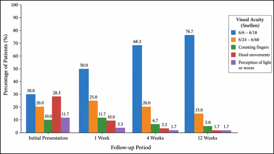
Figure 1. Serial improvement in visual acuity from initial presentation to 1-, 4-, and 12-week follow-up among patients with traumatic hyphema following closed-globe injury
Figure 1 demonstrates a progressive improvement in visual acuity over the 12-week follow-up period. At initial presentation, only 30.0% of patients had visual acuity between 6/6 and 6/18, while a substantial proportion had marked visual impairment, including counting-finger vision, hand movements, or worse. By the first week, the proportion of patients achieving 6/6–6/18 increased markedly, with further improvement observed at 4 weeks. At the final 12-week assessment, 76.7% of patients achieved visual acuity between 6/6 and 6/18, whereas the proportions with severe visual impairment had fallen considerably. Overall, the serial trend indicates substantial functional visual recovery following traumatic hyphema, with most improvement occurring within the first four weeks and further stabilization by 12 weeks.
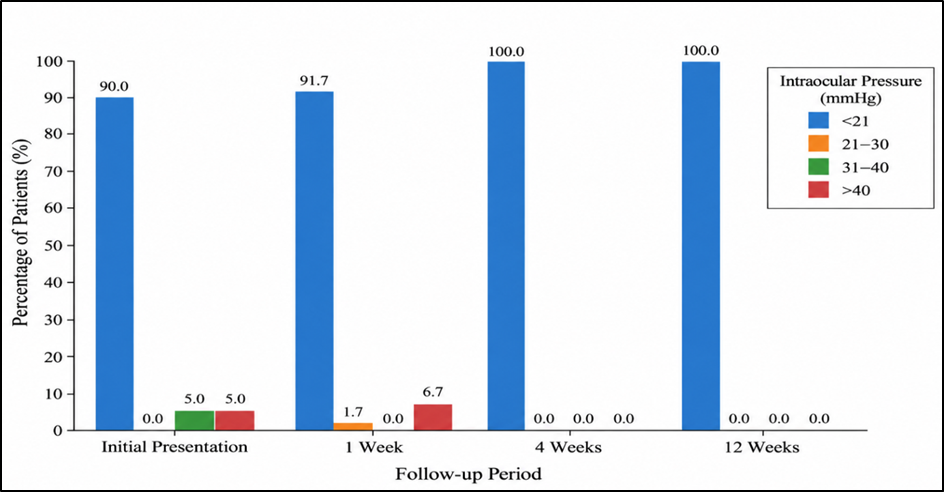
Figure 2. Change in intraocular pressure categories from initial presentation through 12 weeks of follow-up
Figure 2 shows the serial distribution of intraocular pressure (IOP) categories from presentation to the final 12-week follow-up in patients with traumatic hyphema following closed-globe injury. At initial presentation, the majority of patients (90.0%) had an IOP of <21 mmHg, while 5.0% had IOP in the 31–40 mmHg range and another 5.0% had IOP >40 mmHg, indicating raised pressure in a small proportion of cases. At the 1-week follow-up, most patients (91.7%) remained in the <21 mmHg category, while 1.7% had IOP between 21–30 mmHg and 6.7% had IOP >40 mmHg. By the 4-week follow-up, all patients (100%) had IOP values <21 mmHg, and this normal distribution was maintained at the 12-week follow-up. Overall, the figure demonstrates that although a minority of patients had elevated IOP during the early post-traumatic period, intraocular pressure normalized in all cases with follow-up and management.
DISCUSSION:
The present prospective study evaluated the clinical course, visual recovery, and prognostic factors in 60 patients with traumatic hyphema following closed-globe injury. The major finding was the substantial improvement in vision during follow-up, with the proportion of patients achieving a final visual acuity of 6/6–6/18 increasing from 30.0% at presentation to 76.7% at 12 weeks. Most hyphemas resolved within the first week, intraocular pressure normalized during follow-up, and the majority of patients could be managed conservatively. Among the variables evaluated, early presentation and presenting visual acuity emerged as the most important factors associated with final visual outcome, whereas hyphema grade was not significantly associated with final vision. The mechanism of injury showed only a marginal association with outcome.
The demographic profile of the present cohort demonstrated a marked male predominance, with males accounting for 81.7% of cases, while 68.3% of patients were aged 30 years or younger. This pattern is consistent with previous reports demonstrating that traumatic hyphema predominantly affects young males, probably because of greater exposure to outdoor activities, sports, occupational hazards, interpersonal trauma, and other high-risk activities. Nanda et al. reported that 81% of their 76 patients were male and that 85% belonged to the 15–30-year age group [11]. Similarly, Ulagantheran et al. reported male predominance in 93.2% of cases and found that approximately two-thirds of patients were younger than 30 years [12]. Ashaye also documented a predominance of males and younger individuals among 472 cases of traumatic hyphema [13]. These findings collectively underline the importance of preventive strategies specifically directed toward children, adolescents, and young working-age adults.
The mechanism of injury in the present study differed somewhat from that described in several international series. Firecracker-related injury accounted for approximately half of the cases, followed by domestic and sports-related trauma. In contrast, Nanda et al. reported sports-related injury as the most common cause, accounting for 42% of cases [11], while Ulagantheran et al. similarly found sports-related trauma in 38.1% of patients [12]. Galvis et al. reported sports-related and occupational accidents as the major mechanisms in their Colombian cohort [14]. The relatively high contribution of firecracker injuries in our study likely reflects regional and sociocultural exposure patterns. Kurien et al., in an Indian study of firework-related ocular trauma, documented a predominantly young population and found that more than half of the affected individuals were bystanders, demonstrating that firecracker injuries are not restricted to those directly handling explosive devices [15]. The prominence of this mechanism in our population emphasizes the preventable nature of many cases of traumatic hyphema and supports stronger public education, supervision, and eye-safety measures during festivals and celebrations.
A notable finding was the considerable improvement in visual function during follow-up. At initial presentation, 40.0% of patients had vision of hand movements or worse, whereas only two patients remained in this category at 12 weeks. In parallel, the proportion with visual acuity of 6/6–6/18 increased to 76.7%. Such recovery is consistent with the generally favourable prognosis of uncomplicated traumatic hyphema when structural damage to visually important tissues is limited. Nanda et al. reported final BCVA of 6/24 or better in 89% of patients after six weeks [11]. Ulagantheran et al. found that 85.4% achieved a final BCVA of at least 6/18 [12]. In a paediatric cohort, Boese et al. observed substantial recovery during the first 28 days, with 82% attaining 20/30 or better by day 28 and 98% of those with available final follow-up achieving 20/30 or better [16]. Differences in final visual outcomes between studies may reflect variations in injury severity, associated ocular damage, inclusion criteria, mechanisms of trauma, and duration of follow-up.
The initial visual acuity was one of the strongest prognostic factors in the present study. Patients presenting with better vision were considerably more likely to obtain a favourable final outcome, while those presenting with hand movements or worse constituted the largest proportion of patients with persistent visual impairment. The association between initial and final visual acuity was statistically significant (p=0.0004578). This observation is clinically relevant because initial vision represents a readily measurable summary indicator of the severity of the primary trauma and may indirectly reflect corneal injury, extent of hyphema, lens damage, vitreous hemorrhage, macular injury, retinal detachment, or optic nerve involvement. Simanjuntak et al. similarly identified initial visual acuity as a significant risk factor for poor visual outcome in 97 patients with traumatic hyphema [17]. Kurien et al. also demonstrated an association between poor presenting visual acuity and poor final vision in patients with firework-related ocular trauma [15]. Thus, although the anterior chamber hemorrhage may subsequently clear, the initial level of visual impairment remains an important marker of overall ocular injury severity.
The interval between injury and hospital presentation was another important determinant of final outcome. Overall, 71.7% of patients presented within 24 hours, and 83.7% of this group achieved good final vision compared with 58.8% of those presenting after 24 hours. The association between presentation time and final visual outcome was statistically significant. Early ophthalmic evaluation is important because it permits timely recognition of elevated IOP, extensive hyphema, lens damage, retinal complications, and other associated injuries and allows appropriate intervention before secondary complications develop. Simanjuntak et al. also identified onset or timing following injury as a significant factor associated with visual outcome [17]. Nanda et al. emphasized that early presentation, diagnosis, and appropriate treatment could help prevent complications and improve visual recovery following blunt-trauma-associated hyphema [11]. The present findings therefore support prompt ophthalmological assessment following blunt ocular injury, even when external signs initially appear limited.
Grade 1 hyphema was the most frequent presentation in our study, whereas Grade 3 was least common. Hyphema grade showed a numerical relationship with outcome, with good final vision observed in 88.9% of Grade 1 cases compared with 62.5% of Grade 4 cases; however, this difference did not reach statistical significance (p=0.1466). This finding differs from some previous studies. Simanjuntak et al. identified hyphema grade as a significant risk factor for poor visual outcome [17]. In a prospective paediatric study, Rocha et al. reported that greater hyphema severity was associated not only with worse final vision but also with increased IOP and longer hemorrhage absorption time [18]. Amoni similarly reported progressively poorer visual outcome with increasing extent of hyphema [19]. The absence of statistical significance in the current study may relate to the relatively small sample size, limited numbers of Grade 3 and Grade 4 injuries, timely treatment, and the influence of associated structural injuries. Therefore, our findings should not be interpreted as indicating that hyphema severity is clinically unimportant; rather, hyphema grade alone may be less predictive than the overall extent of ocular damage in this cohort.
Hyphema resolution was favourable, with 91.7% of patients demonstrating complete resolution within one week and all cases resolving by one month. Ulagantheran et al. observed disappearance of the hyphema within five days in 66.9% of patients [12], whereas Galvis et al. reported a mean hyphema clearance time of 8.4±3.2 days [14]. The rapid resolution observed in the present study is therefore broadly compatible with existing literature, although direct comparison is affected by differences in follow-up intervals and methods used to define complete clearance. Importantly, clearance of anterior chamber blood should not be equated automatically with full ocular recovery, because persistent visual impairment may be caused by associated lens, vitreous, retinal, choroidal, or optic nerve injury.
Intraocular pressure represented another clinically important component of assessment. At presentation, 10% of patients had an IOP of 21 mmHg or greater. All patients had an IOP below 21 mmHg by four weeks, and this normalization was maintained at the 12-week assessment. Rocha et al. reported IOP exceeding 24 mmHg in 48.6% of children in their prospective traumatic-hyphema series and demonstrated an association between higher hyphema grade and ocular hypertension [18]. The lower frequency of elevated IOP in the present study may partly reflect the greater proportion of lower-grade hyphemas. Nevertheless, the occurrence of IOP elevation even in a minority of patients supports careful tonometric monitoring during the acute phase. A 12-week study period is insufficient to assess the complete long-term burden of post-traumatic glaucoma, particularly in eyes with angle recession; therefore, longer surveillance may be required in selected patients.
Associated anterior segment abnormalities were common. Traumatic anterior uveitis was identified in 38.3% of eyes, traumatic cataract in 31.7%, and iridodialysis in 11.7%. Other findings included secondary glaucoma, corneal blood staining, and lens subluxation. These findings indicate that traumatic hyphema frequently represents one component of a wider spectrum of blunt ocular damage. Ulagantheran et al. also observed frequent iris abnormalities, including sphincter tears, mydriasis, and iridodialysis [12]. Simanjuntak et al. reported that traumatic cataract and iridodialysis were among the associated abnormalities contributing to poor visual outcomes [17]. Therefore, assessment should extend beyond the anterior chamber blood level and include detailed evaluation of the iris, lens, angle, and other ocular structures.
Posterior segment involvement was comparatively less frequent but had important implications for visual prognosis. Vitreous hemorrhage was the most common posterior segment abnormality, while retinal detachment, choroidal rupture, and macular hole occurred less frequently. Although the numbers were too small to demonstrate a statistically robust association between individual posterior segment lesions and final outcome in our cohort, such injuries are clinically important because they may result in persistent visual impairment despite complete resolution of the hyphema. Kearns, in a series of 314 traumatic hyphema cases, found that poor final visual outcome was frequently attributable to retinal pathology rather than the anterior chamber hemorrhage itself [20]. Similarly, Simanjuntak et al. identified vitreous hemorrhage and choroidal rupture among the lesions associated with poor vision [17]. Rocha et al. found posterior segment lesions to be significantly associated with unsatisfactory final visual acuity in children with traumatic hyphema [18]. These observations emphasize the necessity of careful fundus examination and B-scan ultrasonography when direct visualization of the posterior segment is compromised.
Most patients in our study were managed conservatively, while surgical intervention was reserved for selected cases. Cataract surgery represented the most frequent operative procedure during follow-up, reflecting the substantial occurrence of traumatic lens damage rather than failure of hyphema resolution itself. Only one patient required anterior chamber washout at presentation. The generally successful conservative approach is consistent with reports showing favourable outcomes in appropriately selected traumatic hyphema patients. Shiuey and Lucarelli reported that 96% of patients in their traumatic-hyphema series achieved final BCVA of 20/30 or better and concluded that close outpatient management could be effective in selected cases [21]. Galvis et al. similarly reported that only one patient with Grade 4 hyphema required anterior chamber washout in their cohort [14]. Surgical intervention should therefore be directed by the overall clinical condition—including persistent or total hyphema, uncontrolled IOP, corneal blood staining, and associated structural injuries—rather than hyphema presence alone.
The present findings have practical prognostic implications. A patient presenting early with relatively preserved initial vision, limited associated ocular damage, and controlled IOP is likely to experience substantial visual recovery. Conversely, severely reduced presenting vision, delayed presentation, and structural anterior or posterior segment injury should alert the clinician to a potentially less favourable outcome and the need for closer surveillance. Although the grade of hyphema did not independently demonstrate statistical significance in this cohort, it remains clinically relevant because larger hyphemas may coexist with greater ocular trauma and may be associated with IOP elevation and delayed clearance.
This study has certain limitations. The sample size was relatively modest, particularly for uncommon complications such as retinal detachment, choroidal rupture, and macular hole, limiting meaningful subgroup analysis. The study was conducted at a single tertiary-care centre, and the high proportion of firecracker-related injuries may limit generalizability to populations with different trauma patterns. Follow-up was limited to 12 weeks and therefore could not adequately evaluate long-term consequences such as angle-recession glaucoma. In addition, the limited number of adverse outcomes restricted the ability to perform robust multivariable prognostic modelling. Nevertheless, the prospective design, standardized serial assessment, and simultaneous evaluation of initial vision, presentation time, hyphema grade, IOP, associated ocular injuries, and final vision provide clinically useful evidence regarding recovery following traumatic hyphema.
Overall, the study demonstrates that visual recovery following traumatic hyphema in closed-globe injury is generally favourable when patients receive timely evaluation and appropriate management. More than three-fourths of patients attained BCVA of 6/18 or better at 12 weeks. Presenting visual acuity and time to hospital presentation were the most important prognostic factors identified, while the presence of associated ocular injury remained clinically relevant to persistent visual impairment. These findings reinforce the importance of early referral, comprehensive anterior and posterior segment evaluation, careful IOP monitoring, and prevention of avoidable blunt ocular trauma, particularly firecracker-related injuries.
CONCLUSION:
Traumatic hyphema following closed-globe injury demonstrated an overall favourable visual prognosis, with 76.7% of patients achieving a final visual acuity of 6/6–6/18 at 12 weeks. Visual acuity improved significantly during follow-up, most hyphemas resolved within the first week, and intraocular pressure normalized in all patients by 4 weeks. The majority of patients were managed successfully with conservative treatment, although selected cases required surgical intervention for associated ocular injuries.
REFERENCES:
1. Kuhn F, Morris R, Witherspoon CD. Birmingham Eye Trauma Terminology (BETT): terminology and classification of mechanical eye injuries. Ophthalmol Clin North Am. 2002;15(2):139-143.
2. Bansal S, Gunasekeran DV, Ang B, Lee J, Khandelwal R, Sullivan P, et al. Controversies in the pathophysiology and management of hyphema. Surv Ophthalmol. 2016;61(3):297-308.
3. Walton W, Von Hagen S, Grigorian R, Zarbin M. Management of traumatic hyphema. Surv Ophthalmol. 2002;47(4):297-334.
4. Woreta FA, Lindsley KB, Gharaibeh A, Ng SM, Scherer RW, Goldberg MF. Medical interventions for traumatic hyphema. Cochrane Database Syst Rev. 2023;3(3):CD005431.
5. Razeghinejad R, Lin MM, Lee D, Katz LJ, Myers JS. Pathophysiology and management of glaucoma and ocular hypertension related to trauma. Surv Ophthalmol. 2020;65(5):530-547.
6. Ng CS, Sparrow JM, Strong NP, Rosenthal AR. Factors related to the final visual outcome of 425 patients with traumatic hyphema. Eye (Lond). 1992;6(Pt 3):305-307.
7. Cho J, Jun BK, Lee YJ, Uhm KB. Factors associated with the poor final visual outcome after traumatic hyphema. Korean J Ophthalmol. 1998;12(2):122-129.
8. Joseph E, Zak R, Smith S, Best WR, Gamelli RL, Dries DJ. Predictors of blinding or serious eye injury in blunt trauma. J Trauma. 1992;33(1):19-24.
9. Yılmaz YC, Çiloğlu Hayat Ş, Kelebek M, Gözel ME. Visual outcomes after blunt ocular trauma: clinical findings, management, and prognostic factors. Int Ophthalmol. 2026;46(1):134.
10. Liebman DL, Aboobakar IF. Traumatic hyphema: diagnostic and management considerations. Int Ophthalmol Clin. 2024;64(2):49-61.
11. Nanda PK, Meher J, Rath B. Visual outcomes of hyphema in closed globe injury, managed at a tertiary care institute. J Evid Based Med Healthc. 2020;7(2):68-72. doi:10.18410/jebmh/2020/15.
12. Ulagantheran V, Ahmad Fauzi MS, Reddy SC. Hyphema due to blunt injury: a review of 118 patients. Int J Ophthalmol. 2010;3(3):272-276. doi:10.3980/j.issn.2222-3959.2010.03.22.
13. Ashaye AO. Traumatic hyphaema: a report of 472 consecutive cases. BMC Ophthalmol. 2008;8:24. doi:10.1186/1471-2415-8-24.
14. Galvis V, Pedraza-Concha A, Tello A, Plata ML, Escaf CL, Berrospi DR. Clinical features, management and visual outcomes on patients with traumatic hyphema in a reference ophthalmological clinic in Colombia. Rom J Ophthalmol. 2020;64(1):28-34. doi:10.22336/rjo.2020.7.
15. Kurien NA, Peter J, Jacob P. Spectrum of ocular injuries and visual outcome following firework injury to the eye. J Emerg Trauma Shock. 2020;13(1):39-44. doi:10.4103/JETS.JETS_62_18.
16. Boese EA, Karr DJ, Chiang MF, Kopplin LJ. Visual acuity recovery following traumatic hyphema in a pediatric population. J AAPOS. 2018;22(2):115-118. doi:10.1016/j.jaapos.2017.11.004.
17. Simanjuntak GWS, Farinthska G, Simanjuntak GAM, Artini W, Natali R. Risk factors for poor visual outcome in traumatic hyphema: Jakarta eye trauma study. Niger J Clin Pract. 2018;21(7):921-924. doi:10.4103/njcp.njcp_251_17.
18. Rocha KM, Martins EN, Melo LAS Jr, Moraes NS. Outpatient management of traumatic hyphema in children: prospective evaluation. J AAPOS. 2004;8(4):357-361. doi:10.1016/j.jaapos.2004.04.001.
19. Amoni SS. Traumatic hyphaema in Kaduna, Nigeria. Br J Ophthalmol. 1981;65(6):439-444. doi:10.1136/bjo.65.6.439.
20. Kearns P. Traumatic hyphaema: a retrospective study of 314 cases. Br J Ophthalmol. 1991;75(3):137-141. doi:10.1136/bjo.75.3.137.
21. Shiuey Y, Lucarelli MJ. Traumatic hyphema: outcomes of outpatient management. Ophthalmology. 1998;105(5):851-855. doi:10.1016/S0161-6420(98)95025-4.