Morphometric Evaluation of the Foramen Rotundum and Its Spatial Relationship to the Hypophyseal Fossa and Middle Clinoid Process: A Dry Skull Study with Clinical Implications.
- Dr. S. Manonmani , Associate Professor, Department of Anatomy, MVJ Medical College and Research, Bengaluru, India.
- Dr. Priyanka Jainapur , Assistant Professor, Department of Anatomy, MVJ Medical College and Research, Bengaluru, India.
- Dr. Shashi Bhushan G , Professor, Department of Anatomy, Saptagiri institute of Medical Sciences, Bengaluru, India.
- Dr. Sangetha M , Professor and Head, Department of Anatomy, MVJ Medical College and Research, Bengaluru, India.
Article Information:
Abstract:
Background: The foramen rotundum and hypophyseal fossa are anatomically adjacent structures within the sphenoid bone with significant clinical relevance for skull base pathology and neurosurgery. This study aims to provide comprehensive morphometric data and elucidate the anatomical relationships between these structures. Material and Methods: Thirty-five well-preserved dry human skulls of unknown sex were examined. The foramen rotundum was measured bilaterally using digital Vernier callipers in two dimensions: antero-posterior and transverse diameters. The distance from the middle clinoid process to the foramen rotundum was also measured to establish spatial relationships. Results: The foramen rotundum demonstrated predominantly circular to oval morphology. Mean transverse diameter (right: 4.07 ± 0.79 mm; left: 4.62 ± 0.81 mm) and antero-posterior diameter (right: 3.26 ± 0.75 mm; left: 3.33 ± 0.74 mm) showed significant asymmetry, with the right foramen being consistently smaller. Distance from middle clinoid process to foramen rotundum measured 12.44 ± 1.8 mm (right) and 13.47 ± 1.94 mm (left). Anatomical variations included accessory foramina (5.7%) and foramen duplication (2.85%). Conclusion: These morphometric data provide clinically relevant reference standards for skull base procedures and interventional pain management. The documented right-sided foramen narrowing offers a plausible morphological basis for the epidemiologically observed higher incidence of right-sided trigeminal neuralgia. The anatomical variations emphasize the importance of individual morphological assessment in surgical planning. These measurements contribute significantly to skull base anatomy literature and have direct applications for neurosurgeons, otolaryngologists, radiologists, and pain specialists.
Keywords:
Article :
INTRODUCTION:
The foramen rotundum and hypophyseal fossa are crucial anatomical structures located within the sphenoid bone of the middle cranial fossa. The hypophyseal fossa, the deepest compartment of the sella turcica, serves as the bony bed for the pituitary gland4. The anterior boundary of the sella turcica is demarcated by the middle clinoid process. The foramen rotundum is situated on the cerebral surface of the greater wing of the sphenoid bone4, positioned just below and posterior to the medial aspect of the superior orbital fissure1. This foramen opens anteriorly into the pterygopalatine fossa and transmits the maxillary nerve1.
The maxillary nerve is the second division of the trigeminal nerve and is purely sensory. It originates from the trigeminal ganglion, traverses the middle cranial fossa, and exits the skull through the foramen rotundum to enter the pterygopalatine fossa2. The anatomical dimensions and depth of the foramen rotundum are of considerable clinical significance. Pain management specialists utilize these measurements to precisely target the maxillary nerve with local anaesthetics in the treatment of severe trigeminal neuralgia15. Additionally, malignant tumours, particularly nasopharyngeal carcinomas, frequently exploit the foramen rotundum as a pathway for invasion into the middle cranial fossa and potentially into the intracranial space14.
The hypophyseal fossa exhibits considerable morphological variation in size and shape, a phenomenon that has been documented extensively through morphometric studies13. Large pituitary macroadenomas may extend beyond the fossa and invade the cavernous sinus, potentially compressing the maxillary nerve and causing facial pain, numbness, or other trigeminal symptoms prior to surgical intervention15. Extended surgical approaches, such as trans-pterygoid approaches used for accessing deep lateral tumours, require precise anatomical identification of the maxillary nerve to avoid iatrogenic neural injury15. Accidental traction, compression, or localized oedema near the foramen rotundum can result in transient postoperative maxillary nerve deficits, characterized by numbness in the cheek, upper teeth, or hard palate14.
In rare cases, large or refractory pituitary tumours invading the cavernous sinus may precipitate intractable trigeminal headaches that may necessitate advanced interventions such as rhizotomy or ganglion blocks12. The foramen rotundum serves as an essential surgical landmark for endoscopic approaches to both the pterygopalatine fossa and the infratemporal fossa9. Bony anomalies or strictures affecting the foramen may lead to compression of the maxillary nerve, resulting in trigeminal neuralgia or compressive neuropathy. Precise anthropometric measurements are crucial for radiologists to differentiate normal anatomical variations from pathological enlargement caused by malignancy13.
This study aims to provide comprehensive morphometric data regarding the foramen rotundum and to elucidate the anatomical and clinical relationships between this foramen and the hypophyseal fossa. Given their close anatomical proximity within the central skull base and their shared embryological origin from the sphenoid bone, understanding these relationships is essential for clinicians and surgeons involved in skull base pathology.
MATERIALS AND METHODS:
A total of thirty-five dry human skulls of unknown sex were obtained from the Department of Anatomy, MVJ Medical College and Research, Bengaluru. Selection criteria included skulls that were well-preserved and anatomically intact, particularly in the region of the cranial base. Skulls demonstrating damage or erosion at the cranial base were excluded from the study to ensure the accuracy and reliability of measurements. For each skull, the anatomical features and morphological variations of the foramina of the greater wing of the sphenoid bone were carefully examined and documented. Special care was taken to observe the shape and dimensional characteristics of the foramen rotundum. The features of middle cranial fossa are shown in picture-1.
The foramen rotundum was measured in two dimensions using a digital Vernier calliper: (1) antero-posterior diameter as shown in picture-5 and (2) transverse diameter as shown in picture-6. The distance from the foramen rotundum to the middle clinoid process was measured to establish the spatial relationship between the foramen and the hypophyseal fossa as shown in picture-4. All measurements were obtained bilaterally to assess for asymmetry. The measured data were systematically tabulated shown in table-1, and subsequently compared with measurements reported in relevant contemporary literature as shown in Bar chart Figure -1.
RESULTS:
Morphological Observations
The foramen rotundum demonstrated a predominantly oval(19) to circular(16) shape across the sample. Anatomical variations were documented: accessory foramina adjacent to the primary foramen rotundum were observed in 2 out of 35 skulls (5.7%) shown in picture-2, and duplication of the foramen rotundum was identified in 1 out of 35 skulls (2.85%) shown in picture-3.
Table 1: Morphometric Data of FR Dimensions and Distance from MCP
|
NO |
TD (R) |
TD (L) |
AP (R) |
AP (L) |
MCP-FR (R) |
MCP-FR (L) |
|
1 |
4.2 |
4.9 |
3.6 |
2.5 |
10.8 |
11.3 |
|
2 |
4.9 |
4.2 |
2.3 |
2.7 |
12.4 |
14 |
|
3 |
3.9 |
5.2 |
3.9 |
3.5 |
14.3 |
14.5 |
|
4 |
3.3 |
4 |
3.3 |
3.5 |
15.4 |
12.5 |
|
5 |
2.7 |
2.8 |
2.2 |
3.6 |
10.8 |
10.6 |
|
6 |
4.1 |
4.1 |
3.2 |
2.6 |
10.2 |
12.9 |
|
7 |
5 |
5.6 |
4.2 |
4.9 |
12.2 |
12.2 |
|
8 |
2.3 |
5.6 |
2.5 |
2.9 |
11.9 |
15.3 |
|
9 |
3.9 |
4.4 |
2.33 |
3 |
12.1 |
12.5 |
|
10 |
4.7 |
6.9 |
4 |
2.4 |
13.4 |
17.6 |
|
11 |
3.5 |
4.8 |
3 |
3.6 |
10.9 |
10.9 |
|
12 |
4.7 |
4.3 |
4.8 |
2.99 |
10.7 |
12.6 |
|
13 |
5.2 |
3.2 |
3.5 |
3.3 |
16.4 |
13.6 |
|
14 |
5.1 |
4.5 |
3.9 |
4.7 |
15.6 |
14.4 |
|
15 |
4.2 |
4.2 |
3.2 |
4.4 |
10.7 |
13.5 |
|
16 |
3.7 |
4.7 |
2.8 |
3.6 |
11.2 |
12.8 |
|
17 |
3.4 |
5.2 |
2.9 |
4.8 |
12.1 |
13.1 |
|
18 |
4.2 |
4.5 |
3.1 |
3.2 |
11 |
11.4 |
|
19 |
4 |
4.1 |
3 |
2.5 |
12.2 |
13.9 |
|
20 |
3.5 |
4.5 |
3 |
3.4 |
11.5 |
15.2 |
|
21 |
4.5 |
4.2 |
3.8 |
3 |
13.5 |
17.2 |
|
22 |
5 |
5.4 |
4.2 |
4.8 |
10.4 |
12.5 |
|
23 |
2.8 |
3 |
2.1 |
2.5 |
15.2 |
14.2 |
|
24 |
4.8 |
5.6 |
4.2 |
3.4 |
12.2 |
15.2 |
|
25 |
3.8 |
4.2 |
2.1 |
3 |
10.8 |
10.9 |
|
26 |
5.2 |
4.5 |
4.5 |
4.5 |
11.8 |
15.5 |
|
27 |
2.5 |
4.1 |
2.1 |
2.6 |
15.4 |
15.6 |
|
28 |
4.3 |
4.9 |
3.8 |
2.4 |
10.1 |
10 |
|
29 |
3.6 |
4.3 |
2.8 |
3.1 |
11.8 |
15.2 |
|
30 |
3.9 |
4.8 |
2.3 |
3.5 |
12 |
12.4 |
|
31 |
4 |
4.5 |
3.1 |
2.4 |
13.5 |
17.6 |
|
32 |
4.2 |
5.9 |
3.6 |
3.5 |
11 |
11.1 |
|
33 |
3.3 |
4.8 |
3.2 |
3.6 |
14.4 |
13.6 |
|
34 |
4.9 |
5.4 |
3.6 |
3.2 |
12 |
13.1 |
|
35 |
5 |
4.3 |
4.1 |
2.9 |
15.5 |
12.6 |
TD = Transverse Diameter (mm); AP = Antero-posterior Diameter (mm); MCP-FR = Distance from Middle Clinoid Process to Foramen Rotundum (mm); R = Right; L = Left
Morphological Variations and Measurement Methodology
Summary Statistics
Table 2: Summary statistics showing mean values and standard deviations for all measurements.
|
Parameter |
TD (R) |
TD (L) |
AP (R) |
AP (L) |
MCP-FR (R) |
MCP-FR (L) |
|
Mean |
4.07 |
4.62 |
3.26 |
3.33 |
12.44 |
13.47 |
|
SD |
0.79 |
0.81 |
0.75 |
0.74 |
1.80 |
1.94 |
TD = Transverse Diameter AP = Antero-posterior Diameter
MCP-FR = Distance from Middle Clinoid Process SD = Standard Deviation
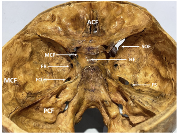
Picture 1: Middle Cranial Fossa (MCF) showing Foramen Rotundum (FR), Foramen Ovale (FO), Hypophyseal Fossa (HF), Foramen Spinosum (FS), and Superior Orbital Fissure (SOF)
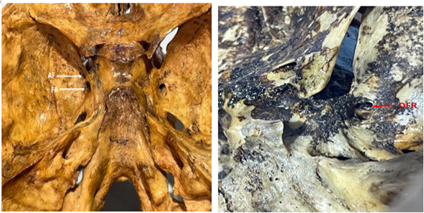
Picture 2: Unilateral Accessory Foramen (AF) adjacent to primary Foramen Rotundum.
Picture 3: Unilateral Duplicated Foramen Rotundum (DFR)
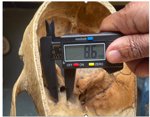
Picture 4: Measurement demonstrating the distance between Middle Clinoid Process (MCP) and Foramen Rotundum (FR)
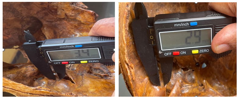
Picture 5 Picture 6
Picture 5: Measurement of antero-posterior diameter FR
Picture 6: Measurement of transverse diameter of FR
Comparative Analysis with Literature
Table 3: Comparative analysis of morphometric measurements from present study with contemporary literature. AP = Antero-posterior diameter; TD = Transverse diameter; FR to MCP = Distance from Foramen Rotundum to Middle Clinoid Process.
|
Authors & Year |
AP (R) |
AP (L) |
TD (R) |
TD (L) |
FR to MCP (R) |
FR to MCP (L) |
|
Khursheed Raza et al. 202613 |
3.54±0.77 |
3.82±0.91 |
3.59±0.65 |
3.82±0.99 |
— |
— |
|
Ajay Kumar Misra et al. 202611 |
4.42±0.66 |
3.97±0.55 |
3.91±0.53 |
4.41±0.59 |
— |
— |
|
Dr. Purohit B et al. 202412 |
— |
2.70±0.82 |
— |
2.74±0.90 |
13.07±1.99 |
12.91±1.88 |
|
Dr. K. Manivannan et al15. 2024 |
5.23±0.57 |
— |
5.15±1.14 |
— |
— |
— |
|
Dr. W.R. Reema et al16. 2024 |
3.27-6.98 |
— |
2.54-9.9 |
— |
— |
— |
|
Edward et al.2022 14(Radiology) |
3.99 ± 1.80 |
4.12 ± 1.89 |
4.61 ± 1.62 |
4.73 ± 1.64 |
— |
— |
|
Current Study |
3.26±0.75 |
3.33±0.74 |
4.07±0.79 |
4.62±0.81 |
12.44±1.8 |
13.47±1.94 |
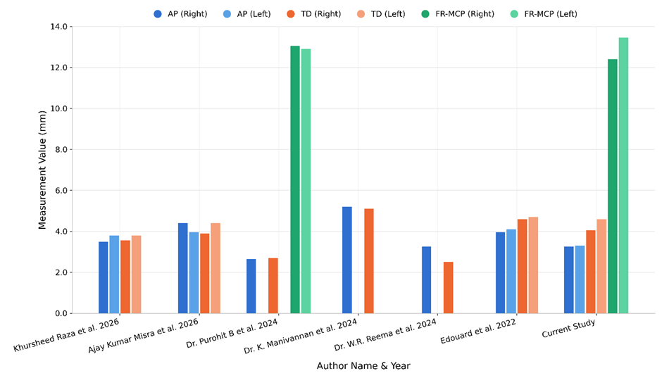
Figure - 1
Embryological Basis
The neurocranium of the skull develops from mesenchymal cells originating from neural crest cells and paraxial mesoderm5. The sphenoid bone undergoes both intramembranous and endochondral ossification, resulting from the fusion of pre-sphenoid and post-sphenoid ossification centers9.
The foramen rotundum and hypophyseal fossa share a common embryological origin, both from the developing sphenoid bone through combined endochondral and intramembranous ossification18. The foramen rotundum develops as the maxillary division of the trigeminal nerve becomes progressively enclosed by the ossifying greater wing of the sphenoid bone. As ossification advances, the nerve becomes sequentially enclosed, with the foramen rotundum ultimately representing the final bony canal through which the maxillary nerve passes18.
The hypophyseal fossa forms as a depression within the developing body of the sphenoid bone, sculpted by the growth of the pituitary gland. The pituitary gland itself develops from the fusion of two distinct embryological components: Rathke's pouch, an ectodermal outpouching from the stomodeum, and the infundibulum, a downward extension of the diencephalon5. This shared embryological relationship underscores the anatomical interdependence of these structures.
DISCUSSION:
The foramen rotundum is a consistently identifiable landmark within the middle cranial fossa, serving as the path for the maxillary nerve as it passes from the intracranial space into the pterygopalatine fossa. The sella turcica, a saddle-shaped concavity on the superior surface of the sphenoid body, houses the hypophyseal fossa-its deepest central component. Laterally, the hypophyseal fossa is bounded by the middle clinoid process3, which serves as an important anatomical reference point for skull base surgeons.
Morphological Characteristics and Variations
In the present study, the foramen rotundum was predominantly circular to oval in shape, consistent with the findings of numerous researchers and contemporary anatomical literature. Anatomical variations, including unilateral accessory foramina and foramen duplication, were observed in the sample population, with incidences of 5.7% and 2.85%, respectively. These findings align generally with reported literature, though our observed incidence of accessory foramina (5.7%) is notably higher than the 0.17% reported by Dr. Purohit et al., suggesting possible geographic or populational variation. The duplication rate of 2.85% in the present study exceeds the 0.01% documented by the same author, further emphasizing the importance of morphological assessment in individual populations.
Piagkou MN, et al Trigeminal Pain sited in their study right side of the face is the most effected due to the narrower foramina rotundum and ovale 19.
Such anatomical variations have important clinical implications. Accessory foramina or duplicated foramina may provide alternative pathways for neurovascular structures or infectious processes. These anomalies could potentially affect the distribution of anaesthetic solutions during maxillary nerve blocks or influence the pathophysiology of compressive neuropathies affecting the maxillary nerve.
Dimensional Analysis and Asymmetry
The morphometric data reveals distinct asymmetry between the right and left foramina rotunda. accessory foramina adjacent to the primary foramen rotundum were observed in 2 out of 35 skulls (5.7%) shown in picture-2, and duplication of the foramen rotundum was identified in 1 out of 35 skulls (2.85%) shown in picture-3.
Edouard et al in their radiological study has recorded the same shown in table-2
Notably, the smaller dimensions of the right foramen rotundum correlate epidemiologically with the documented higher incidence of right-sided trigeminal neuralgia in the general population19. While morphometric differences alone cannot definitively establish causation, the smaller cross-sectional area of the foramen on the right side may create a relatively narrower channel through which the maxillary nerve passes19. This anatomical constraint could theoretically predispose the right maxillary nerve to greater vulnerability to compression by expanding masses, inflammation, or vascular structures.
Comparative Analysis with Literature
The dimensional measurements obtained in the present study are comparable to those reported in contemporary morphometric literature, though subtle variations exist, reflecting the diversity of anatomical measurements across different populations and skull samples. These dimensions are tabulated in table – 2The measurement of the distance from the middle clinoid process to the foramen rotundum (right: 12.44 ± 1.8 mm; left: 13.47 ± 1.94 mm) represents a novel contribution to the morphometric literature, as this particular measurement has been previously documented by only one research group (Dr. Purohit et al., 2024). Rusu, M. C et al in their study found the Mean canal length was 4.07 ± 1.78 mm with a coefficient of variation of 43.8%.The left side demonstrated significantly longer canals than the right (4.33 ± 1.77 mm vs. 3.81 ± 1.77 mm; p = 0.013)18.This study shows almost similar value with the mean transverse diameter (right: 4.07 ± 0.79 mm; left: 4.62 ± 0.81 mm) and antero-posterior diameter (right: 3.26 ± 0.75 mm; left: 3.33 ± 0.74 mm).
Clinical Significance and Surgical Implications
Knowledge of the precise morphometry and anatomical relationships of the foramen rotundum is indispensable for multiple medical specialties. Otolaryngologists, neurosurgeons, and interventional radiologists frequently encounter this foramen during skull base procedures. The foramen serves as a critical surgical landmark for endoscopic approaches to both the pterygopalatine fossa and the infratemporal fossa9.
For pain management specialists, understanding the dimensions and spatial orientation of the foramen rotundum enables precise targeting of the maxillary nerve with anaesthetic agents and neurolytic procedures in the management of severe trigeminal neuralgia15. The observed asymmetry may necessitate careful attention to the dimensions of the foramen on each side during planning of such interventions.
Pituitary tumours, particularly macroadenomas, present unique challenges due to the intimate anatomical relationship between the hypophyseal fossa and the adjacent foramen rotundum. Tumour expansion may directly compress the maxillary nerve or cause secondary effects through mass effect on surrounding structures. Advanced surgical approaches, such as extended endonasal endoscopic approaches or trans-pterygoid corridors, require precise anatomical understanding to minimize iatrogenic neural injury.
Embryological Significance
The shared embryological origin of the foramen rotundum and hypophyseal fossa from the developing sphenoid bone provides insight into their anatomical interdependence. Phylogenetic analyses have demonstrated that the basic configuration of the sphenoid bone and its foramina is highly conserved across mammalian species, reflecting the fundamental importance of these structures for cranial nerve passage and pituitary gland housing18. This evolutionary conservation highlights the biological significance of maintaining precise anatomical relationships within the central skull base.
Limitations of the Present Study
The present study was conducted on dry bone specimens with unknown demographic variables, including age, sex, and ethnic origin. These factors may influence morphometric measurements. The sample size of 35 skulls, while adequate for descriptive morphometry, may not capture the full spectrum of anatomical variation in larger populations.
CONCLUSION:
This morphometric analysis of the foramen rotundum and its spatial relationship to the hypophyseal fossa and middle clinoid process provides quantitative anatomical data with substantial clinical relevance. The predominant circular to oval morphology observed is consistent with existing anatomical literature. The identification of anatomical variations, including accessory foramina (5.7%) and foramen duplication (2.85%), emphasizes the importance of individual anatomical assessment in surgical planning.
The documented asymmetry between the right and left foramina rotunda, with the right side demonstrating consistently smaller dimensions, offers a plausible morphological explanation for the epidemiologically documented higher incidence of right-sided trigeminal neuralgia. The measurements presented contribute to the existing body of morphometric knowledge and establish a reference standard for the population studied.
The dimensions and spatial relationships documented in this study provide valuable reference data for radiologists interpreting imaging studies, for neurosurgeons planning surgical approaches, and for pain management specialists performing interventional procedures targeting the maxillary nerve. Furthermore, the novel measurement of the distance from the middle clinoid process to the foramen rotundum adds to the contemporary literature regarding skull base anatomy.
Acknowledgement: I sincerely thank my daughter and the attenders of anatomy department of MVJ medical college for their contribution to complete this study.
REFERENCES:
1. Standing, S. (Ed.). (2008). Gray's anatomy: The anatomical basis of clinical practice (40th ed.). Churchill Livingstone.
2. Dutta, A. K. (2013). Essentials of human anatomy (5th ed., Vol. 2). Current Books International.
3. Kulkarni, N. V. (2012). Clinical anatomy (2nd ed., Vol. 3). Jaypee Brothers Medical Publishers.
4. Faruki, N. A. (2012). Human osteology: A clinical orientation (3rd ed.). CBS Publishers & Distributors.
5. Sadler, T. W. (2014). Langman's medical embryology (13th ed.). Wolters Kluwer Health.
6. Shapiro, R., & Janzen, F. (1967). The foramina of the middle fossa: a phylogenetic, anatomic and pathologic study. American Journal of Roentgenology, 101(4), 779–794.
7. Khan, A. A., Asari, M. A., & Hassan, A. (2012). Anatomic variants of foramen ovale and spinosum in human skulls. International Journal of Morphology, 30(2), 445–449.
8. Kumar, A., Sehgal, R., & Roy, T. S. (2016). A morphometric analysis and study of variations of foramina in the floor of the middle cranial fossa. Journal of the Anatomical Society of India, 65, S35–S41.
9. Gurcan, O., Gurcay, A. G., Kazancı, A., Yıldırım, A. E., Turkoglu, O. F., Komurcu, H. F., & Beşkonakli, E. (2016). Trigeminal neuralgia as an unusual isolated symptom of pituitary adenoma: Case report and review of the literature. Turkish Neurosurgery, 26(1), 180–183.
10. Mishra, A. K., Paiker, M., Khan, A. Z., Kirti, S., Jah, M., & Sarish, K. (2020). Morphological and morphometric study of the foramina of the middle cranial fossa in dried human skulls. International Journal of Anatomy, Radiology and Surgery, 9(3), AO14–AO18.
11. Luz, M. A. M., Domingos, M. B., Queiroz, G. F. R., Cunha, J. V. P., & Santo Neto, H. (2021). Asymmetry of the foramen rotundum and of the foramen ovale. Journal of Morphological Sciences, 38, 268–272.
12. Purohit, B., Rajguru, J., Shah, S., & Vipra, D. (2021). Anatomical variations of foramen rotundum and its clinical significance in Gujarat region - A cross sectional study. Journal of Cardiovascular Disease Research, 12(3), 134–140.
13. Raza, K., Xalxo, N., Shivshankar, S., Patil, K. S., Patel, R. B., & Patel, A. K. (2022). Foramen rotundum dimensions and landmarks in Indian adults: Anatomical insight for skull base surgery. Journal of Morphological Sciences, 39, 162–167.
14. Edouard, A., Ibrahim, A. A., Shama, S. A., Abdelnabi, M. M., Birindwa, P. B., & Elwany, S. (2023). Radiological anatomy of foramen rotundum and its surgical implications. The Egyptian Journal of Otolaryngology, 39(1), 45.
15. Manivannan, K., Rao, W. R. R., & Sasikumar, R. (2024). Incidence and morphological variations of foramen vesalius, foramen rotundum, and foramen lacerum in 173 human skulls from Andhra Pradesh, India. Journal of Cardiovascular Disease Research, 15(1), 1332–1341.
16. Rao, W. R. R., Manivannan, K., & Sasikumar, R. (2024). Morphological and morphometric analysis of middle cranial fossa foramina in adult human skulls: Implications for neurosurgical procedures. Journal of Cardiovascular Disease Research, 15(1), 1342–1351.
17. Rusu, M. C., Bănică, D. A., Dumitru, C. C., Jianu, A. M., & Tudose, R. C. (2026). Foramen rotundum versus canal of the maxillary nerve. Surgical and Radiologic Anatomy, 48(1), 172.
18. Rusu, M. C., Bănică, D. A., Dumitru, C. C., Jianu, A. M., & Tudose, R. C. (2026). Foramen rotundum versus canal of the maxillary nerve. Surgical and Radiologic Anatomy, 48(1), 172.
19. Piagkou MN, Skandalakis P, Piagkos G, Demesticha T. Trigeminal Pain and Its Distribution in Different Trigeminal Nerve Branches. Anesth Pain. 2012: 1(4):271-2. DOI: 10.5812/aapm.3764