An alternative minimally invasive procedure to combact maxillary constriction with marpe-a case report.
- Dr. Samit Mondal , Associate Professor, MDS, Department of Orthodontics & Dentofacial Orthopedics, Burdwan Dental College Hospital, West Bengal
- Dr. Prasenjit Banerjee , Prof. & HOD, MDS, Department of Orthodontics & Dentofacial Orthopedics, Guru Nanak Institute of Dental Sciences & Research
- Dr. Debrup Halder , Dental surgeon, (Mo supy), MDS (Orthodontics & Dentofacial Orthopedics) Swasthya Bhawan, Dept of Health and family welfare, Govt of west Bengal.
- Dr. Nilanjan De , PGT, MDS, Department of Orthodontics & Dentofacial Orthopedics, Guru Nanak Institute of Dental Sciences & Research
- Dr. Sangeeta Sen , PGT, MDS, Department of Orthodontics & Dentofacial Orthopedics, Guru Nanak Institute of Dental Sciences
Article Information:
Abstract:
Background: The incorporation of miniscrews into conventional rapid palatal expansion (RPE) appliances forms the Miniscrew-Assisted Rapid Palatal Expansion (MARPE) system. While conventional RPE relies on tooth support, MARPE uses bone-anchored miniscrews to transmit expansion forces, distributing stress along key maxillary buttresses (zygomaticomaxillary, nasomaxillary, and pterygomaxillary). Objective / Rationale: Conventional RPE is associated with several adverse effects, including tipping of anchor teeth, alveolar bone bending, root resorption, bony dehiscence, fenestrations, limited skeletal expansion, and post-expansion relapse. MARPE was developed to overcome these dental limitations through enhanced skeletal anchorage. Key Advantages: By direct anchorage to bone (exclusively bone-borne or tooth-bone-borne via two or four miniscrews), MARPE achieves maximum skeletal expansion with minimal dental tipping. It minimizes tooth extrusion and undesirable clockwise mandibular rotation, thereby improving vertical control during orthodontic treatment. Conclusion: MARPE serves as an effective, minimally invasive alternative for young adults with moderate sutural resistance, avoiding invasive surgical expansion procedures and positively impacting patient treatment acceptance.
Keywords:
Article :
INTRODUCTION:
The advent and incorporation of mini screws with conventional rapid palatal expander transforms it into a miniscrew-assisted rapid palatal expansion (MARPE) appliance [1]. Bone-anchored mini screws can help to achieve maximum skeletal expansion with a minimum amount of dental tipping [2]. Various designs have been recommended by many authors [3,4] without any dental support (exclusively bone borne), with support from teeth (teeth-bone borne) and two/ four mini screws. Stress distribution and force trajectories are directed mainly along three buttresses in the maxilla; namely zygomaticomaxillary, nasomaxillary and pterygomaxillary. Major disadvantages of conventional RPE appliances include tipping of anchor teeth [5], limited skeletal movement [6], undesirable tooth movement [7], root resorption [8], bony dehiscence’s and fenestrations as well as post expansion relapse [9]. As, conventional RPE appliances transmit the expansion forces through the teeth, alveolar bone bending and tipping of buccal segments are the major concern.
With significant modification of bone anchored screws also reduces clockwise rotation of mandible by reducing extrusion of dental components, thus affects vertical control during orthodontic therapy. MARPE appliance is beneficial in young adult patients with moderate sutural resistance for skeletal expansion and invasive surgical procedure can be avoided that provide a positive psychological impact on acceptance of orthodontic treatment.
CASE REPORT:
A 17-year-old female was referred to dept. department orthodontics and dentofacial Orthopaedics. She presented with Angle Class II div-1 malocclusion, with proclined upper anteriors and increased overjet, and maxillary arch constriction. It was predicted that maxillary alveolar arch expansion through orthodontic treatment would be necessary to address the transverse discrepancy of maxillary arch. A treatment plan was established to perform miniscrew-assisted rapid palatal expansion (MARPE) treatment to correct the transverse dimension. Comprehensive blood tests, including biochemistry, complete blood count, coagulation tests, infection screening, and imaging (electrocardiogram, chest X-ray), revealed no abnormalities. There were no findings suggestive of musculoskeletal metabolic disorders were diagnosed before starting of treatment.
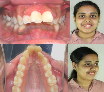
Fig-1
Location of Mini Screws Factors like convenient access, low risk of damage to the surrounding anatomical structures [10-12], high-quality cortical bone and thin mucosa confirming adequate stability [13,14]. makes the paramedian area (3 mm lateral to the suture in the 1st premolar region) the most appropriate site for placement of mini screws [15-17].
Mini Screw Configuration
Dimensions of four mini screws as per the design were selected as follows;
Total 4 miniscrews (2 Anterior and 2 posterior screws): 2 x 10 mm (FavAnchorTM SAS, India) SAS, India)
The lengths were chosen considering the height of insertion slot, space between the appliance and the palate, thickness of the palatal mucosa and desired 5-7 mm of bone engagement .A pretreatment CBCT scan of the maxillary arch was done for assessment of palatal cortical bone depth ,Intention was to achieve bicortical engagement [18], thus aiding for better stability of the mini screws as well complete bony separation of mipalatal suture. TAD placement with a conventional straight driver or an engine-mounted driver is problematic sometimes for the reasons of directional control and lack of torque to drive the implant in hard palatal bone.
A dedicated palatal driver [(L’il One, Fav Anchor TM SAS, India)] is used to maintain adequate insertion angulation and torque while placing the mini screws. This unique design of the driver makes it very convenient to place the palatal implants with great ease and precision.
Appliance design & Fabrication
An intraoral scan of the maxillary arch was taken, along with a CBCT scan of maxillary arch, which was done and converted to STL files. These files were sent to Dr Diggant THakkars' lab to fabricate direct 3d printed customised MARPE design. The size of the expansion screw was selected taking into consideration it’s close adaptation ,a hyrax screw (Leone SPE, 9 mm) was chosen. Two mini screws (2 x 12mm, Fav Anchor TM SAS, India) were placed in the paramedian region, and the posterior arms of the appliance were anchored to the molar. MARPE expander was fabricated by CAD-CAM technique, directly 3D printed with a digitalised stereolithographic model from intraoral and CBCT scan.
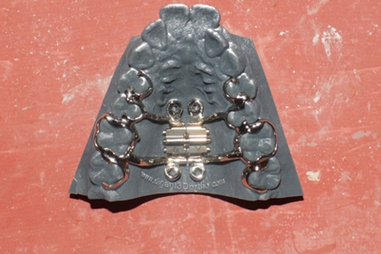
fig.2
Activation Protocol/ Schedule
Activation was initially done for 2 turns/ day (360° x 2) till the development of diastema, followed by 1 turn/ day till sufficient expansion has been achieved and stopped when the palatal cusp of maxillary molars and premolars had just an edge-to-edge contact of buccal cusp of lower molars and premolars . Activation schedule was followed as described (Table 1).
|
Age of the patients |
Initial expansion rate |
Expansion rate after opening of the diastema |
|
Early teens |
3 turns /week |
3 turns /week |
|
Late teens |
1 turn/day |
1 turn /day |
|
adults |
2 turns /day |
1 turn/day |
Treatment Objectives
The treatment objectives were to as follows:
1. To correct transverse maxillary deficiency
2. To maximizing skeletal expansion
3. To minimizing buccal tipping
4. To establish acceptable buccal occlusion and
5. To maintain sound periodontal and bone support
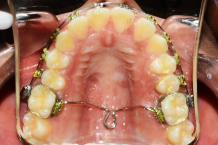
Treatment progress and result: after taking measurement of pretreatment and post treatment photographs and study model it can be concluded that total expansion of 4.5 mm in premolar region and 5mm in molar region was achieved respectively (Table-2). side by side orthodontic treatment was initiated, appliance was left remain intraorally for 4months to ensure midpalatal sutural callus formation , and after removal retention was given with trans-palatal arch along with thicker .019x.025 ss arch wire that is manually pre-fabricated with 2mm wider arch form in both molar and premolar region.
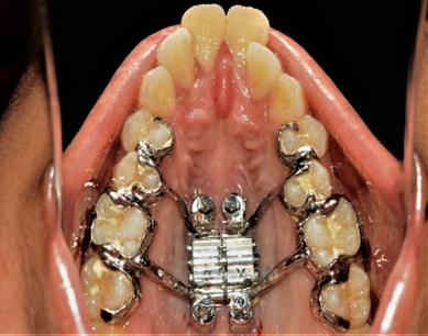
after placement of MARPE
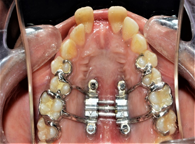
after initial expansion is achieved
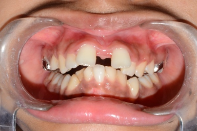
intra-oral pic after expansion
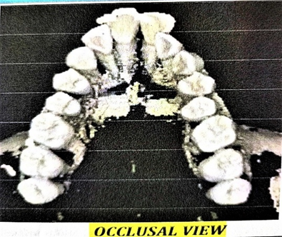
pretreatment cbct
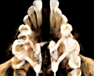
cbct of maxilla after expansion
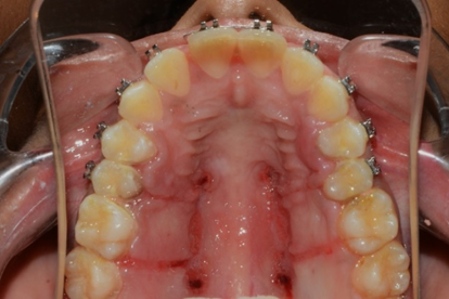
after removal of appliance MARPE
treatment in progress with retention
Table-2
|
Maxillary width(in mm) |
Pre treatment |
Post treatment |
Expansion achieved (in mm) |
|
Inter premolar |
32mm |
36.5mm |
4.5mm |
|
intermolar |
38.5mm |
43.5mm |
5mm |
DISCUSSION:
MARPE effectively achieved skeletal expansion by the separation of the midpalatal suture. Park et al. [19] reported a success rate of 84.2% in cases treated with a similar protocol. Skeletal expansion observed in the present study included the expansion of the zygomatic arch as well as nasal cavity. While the zygomatic arch expanded to a lesser extent, expansion of nasal cavity was much more evident, and thus can result in improvement of nasal breathing owing to increased air flow. Thus, by effectively increasing the nasal cavity volume, treatment with a MARPE appliance can improve the constricted airway, thus aiding in long-term stability. Garib G et al. [20], Gurgel JA et al. [21], also reported some amount of buccal tipping. Previous studies have reported greater changes in the degree of molar inclination than that of premolar inclination . The higher density of the buccal cortical bone in the maxillary canine and premolar regions might have resulted in the greater buccal inclination of the first molar in comparison with that of the first premolar [22,23]. Using a MARPE appliance, some amount of buccal tipping is inevitable, though much less as compared to RPE, as teeth are still used as anchor units alongside miniscrews. Tendency buccal tipping is directly proportional to the resistance exerted by the midpalatal suture.
CONCLUSION:
Expansion achieved in the cases treated by MARPE are majorly skeletal expansion, as the appliance is a tooth-and-tissue-borne appliance. It can be used in young adults from late teens to midtwenties and exhibits a high success in this particular age group [24 ,25,26]. Skeletal maxillary expansion was effectively done with MARPE, and clinical observations suggest that MARPE prevents many of the adverse effects of RPE and should be considered as a preferred and effective alternative to the invasive procedure like surgical skeletal expansion.
REFERENCES:
1. Lee KJ, Park YC, Park JY, Hwang WS. Miniscrew-assisted nonsurgical palatal expansion before orthognathic surgery for a patient with severe mandibular prognathism. American Journal of Orthodontics and Dentofacial Orthopedics. 2010 Jun 1;137(6):830-9.
2. MacGinnis M, Chu H, Youssef G, Wu KW, Machado AW, Moon W. The effects of micro-implant assisted rapid palatal expansion (MARPE) on the nasomaxillary complex—a finite element method (FEM) analysis. Progress in orthodontics. 2014 Aug 29;15(1):52..
3. Kim KB, Helmkamp ME. Miniscrew implant-supported rapid maxillary expansion. Journal of Clinical Orthodontics: JCO. 2012 Oct 1;46(10):608-12.
4. Carlson C, Sung J, McComb RW, Machado AW, Moon W. Microimplant-assisted rapid palatal expansion appliance to orthopedically correct transverse maxillary deficiency in an adult. American Journal of Orthodontics and Dentofacial Orthopedics. 2016 May 1;149(5):716-28..
5. Shapiro PA, Kokich VG. Uses of implants in orthodontics. Dental Clinics of North America. 1988 Jul 1;32(3):539-50.
6. Smalley WM, Shapiro PA, Hohl TH, Kokich VG, Brånemark PI. Osseointegrated titanium implants for maxillofacial protraction in monkeys. American Journal of Orthodontics and Dentofacial Orthopedics. 1988 Oct 1;94(4):285-95.
7. Erverdi N, Okar I, Kücükkeles N, Arbak S. A comparison of two different rapid palatalexpansion techniques from the point of root resorption. American Journal of Orthodontics and Dentofacial Orthopedics. 1994 Jul 1;106(1):47-51.
8. Parr JA, Garetto LP, Wohlford ME, Arbuckle GR, Roberts WE. Sutural expansion using rigidly integrated endosseous implants: an experimental study in rabbits. The Angle Orthodontist. 1997 Aug 1;67(4):283-90.
9. Harzer W, Reußer L, Hansen L, Richter R, Nagel T, Tausche E. Minimally invasive rapid palatal expansion with an implant-supported hyrax screw/Minimalinvasive forcierte Gaumennahterweiterung mit einer implantatgetragenen Hyraxschraube. Biomedical Engineering/Biomedizinische Technik. 2010 Feb 1;55(1).
10. Marquezan M, Nojima LI, Freitas AO, Baratieri C, Alves Júnior M, Nojima MD, Araújo MT. Tomographic mapping of the hard palate and overlying mucosa. Brazilian oral research. 2012 Feb;26(1):36-42.
11. Lombardo L, Gracco A, Zampini F, Stefanoni F, Mollica F. Optimal palatal configuration for miniscrew applications. The Angle Orthodontist. 2010 Jan 1;80(1):145-52.
12. Kyung SH, Hong SG, Park YC. Distalization of maxillary molars with a midpalatal miniscrew. Journal of clinical orthodontics: JCO. 2003 Jan;37(1):22-6.
13. Kang S, Lee SJ, Ahn SJ, Heo MS, Kim TW. Bone thickness of the palate for orthodontic mini-implant anchorage in adults. American Journal of Orthodontics and Dentofacial Orthopedics. 2007 Apr 1;131(4):S74-81.
14. King KS, Lam EW, Faulkner MG, Heo G, Major PW. Vertical bone volume in the paramedian palate of adolescents: a computed tomography study. American Journal of Orthodontics and Dentofacial Orthopedics. 2007 Dec 1;132(6):783-8.
15. Moon SH, Park SH, Lim WH, Chun YS. Palatal bone density in adult subjects: implications for mini-implant placement. The Angle Orthodontist. 2010 Jan 1;80(1):137-44.
16. Kim HJ, Yun HS, Park HD, Kim DH, Park YC. Soft-tissue and cortical-bone thickness at orthodontic implant sites. American Journal of Orthodontics and Dentofacial Orthopedics. 2006 Aug 1;130(2):177-82.
17. Motoyoshi M, Yoshida T, Ono A, Shimizu N. Effect of cortical bone thickness and implant placement torque on stability of orthodontic mini-implants. International Journal of Oral & Maxillofacial Implants. 2007 Sep 1;22(5).
18. Yılmaz A, Arman-Özçırpıcı A, Erken S, Polat-Özsoy Ö. Comparison of short-term effects of mini-implant-supported maxillary expansion appliance with two conventional expansion protocols. European journal of orthodontics. 2015 Jan 6;37(5):556-64.
19. Park JJ, Park YC, Lee KJ, Cha JY, Tahk JH, Choi YJ. Skeletal and dentoalveolar changes after miniscrew-assisted rapid palatal expansion in young adults: a cone-beam computed tomography study. The korean journal of orthodontics. 2017 Mar 1;47(2):77-86.
20. Garib DG, Henriques JF, Janson G, de Freitas MR, Fernandes AY. Periodontal effects of rapid maxillary expansion with tooth-tissue-borne and tooth-borne expanders: a computed tomography evaluation. American journal of orthodontics and dentofacial orthopedics. 2006 Jun 1;129(6):749-58.
21. Gurgel JD, Tiago CM, Normando D. Transverse changes after surgically assisted rapid palatal expansion. International journal of oral and maxillofacial surgery. 2014 Mar 1;43(3):316-22.
22. Christie KF, Boucher N, Chung CH. Effects of bonded rapid palatal expansion on the transverse dimensions of the maxilla: a cone-beam computed tomography study. American journal of orthodontics and dentofacial orthopedics. 2010 Apr 1;137(4):S79-85.
23. Rungcharassaeng K, Caruso JM, Kan JY, Kim J, Taylor G. Factors affecting buccal bone changes of maxillary posterior teeth after rapid maxillary expansion. American Journal of Orthodontics and Dentofacial Orthopedics. 2007 Oct 1;132(4):428-e1.
24. Corbridge JK, Campbell PM, Taylor R, Ceen RF, Buschang PH. Transverse dentoalveolar changes after slow maxillary expansion. American Journal of Orthodontics and Dentofacial Orthopedics. 2011 Sep 1;140(3):317-25.
25. Haas AJ. Palatal expansion: just the beginning of dentofacial orthopedics. American journal of orthodontics. 1970 Mar 1;57(3):219-55.
26. Wehrbein H, Yildizhan F. The mid‐palatal suture in young adults. A radiological‐histological investigation. The European Journal of Orthodontics. 2001 Apr 1;23(2):105-14.