Histopathological Spectrum Of Breast Lesion In Younger Women: Retrospective Study In A Tertiary Healthcare Centre.
- Dr. Zeel Thakkar , Third Year Md Pathology Resident.
- Dr. Suhani Patel , Third Year Md Pathology Resident.
- Dr. Jignasa Bhalodia , Hod And Dean Gmers Medical College.
- Dr. Chahat Shah , Third Year Md Pathology Resident.
Article Information:
Abstract:
Background: Breast lesions in young women are predominantly benign, although malignancy may occasionally mimic benign masses and lead to delayed diagnosis. This study evaluated the histopathological spectrum of breast lesions in women younger than 40 years. Methods: A retrospective study was conducted at GMERS Medical College and Hospital, Sola, Ahmedabad, including specimens received from January 2023 to December 2025. 260 breast specimens, including core biopsies, excisional biopsies and mastectomy specimens, were reviewed and classified according to WHO criteria. Results: Benign lesions constituted 194 cases (74.6%), inflammatory lesions 34 (13.07%), and malignant lesions 32 (12.30%). Fibroadenoma was the commonest benign lesion (144/194; 74.3%). Invasive ductal carcinoma NST was the predominant malignancy (26/32; 81.25%), with malignant cases occurring in women aged 31–40 years. The upper outer quadrant was the most frequent site. Conclusion: Breast lesions in young women are predominantly benign, but histopathological evaluation remains essential for diagnosis and detection of malignancy.
Keywords:
Article :
INTRODUCTION:
Breast cancer remains the most commonly diagnosed cancer among women worldwide, which makes it essential to continually assess diagnostic trends to enhance early detection and management strategies1 Breast carcinoma is the most common malignant tumor and the second most common cause of carcinoma death in women2 .In India, breast cancer is the second common malignancy in women after cervical cancer and is detected 20 per 100,000 women.3,4,5. Benign lesions of the breast usually present in the second decade of life. Common benign lesions of the breast include fibroadenoma, phyllodes tumour, lactating adenoma and tubular adenoma. Other causes include infection, trauma, and cyst formation.6 Malignant lesions are ductal carcinoma, lobular carcinoma, colloid carcinoma, mucinous carcinoma and medullary carcinoma.7.
Inflammatory diseases of the breast are rare (accounting for less than 1% of breast symptoms) and are caused by infections, autoimmune disease, or foreign body-type reactions to extravasated keratin or secretions. These lesions are important not only in terms of local symptoms and discomfort but also because many may mimic malignancy8. A young woman presenting with a breast mass raises anxiety in both patient and family regarding the possibility of breast cancer.9The types of breast lesions in young female vary markedly from that for adults, with the former lesions being overwhelmingly benign. After the onset of puberty, most cases of breast masses are benign fibro adenoma. Malignant masses of the breast in young females are rare.6. Early presentation of breast carcinoma in a young woman may be very similar to that of fibroadenoma. As a consequence, however, the rare cases of breast carcinoma in adolescents and young women are commonly associated with delayed diagnosis, due to a low index of suspicion on the part of physicians, delayed presentation by the patient, or both physician suspicion and delayed presentation.9
Aim
The primary aim of this study is to retrospectively analyse the complete histopathological spectrum of breast lesions in women younger than 40 years whose specimen was received at GMERS medical college and hospital, sola, Ahmedabad over a defined period.
Objectives:
1. To determine the incidence and relative frequency of various inflammatory, benign, and malignant breast lesions diagnosed in younger women.
2. To determine age vise occurence of various benign and malignant breast lesions.
3. To identify the common histological types and grades of breast carcinoma encountered in this younger patient population.
4. To compare the spectrum and frequency of breast lesions observed in this study with published data from national and international literature.
MATERIALS AND METHODS:
The retrieval of histopathological data of breast lesions done during the 1st January 2023 to 31st December 2025(3 years) was accomplished by reviewing hospital records retrospectively. The patient’s age and histopathological diagnosis were obtained from the medical centers information database. Total of 260 cases were retrieved. The samples for this study included breast trucut biopsies, needle core biopsies, excisional biopsies and mastectomy specimens received in our department. Samples were fixed in 10% formalin and then grossly examined for their size and consistency.
Cut surface were noted for colour, secondary changes like necrosis, haemorrhage, cystic degeneration, fibrosis and infiltration by tumour. H&E slides of tissues were reviewed by two histopathologist and lesions were classified based on standard WHO classification. In cases where stain faded, restaining of slides were done.Results were entered into tabulated form and analysed statistically
RESULTS:
Total 260 cases were recorded between mentioned time period. As shown in table 1, according to histopathological examination, most of the cases were benign (n = 194/260; 74.6%), followed by inflammatory lesions (n = 34/260; 13.07%), and the rest was malignant (n=32/260;12.30%)%).
Table: 1. Spectrum of breast lesions in young females.(Original source)
|
Type |
<10 years |
10-20 years |
21-30 years |
31-40 years |
Total |
|
Inflammatory |
0 |
1 |
18 |
15 |
34 |
|
Benign |
1 |
65 |
76 |
52 |
194 |
|
Malignant |
0 |
0 |
6 |
26 |
32 |
|
Total |
1 |
66 |
100 |
93 |
260 |
Table: 2. Histopathological spectrum of inflammatory breast lesions along with age wise distribution(Original source)
|
Histopathological pattern |
<10 years |
10-20 years |
21-30 years |
31-40 years |
Total |
|
Acute mastitis |
0 |
1 |
4 |
5 |
10(29.41%) |
|
Acute on chronic mastitis |
0 |
0 |
3 |
2 |
5(14.71%) |
|
Granulomatous mastitis |
0 |
0 |
2 |
3 |
5(14.71%) |
|
Lymphocytic mastitis |
0 |
0 |
1 |
0 |
1(2.94%) |
|
Breast abscess |
0 |
0 |
3 |
3 |
6(17.65%) |
|
Non specific inflammation |
0 |
0 |
3 |
0 |
3(8.82%) |
|
Ductal ectasia |
0 |
0 |
2 |
2 |
4(11.76%) |
|
Total |
0 |
1 |
18 |
15 |
34 (100) |
Table: 3. Histopathological spectrum of benign breast lesions along with age wise distribution(Original source)
|
Histopathological pattern |
<10 years |
10-20 years |
21-30 years |
31-40 years |
Total |
|
Fibroadenoma |
1 |
57 |
61 |
25 |
144(74.23) |
|
Fibrocystic disease |
0 |
0 |
6 |
14 |
20(10.30) |
|
Benign phyllodes |
0 |
1 |
3 |
12 |
16(8.24) |
|
Tubular adenoma/ Tubular adenosis |
0 |
4 |
0 |
1 |
5(2.57) |
|
Lactating adenoma |
0 |
0 |
3 |
0 |
3(1.54) |
|
Ductal papilloma |
0 |
0 |
2 |
0 |
2(1.03) |
|
Mammary hemartoma |
0 |
1 |
0 |
0 |
1(0.51) |
|
Galactocele |
0 |
0 |
1 |
0 |
1(0.51) |
|
Cystic hyperplasia |
0 |
0 |
1 |
0 |
1(0.51) |
|
Sclerosing adenosis |
0 |
0 |
1 |
0 |
1(0.51) |
|
Total |
1 |
63 |
78 |
52 |
194(100) |
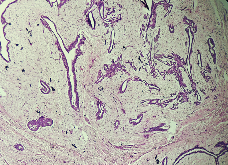
Figure1: Fibroadenoma-H & E Section show Intracanalicular and Pericanalicular pattern of ductal cell proliferation in fibromyxoid stroma. (10x, Low Power View )(Original source)
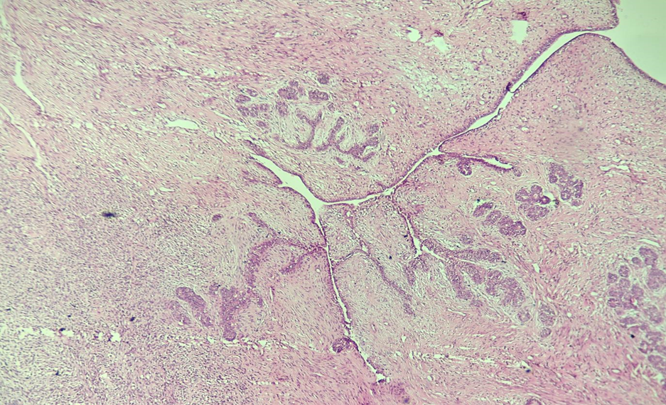
Figure 2: Benign Phyllodes- H & E Section show predominantly intracanalicular pattern of ductal cell proliferation with hyper cellular stroma. (10x, Low Power View )(Original source)
Out of all benign breast lesions, Fibroadenoma was most prevalent and found in 144(74.33%), followed by fibrocystic breast disease in 20(10.30%), benign phyllodes in 16(8.24),tubular adenoma in 5(2.57%),lactating adenoma in 3(1.54), ductal papilloma in 2(1.03), mammary hemartoma in 1(0.51),galactocele in 1(0.51), cystic hyperplasia in 1 (0.51)and sclerosing adenosis in 1(0.51%) cases. In the present study, most cases (n = 142/194; 73.19%) of benign breast lesions were diagnosed before 30 years, and most of the patients (n = 76/194; 39.17%) were in the age group of 21-30 years.
Table: 4. Histopathological spectrum of Malignant breast lesions along with age wise distribution(Original source)
|
Histopathological pattern |
<10 years |
10-20 years |
21-30 years |
31-40 years |
Total |
|
Ductal carcinoma in situ |
0 |
0 |
1 |
3 |
4(12.50%) |
|
Invasive ductal carcinoma NST |
0 |
0 |
4 |
22 |
26(81.25%) |
|
Anaplastic carcinoma |
0 |
0 |
1 |
0 |
1(3.13%) |
|
Medullary carcinoma of breast |
0 |
0 |
0 |
1 |
1(3.13%) |
|
Total |
0 |
0 |
6 |
26 |
32(100%) |
In malignant lesions, Invasive ductal carcinoma NST was most common type accounting for 26/32-81.25% cases followed by Ductal carcinoma insitu(4/32-12.5%),anaplastic carcinoma, medullary carcinoma accounting for one case each. Breast carcinoma is more commonly seen in the age group of 31-40 years accounting for 81.25% of all the cases.
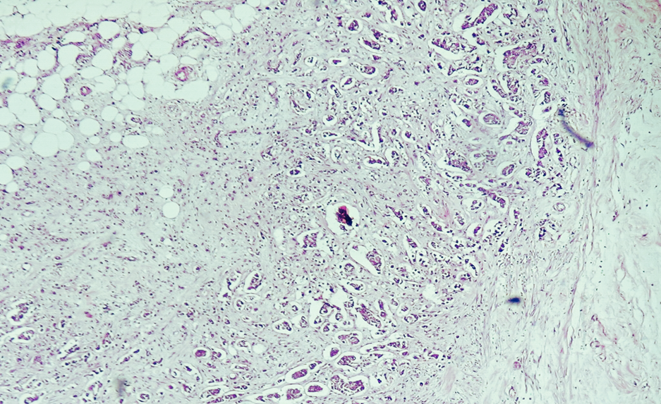
Figure 3: IDC- H & E Section shows nest like proliferation of malignant epithelial cells infiltrating stroma with stromal retraction around nest along with desmoplastic changes (40x, High Power View )(Original source)
Table: 5. Site of breast lesion(Original source)
|
Site of breast lesion |
Number |
Percentage |
|
Upper Outer Quadrant |
102 |
39.23 |
|
Lower Outer Quadrant |
64 |
24.61 |
|
Upper inner Quadrant |
58 |
22.3 |
|
Lower inner Quadrant |
11 |
4.23 |
|
Central quadrant |
8 |
3.07 |
|
Whole breast |
17 |
6.53 |
Table: 6. Incidence of metastases in malignant breast lesions.(Original source)
|
Total malignant cases |
Nodal metastasis |
Percentage |
|
32 |
17 |
53.13 |
As shown in table5, out of total 32 modified radical mastectomy specimens, 17 cases were showing nodal metastasis.
DISCUSSION:
Breast lesions are the leading cause prompting patients with breast concerns to seek surgical consultation. A thorough histopathological assessment of various types of breast biopsies is pivotal for accurate diagnosis, management, and follow-up of patients with diverse breast lesions8. The present study reveals that in the first decade of life breast lesions are extremely rare and only 1(0.38%)out of 260 presented with breast lesions in this age group. Lesion was fibroadenoma diagnosed at age of 9 years. The rarity of breast disease in the first decade of life is reported by Sudhakar et al and Amede Ogechi Nnorom also 10,12.
Incidence of benign breast lesions in the present study was 74.6% of all the breast lesions. The findings were comparable to the studies done by Sudhakar et al (82.9), Khanna R et al (61.36%)and Nikumbh et al (71.05%). 10,4,11This finding reinforces the general clinical principle that a breast mass in a woman under 40 is most likely non-cancerous.10,4. In our study, the most common age group for benign breast lesions was between 21-30 years (39.17%) which is similar to study done by Vilasini P et al , in which he found 38.7% benign breast lesion in age group of 21-30 years.3
In our study, fibroadenoma emerged as the most common benign breast lesion (n = 144/194; 74.3%), with the majority of patients presenting between the ages of 21-30 years (61 cases), followed by the age group of 11-20 years (57 cases). This aligns with global and Indian studies that identify fibroadenoma as the single most frequent breast tumor in this age group, typically peaking in the second and third decades of life 13,14. Additionally, fibrocystic disease represented the second most common benign breast lesion (n = 20/194; 10.30%), with the highest incidence in the age group of 31-40 years (14 cases), which is comparable with Vilasini P et al in which he has found 10% cases of fibrocystic disease majority of which fall into 30-39 years age group.3
This pattern was also observed by Sudhakar et al who reported a higher prevalence (60.3%) of fibroadenoma and identified fibrocystic disease as the second most common benign breast lesion (9.1%) in their study.10. In the present study the incidence of malignancy is only 12.3% and the peak incidence (n=26/32cases-81.25%)of malignancy is 31-40 years age group. Sudhakar et al also found similar results in their study; in which incidence of malignancy was 14.1% and most common age group was 31-40 years(n=38/48cases-79.16%)10
Invasive ductal carcinoma of no special type (IDC-NST) was the most common type of carcinoma encountered (n = 26/32; 81.25%) in our study. Ibrahim E H et al also found IDC NST to be most common(62%) malignant breast tumor in their study.8. Most common site was found to be outer upper quadrant in 102(39.23%) cases followed by 64(24.61%) in lower outer quadrant. Sharma A. K., et al in their study also found that most common location to be upper outer quadrant (28.75) followed by lower outer quadrant (22%).7.
REFERENCES:
1. Sahil SP, Anup AN, Suranjan SM. Study of Histopathological Patterns in Breast Lesions: A 5-Year Data Analysis. Journal of Contemporary Clinical Practice. 2025 Jul;11(7):963-968.
2. Goldblum JR, Lamps LW, McKenney JK, et al, editors. Rosai and Ackerman's Surgical Pathology. 11th ed. Amsterdam, Netherlands: Elsevier; 2018
3. Vilasini Patil, Archana Khandelwal, Kanchanmala G. Ghorpade. Histopathological Spectrum of Benign Breast Lesions, J Res Med Dent Sci, 2017, 5 (4): 9-14, DOI: 10.24896/jrmds.2017543
4. Nikumbh, D. B., Kanthikar, S. N., Suryawanshi, K. H., Jagtap, S. V., Dravid, N. V., & Gondane, S. R. (2016). Histopathological Spectrum of Unusual Breast Lesions: A Seven Year Retrospective Review. Indian Journal of Pathology and Oncology, 3(3), 456–462
5. Jebmh, & Com, & Kumbhakar, Dharmakanta. (2021). Histopathological Patterns of Breast Lesions -A Hospital-Based Study. Journal of Evidence Based Medicine and Healthcare. 8. 10.18410/jebmh/2021/111
6. Alawi et al. Egyptian Journal of Radiology and Nuclear Medicine (2020) 51:96 https://doi.org/10.1186/s43055-020-00209-y
7. Sharma A. K., Thaker, B., Hans, D., Sharma, S., Rasool, I., & Thake, K. S. (2022). Histomorphological spectrum of breast lesions in young females <25 years: 5 years retrospective study. NeuroQuantology, 20(22), 1726–1734. doi: 10.48047/nq.2022.20.22.NQ10162
8. Ibrahim E H, Ali T A, Sharbatti S, et al. (May 16, 2024) Histopathological Profile of Different Breast Lesions: A Single-Center Observational Study. Cureus 16(5): e60408. DOI 10.7759/cureus.60408
9. Simmons, P. S., Jayasinghe, Y. L., Wold, L. E., & Melton III, L. J. (2011). Breast carcinoma in young women. Obstetrics & Gynecology, 117(4), 932–936.
10. Sudhakar and Raja Pramila, World J Pharm Sci 2015; 3(12): 2490-2496
11. Khanna R, Khanna S, Chaturvedi S, Arya NC. Spectrum of breast disease in young females: a retrospective study of 1315 patients. Indian J Pathol Microbiol. 1998 Oct;41(4):397-401. PMID: 9866898.
12. Nnorom, Amede & Ugwa, Okezie. (2025). Histological pattern of breast lesions in children and adolescents: a ten-year review. Journal of Global Medicine. 5. e313. 10.51496/jogm.v5.313.
13. Zhong, L. M. et al. (2013). Comparison of clinicopathological characteristics of breast cancer in patients younger than 40 and older than 40 years. Chinese Journal of Cancer, 32(9): 485–492.
14. Tan, P. H. et al. (2009). The diagnosis of fibroadenoma in breast lesions in young women. Breast Cancer Research and Treatment, 118(3): 481-487.