A Prospective Analytical Observational Study Of Management Of Diaphyseal Fractures Of Femur By Closed Intra Medullary Interlocking Nail In Adults.
- K. Vamsi Krishna , Assistant Professor, Dept of Orthopaedics, Government Medical College Paderu, Alluri Sitharamaraju District, Andhra Pradesh -531024
- Ram Mohan Dora. H , Assistant Professor, Dept of Orthopaedics, Government Medical College Paderu, Alluri Sitharamaraju District, Andhra Pradesh -531024
Article Information:
Abstract:
Background: Fractures of femoral shaft are among the most common fractures that orthopaedist see. Advance in mechanization and acceleration of travel have been accompanied by an increase in the number and severity of fractures. Objective: To study the effectiveness of closed intramedullary nailing of diaphyseal fractures of femur in adults. Methods: This prospective study was done from January 2025 to June 2026 which comprises of 60 cases of fracture shaft of the femur in adults above 16 years of age, admitted in Department of Orthopedics , Government General Hospital / Government Medical college, Paderu, Alluri sita rama raju district , Andhra Pradesh who were treated with closed intramdullary interlocking nail. The patients were followed up for about 12 months. Result: Commonest age group affected was between 16 to 25 years and the mean age was 27.4 years. There was male preponderance (80%) in the present study. Right side (60%) was affected more commonly than left side (40%). Road traffic accident was the commonest mode of injury (83.34%)). Middle third (zone III and IV) was the commonest level of fracture (56.66%). in the present study. Most common pattern of fracture were comminuted and transverse fractures. 73.3% patients were admitted within 24 hours of injury. Surgery was done at an average of 6 days (ranging from 2 to 15 days). Average duration of hospital stay was 18.3 days (ranging from 11 to 33 days). Mean time of bony union was 16.5 weeks. Excellent to good results were seen in 86.6% of cases. Conclusion: The incidence of fracture femur due to road traffic accident is on the increase. Intramedullary nail is loaded less than a plate, making it less likely to fail in fatigue. Cortical osteopenia is not seen with nailing. Closed intramedullary interlocking nailing has now become the treatment of choice for closed diaphyseal fractures of femur in adults especially those with high comminution, long spiral and segmental fractures.
Keywords:
Article :
INTRODUCTION:
Since femur is the largest bone of the body and one of the principal load bearing bones in the lower extremity, femoral shaft fractures are associated with considerable mortality and morbidity whether they are caused by high or low energy trauma. Femoral shaft fractures resulting from high energy trauma are often associated with concomitant injury on internal organs1.
The art and science of fracture management has tremendously advanced over the years. From the use of external splints in the Hippocratic age to the recent sophisticated instrumentation, treatment of fractures has made an impact in the surgical field.
The intramedullary techniques that are in common use today are derived mainly from the work of Sir Gerhard Kuntscher2 in Germany and the Rush family in USA.Kuntscher is no doubt the father of reamed intramedullary nailing. At the beginning of intramedullary nail era, the conventional intramedullary nails by closed methods, gained wide popularity in transverse fracture of the middle third of the femur due to no disturbances of periosteal blood supply, fracture haematoma and rapid healing of fracture with lesser risk of complications like infection, non-union and shortening. But still it had a drawback in reference to comminuted fractures of the femur as in not providing rotational stability and axial length.
The advent of interlocking nailing has widened the spectrum of femoral shaft injuries that could be stabilized by intramedullary technique. Use of interlocking nailing minimizes the incidence of leg length discrepancies, rotation and angulation deformities. Intramedullary nail being close to centre of femur can tolerate bending and torsional loads better than plates and the locking mechanism provides less tensile and shear stress than plates. Intramedullary interlocking nail is a load sharing device. It is less loaded than plates causing less cortical osteopenia of stress shielding, which is a feature of the load bearing plates.
Closed nailing cause no damage to extraperiosteal soft-tissue and the biological environment around the fracture is least disturbed. Another important feature of the closed intramedullary interlocking nail is the chance of early ambulation of the patient which reduces the complications of prolonged bed ridden patient. The following study attempts to analyse the effectiveness of interlocking nail in management of diaphyseal fracture of femur so as to return the patient to functional pre-fracture state as soon as possible.
MATERIALS AND METHODS:
This prospective study was done from January 2025 to June 2026 which comprises of 60 cases of fracture shaft of the femur in adults above 16 years of age, admitted in Department of Orthopedics , Government General Hospital / Government Medical college, Paderu, Alluri sita rama raju district, Andhra Pradesh who were treated with closed intramdullary interlocking nail. The patients were followed up for about 12 months.
Inclusion criteria:
1. All closed diaphyseal femoral fractures above the age group of 16 years treated by closed intramedullary nailing during the study period.
2. Gustilo-Anderson type I open diaphyseal femoral fractures.
Exclusion criteria:
1) All cases of closed diaphyseal femoral fractures presenting under the age of 16 years.
2) Gustilo-anderson type II and III open diaphyseal femoral fractures
3) Pathological fractures.
This study comprises of 66 cases of fracture shaft of femur admitted during the study period and selection of these cases were at random. Among the 66 cases considered for closed intramedullary nailing, closed nailing was not possible in 4 cases due to soft tissue interposition and delayed fixation, so we opened the fracture site and hence these cases were excluded from the study. Among the 62 patients in whom closed intramedullary nailing was done, 2 cases were lost to follow up and were excluded from the study.
Out of 60 fractures included in the study, all were of traumatic origin, where in road traffic accident were responsible in 50 patients, fall from height in 5 patients and industrial accident in 2 patients. Patients in this study varied from 16yrs to 60 yrs of age, out of which 48 cases were males and 12 were females. There were 5 cases of Gustillo-Anderson type I open fracture which were treated with interlocking nail. Most of the cases were seen within 24 hours (44 cases), some were seen between 24-48 hours (12 cases) and remaining 4 cases after 48 hours. The interval between injury and operative intervention ranged from 2 to 15 days with an average of 6 days. The delay was due to poor medical condition or associated head injury.
As soon as the patients were admitted, he/she had undergone standard initial evaluation prior to surgery. A detailed history was taken with special reference to mode of injury, severity of trauma and duration.
Management:
General Examination: After taking history, a thorough clinical examination regarding the general condition of the patient was done. Any associated systemic diseases and associated injuries were ruled out clinically. Special attention was given to the cardiopulmonary status and vital signs.
Pre operative management includes:
1. Stabilization of the patient
2. Diagnosing other associated injuries
3. Timing of surgery
I. Stabilization of the patient: It includes
a. Emergency care
b. Immobilization of the affected limb
c. Investigations
a) Emergency care:
Special attention was given to cardiopulmonary status. Vital signs were monitored from time to time. The arterial status of the limb was under constant supervision, if the fracture was in the distal 1/3. Arterial blood gas levels were monitored for early diagnosis of fat embolism. Blood transfusion was done in few cases preoperatively because fracture shaft of the femur is associated with loss of 1200ml of blood into the soft tissues.
b.) Immobilization of the affected limb :
The affected limb was inmiobilized in Thomas's splint to prevent soft tissue damage, to decrease pain, to distract the fragments and for easy mobilization of the patient.
c) Investigations
Blood: Hb%, TC, DC, Blood urea, Serum creatinine, Blood grouping
· Urine: routine. Microscopy
· HIV,HBsAg
· ECG, Chest X-ray
· Roentgenograms includes
1. AP and lateral radiograph of thigh to diagnosis the and pattern of fracture
2. Pelvis with both hip joints AP view to rule out fracture neck of femur and posterior dislocation of hip.
3. Ipsilateral knee to rule out patellar fracture.
II. Diagnosing other associated injuries:
• Ipsilateral knee ligamentous injuries
• Ipsilateral fracture neck of the femur
• Injury to the abdomen
III. Timing of surgery10:
Timing of surgery was considered carefully for each patient. Definition are lacking for timing. If the fracture shaft of the femur is associated with some disastrous complications like ARDS, fat embolism, chest injury etc., then the surgery was postponed.
If the osteosynthesis is performed within 24 hours - primary fracture fixation.
If the osteosynthesis is performed within 2nd week - delayed primary fracture fixation.
If the osteosynthesis is performed after 2nd weeks - delayed fracture fixation
The routine procedures which were followed in patients who were posted for surgery includes:
· Improvement of general condition of the patients and making him fit for anaesthesia
· Skeletal traction through the upper tibia, if surgery is delayed more than a week
· Shaving and scrubbing of the entire limb on the day before surgery
· Intravenous generation cephalosporin on the day before surgery, and just before, incision
· One bottle of compatible blood was kept ready and used where necessary.
Pre-operative planning27,28:
The clinical results of closed interlocking nailing are largely a function of meticulous pre operative and intra operative planning. Pre operative anticipation of technical problem can save the surgeon considerable time in the operation room.
• At first, the geometry of the fracture dimensions of femur should be assessed accurately on plain radiography.
• Pre operative roentgenogram of the uninjured femur can be used to estimate proper nail diameter, expected amount of reaming required.
• Radiographic measurement of the contralateral femur with a nail of known length taped to the lateral surface of the thigh yields a more precise determination of normal femoral length. Alternatively, kuntscher ossimeter, which corrects the radiographic magnification, can be used.
A stock of standard interlocking nails of at least 4cm greater and lesser in length, and 2 mm lesser and greater in diameter than pre-operative measurement should be made available in the operating room.
Operative Protocol:
1) Patient positioning
2) Reduction of fracture under c-arm guidance
3) Incision and entry point location
4) Opening of the medullary canal
5) Passing of curved tip guide wire
6) Reaming
7) Exchange of curved tip guide wire with plain tip guide wire
8) Selection of the nail
9) Assembly for inserting the femoral nail
10) Nail insertion
11) Locking the nail
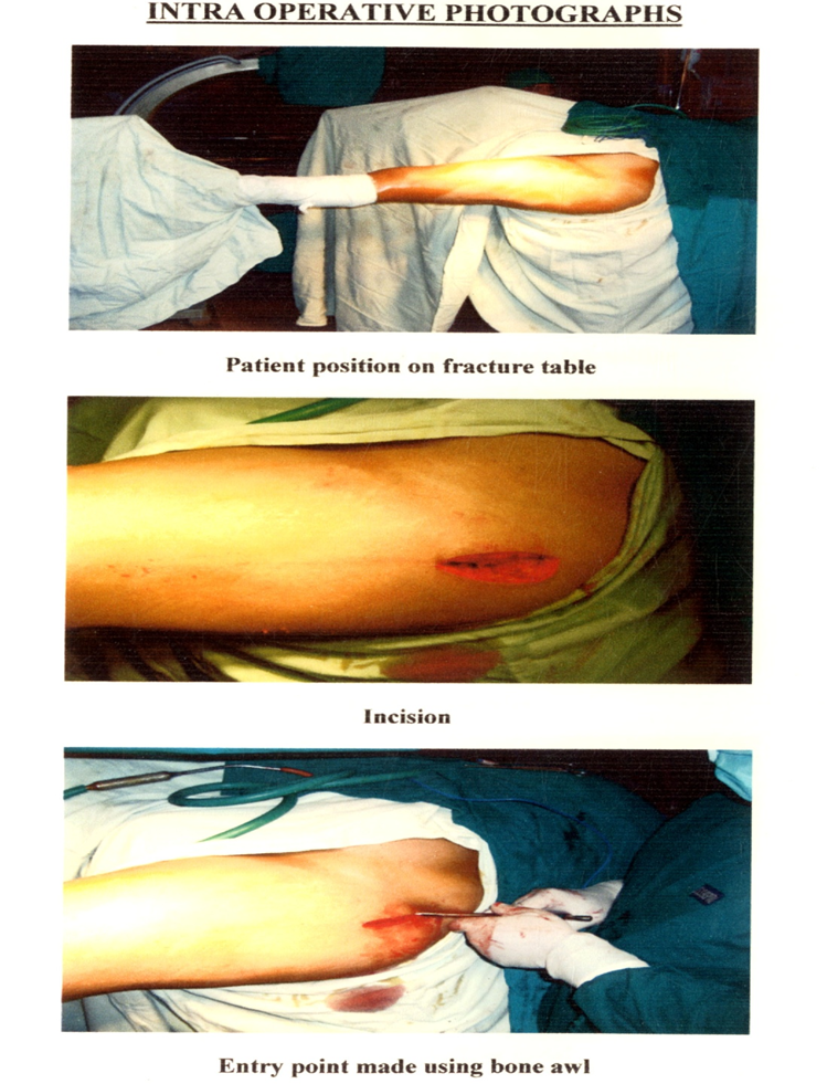
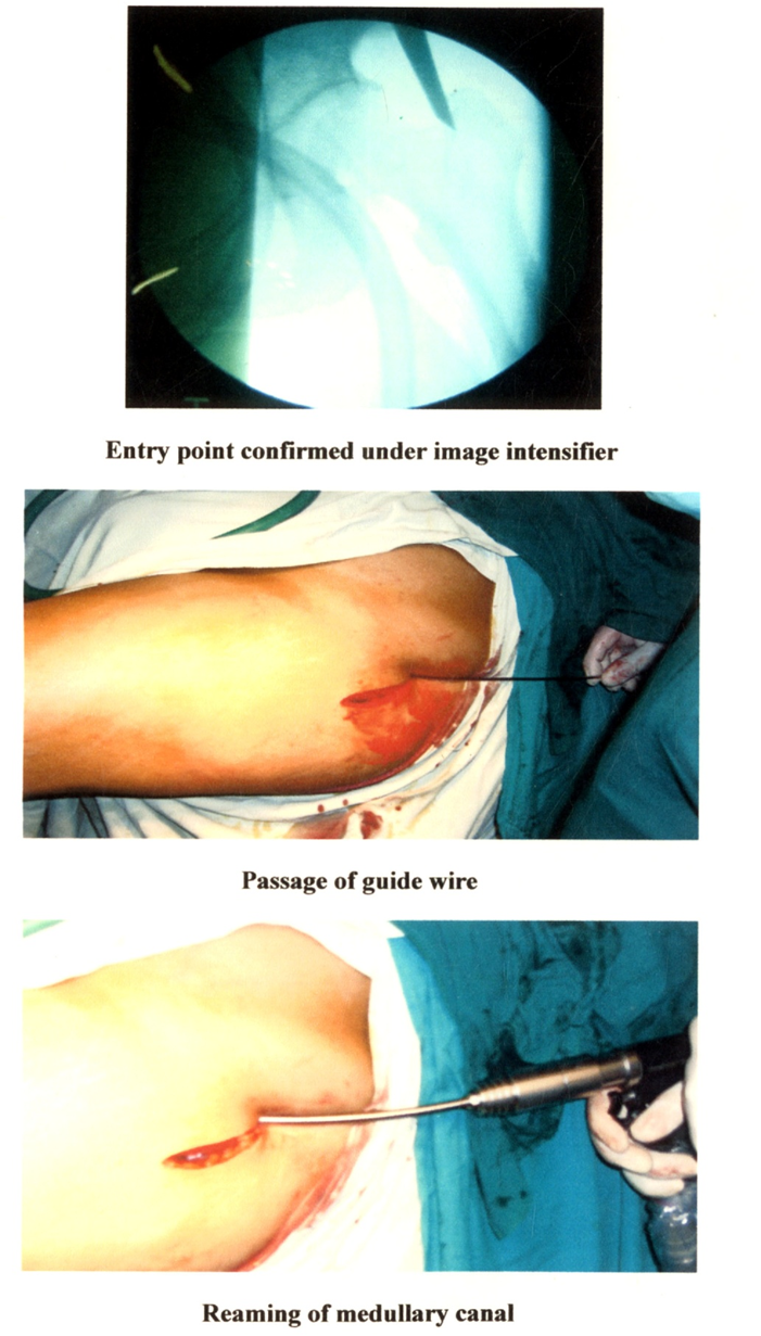
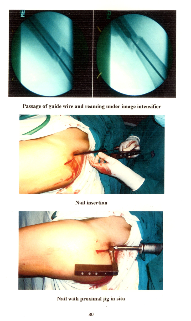
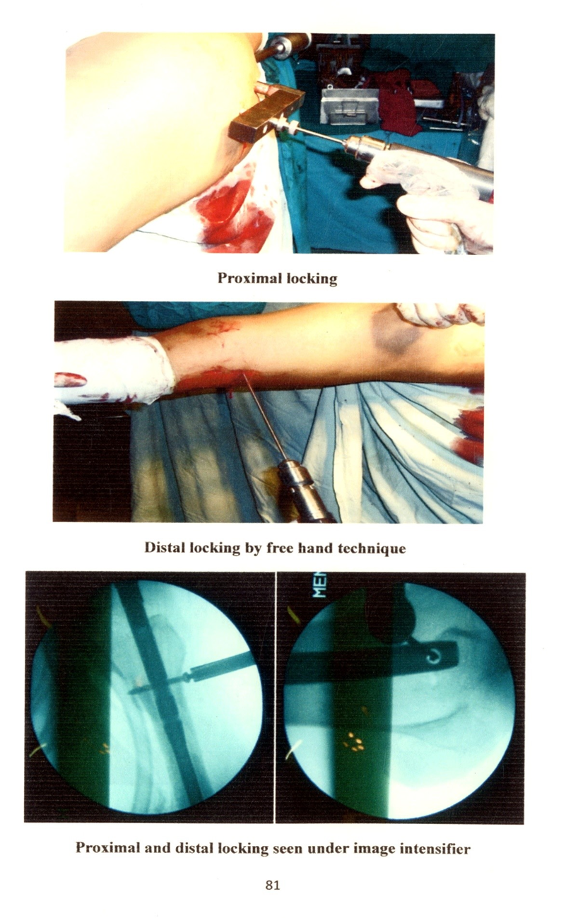
RESULTS :
The study consists of 60 cases of diaphyseal fractures of femur treated by closed intramedullary interlocking nailing during the study period January 2025 to June 2026.
The age ranges from 16-60 years with mean age of 20 yrs. Maximum incidence of fracture was between 16-25 yrs age group. Most of the patients were either manual labourers or agriculturists. 48 patients were males (80%) 12 patients were females (20%)
Table 1: Showing age and Sex distribution in present study
|
Age |
Males |
Females |
Total |
Percentage |
|
16-25 |
32 |
4 |
36 |
60 |
|
26-35 |
10 |
2 |
12 |
20 |
|
36-45 |
4 |
4 |
8 |
13.33 |
|
46-55 |
2 |
2 |
4 |
6.67 |
|
Total |
48 |
12 |
60 |
100 |
CC (Age*Sex) = 8.611;p=0.035 (HS), X2 (Age) = 41.33 ; p<.000 (HS), X2 (Sex) = 21.600; p <.0000 (HS)
Right sided fractures were seen in 36 patients (60%), of which 28 were males and 8 females. Left sided fractures were seen in 24 patients (40%), of which 20 were males and 4 were females.
Mode of Injury:50 patients (83.34%) sustained fracture because of road traffic accident, 8 patients (13.33%) from fall from height and 2 (3.33%) patient from industrial accident
Table 2: Showing Mode of injury in present study
|
Mode of Injury |
No. of cases |
Percentage |
|
Road traffic accident |
50 |
83.34 |
|
Fall from height |
8 |
13.33 |
|
Industrial accident |
2 |
3.33 |
X2 (Mode of injury) = 68.400; p < .000 (HS)
Type of fracture:
50 cases were closed fracture and there were 10 cases of Gustilo – Anderson type I open fracture [X2 (Type) = 26.667; p <000 (HS)]
Location of fracture:
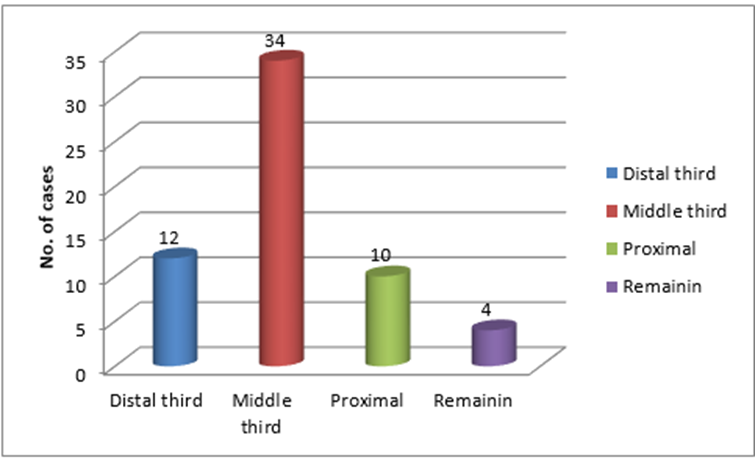
10 fractures were in proximal third (16.67%), 34 fractures were in middle third (56.66%) and 12 fractures were in lower third (20%). Remaining 4 fractures (6.67%) were segmental which involved more than two zones. [ X^ (Location)= 34.400; p < .000 (HS) ]
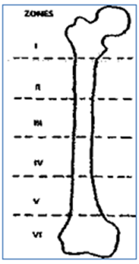
For the purpose of classification, the femur was divided into 6 segments from the tip of greater trochanter to the knee joint as shown in the figure.
Table 3: showing location of fracture in present study
|
Zone involved |
No. of cases |
Percentage |
|
Zone 1 |
0 |
0 |
|
Zone 2 |
5 |
16.67 |
|
Zone 3 |
7 |
23.33 |
|
Zone 4 |
10 |
33.33 |
|
Zone 5 |
6 |
20 |
|
Zone 6 |
0 |
0 |
Pattern of fracture:
Comminuted fractures was the most commonest type in our study and it constituted 36.66% followed by transverse fractures of 26.67%, spiral fractures of 16.67%, oblique fractures of 13.33% and segmental fractures of 6.67%.
Among comminuted fractures, according to Winquist and Hansen classification:
Type I - 1 Fracture (3.33%) Type II - 6 fractures (20%)
Type III - 3 Fractures (10%)) Type IV - 1 Fracture (3.33%)
Table 4: Showing pattern of fracture in present study
|
Pattern |
No of fractures |
Percentage |
|
Transverse |
16 |
26.67 |
|
Spiral |
10 |
16.67 |
|
Oblique |
8 |
13.33 |
|
Segmental |
4 |
6.67 |
|
Comminuted |
22 |
36.66 |
X2 (Pattern) = 16.667; p < .0002 (HS)
26 patients (43.33%) out of 30 had associated injuries and 56.67% of patients had only femoral shaft fractures. [X2 (Associated injuries) = 3.077; p < 0.799 (NS)]
Duration of time between injury and admission:
44 patients (73.3%) came to the hospital within 24 hrs after injury, while 12 patients (20.0%) came in between 24-48 hrs and 4 patients (6.7%) after 48 hrs.
Table 5: Showing duration of time between injury and admission
|
Duration |
No. of cases |
Percentage |
|
<24hrs |
44 |
73.3 |
|
24-48 hrs |
12 |
20.0 |
|
>48hrs |
4 |
6.7 |
X (Time between injury and admission) = 44.833 ; p < .000 (HS)
Statistics of surgery:
· The surgery was done on an average of 6 days (range 2-15 days) after patient's
admission. Delay was due to poor general condition of the patient or associated lead injury
· Spinal anaesthesia was used 54 patients and general anaesthesia in 6 patients.
· Average duration of surgery was around 120 mins. (Range – 90 to 150 min)
Duration of Hospital stay:
Duration of hospital stay ranged from 11 to 33 days. (Average -18.3 days)
Table 6: Showing the duration of hospital stay in present study
|
Duration |
No. of cases |
Percentage |
|
10-20 |
42 |
70 |
|
21-30 |
10 |
16.7 |
|
>30 |
8 |
13.3 |
X2 (Duration of hospital stay) = 36.400; p<.000 (HS)
Follow up period:
The patients were followed up in the outpatient department and were assessed clinically and radiologically at 6 weeks, 10 weeks, 12 weeks, 4 months and 6 months. Average duration of followup was 5.2 months with a range of 4 to 6 months.
Fracture Union:
Fracture union was defined as the period between operation ie., interlocking nailing and painless full weight bearing without external support and a radiologically healed fracture (trabecullation seen to cross the fracture in three of the four cortices).
Goessens stated that fracture gap should be filled with structured callus and not just the presence of bridging callus. The appearance of this bridging callus is an dynamisation is possible. Delayed union was considered when the radiographs failed to demostratate progressive consolidation between 16-24 weeks after nailing.
Un-united fracture was defined by the presence of pain on motion at the fracture site, without radiographic evidence of progressive healing 26 weeks following nailing.
Malunion was considered when there was shortening of limb length of more than 1cm, angulatiion in any plane of more than 70 and malrotation more than 100.
The mean time of fracture union in our study was 16.5 wks (range 13 to 28 wks)
Table 7: Showing fracture union in different age groups
|
Age |
No. of Patients |
Weeks |
|
16-25 |
18 |
14.83 |
|
26-35 |
6 |
15.66 |
|
36-45 |
4 |
22.25 |
|
46-55 |
2 |
22.5 |
Assessment of functional outcome:
Functional outcome was assessed based on modified klaus and klemm criteria. Excellent result were obtained in 40 cases (66,6%)), good result in 12 cases (20%), fair in 6 cases (10%) and poor in 2 case (3.4%).
DISCUSSION:
The treatment of fracture shaft of femur has evolved from the old conservative management to the most recent methods of interlocking nails, and continues to pose vexing problems for orthopaedic surgeons. This is the era of biological fixation. Interlocking nails have greatly expanded the indications for closed intramedullary nailing of femoral fractures. Early mobilization following fractures of the femoral shaft has been shown to have a significant advantage in terms of both joint mobility and economic impact, which are very well attained by the use of interlocking nails. The average age in the present series was found to be 27.4 years and the most common age group was between 16-25 years. This result is well comparable with other series. 10,33,14,15 Most of the patients in our study were males (80%) and females formed about 20% of study group. Male to female ratio was 4:1. This reflects the fact that under Indian circumstances males were more exposed to trauma. The involvement of right side (60%) was found to be more than left side (40%) which is in well accordance with other series. We did not encounter any bilateral fracture shaft femur. The most common mode of injury in our series is that by road traffic accident (83.34%) followed by fall from height (13.33%) and industrial accidents (3.33%). This is well comparable with other series. 10,34
We encountered fractures at all levels of shaft of the femur, commonest site being middle third (56.66%) of the femur, which is well comparable with Bjom.O.Thoressen Series. 33,34,15 Out of 60 fractures, majority were closed fractures (83.33%), while the remaining (16.67%) were open fractures. This is well in accordance with Robert A Winquist series. The most commonest pattern of fracture encountered in our study was comminuted fractures (36.66%), followed by transverse fractures (26.67%), spiral fractures (16.67%), oblique fractures (13.33%) and segmental fractures (6.67%). Associated injuries were seen in 43.33% of patients, where as 56.67% had only fracture femur, which is comparable with other series. 10,14,35 Associated injuries were treated accordingly during the management of femoral shaft fractures, if surgery was needed. In our study, 73.3% of patients came to hospital within 24 hours of injury, 20% between 24-48 hours and 6.7% after 48 hours due to delayed refen-al. We encountered one case of refracture of femur with broken intramedullary interlocking nail in situ due to fall from height, where exchange nailing was done.
All fractures were internally fixed with Indian version of AO femur nail . We came across 2 cases of HBsAg positive and one HIV positive patieiit where closed nailing was done successfully under universal precautions. All fractures were fixed using piriform fossa as entry point except in two cases where junction of anterior 1/3 and posterior 2/3 of greater trochanter was used as entry point. Medullary cavity of all the fractures were reamed using flexible reamers with an average diameter upto 11mm. The advantages of reaming are to create uniform medullary surface area, to promote osteogenesis, to place larger nails which can overcome bending or fatigue failure and overall to provide high union rates. When cortical chatter was encountered, reaming was stopped and the nail of size 1mm lesser the diameter of last reamer used, was inserted. Different size nails were used ranging from 8mm to 11mm (average: 10mm). In our study, we used lesser diameter nails compared to others as the skeletal framework of Indian population is on the lower side compared to the western population.
The average duration of surgery was 120 mins (range - 90 mins to 150 min). Prolonged duration of surgery was due to difficulty in fracture reduction and distal locking. In Robert A Winquist series, the average duration of surgery was 80 minutes and in Kenneth D Johnson series, it was between 120 to 160 minutes.
The average time taken for bony union in our study was 16.5 weeks. The delayed union was seen in 2 cases (6.6%), but one case united at the end of 7 months without any further surgical intervention and the remaining one case needed dynamization. The incidence of delayed union in our series is comparable with Robert.A. Winquist. 10,33, The shortening of the limb was seen in 2 cases (6.66%), of which 1 had 1 cm shortening and 1 cases had 0.5cm shortening. None of the patients had more than 1.0 cm shortening. The incidence is well comparable with other series. 33,14,1 The union rate in our series was 96.6%, which is comparable with other series10,33,14,15, 38 The high union rate in our study can be attributed to the preservation of fracture haematoma, using closed technique, early surgical intervention, early mobilization and early weight bearing.
Functional outcome in all cases were assessed using modified Idaus and klemm criteria32, at the end of final follow up.
The results can be compared with that of Kluas and Klemm series (1985) in which excellent results were obtained in 79.3%, good results in 17.7%, fair in 13.3% and poor in none
CONCLUSION:
The incidence of fracture femur due to road traffic accident is on the increase. Intramedullary nail is loaded less than a plate, making it less likely to fail in fatigue. Cortical osteopenia is not seen with nailing. Closed intramedullary interlocking nailing has now become the treatment of choice for closed diaphyseal fractures of femur in adults especially those with high comminution, long spiral and segmental fractures. Restoration of anatomical length and alignment of comminuted fractures is possible with interlocking nailing. Interlocking nail offers the added advantages of early joint mobilization, early muscle rehabilitation, shortened hospital stay and most importantly early return to work and prefracture state. Closed nailing constitutes a form of biological fixation resulting in high union rates. Interlocking nail reduces the incidence of complications like infection, malunion and non union. It achieves the strength for femoral shaft fracture in all three planes of loading-bending, compression and torsion. There is no significant change in union rate when compared with age, sex, level and pattern of fracture.
REFERENCES:
1. Adili A, Bhandari M, Lachowski RJ, Kwok DC, Dunlop RB. Organ injuries associated with femoral fractures: implication for severity of injury in motor vehicle collisions. Journal of trauma. 1999;46:386-391
2. Gerhard EG Kuntscher. The kuntscher's method of intramedullary fixation. Journal of bone and joint surgery. Jan 1958;40-A
3. Rockwood and greens, adult orthopaedics, ed 3,vol.2.1653
4. Farrell J. Treatment of non unions of long bones. Journal of bone and joint surgery. 1956;30-A:506
5. Dana M Street. The evolution of intramedullary nailing, intramedullary fixation of long bones. 1-16
6. Street DM. IM nailing of the femur- in AAOS instructional course lectures. Ann Arbor. 1958:15
7. David seligson MD. Concepts in intramedullary nailing. 1985 edition
8. D Kay Clawson, Robert F Smith and SigvardT Hansen. Closed intramedullary nailing of femur. Journal of bone and surgery. 1971;53-A(9):681-692
9. Grosse A, I Kempf and Beck G. Closed locked intramedullary nailing: its application to comminuted fractures of femur. Journal of bone and joint surgery. 1985;67-A(5):70-71
10. Robert A Winquist. Comminuted fractures of femoral shaft treated by IM nailing.
Orthopaedic clinics of north America. July 1980; 11(3)
11. Zimmerman KW, Klassen HJ. Mechanical failure of intramedullary nails after fracture union. Joumal of bone and surgery. 1983;65-B:274-284
12. RA Winquist, ST Hansen and DK Clawson. Closed intramedullary nailing of femoral fractures. A report of five hundred and twenty cases-1984. Joumal of bone and joint surgery. 2001;83-A(12): 1912
13. Charles taylor J. Fractures of lower extremity. Chapter 23 in Campbells operative orthopaedics, Crenshaw AH, St. Louis Mosby year book, 1992:758-894
14. Kenneth D Johnson, MD. Comminuted femoral shaft fractures. Orthopaedics clinics of north America. Jan 1987;18(1)
15. Bjom O thoresen et al. Interlockng intramedullary nailing in femoral shaft fractures- a report of forty eight cases. Joumal of bone and joint surgery. 1985;67-A(9): 1313-20
16. Donald A wiss et al. Comminuted and rotationally unstable fractures of the femur treated with an interlocking nail. Clinical orthopaedics. Nov 1986;212:35-47
17. Wiss DA, William W brien and William B stenson. Interlocking nailing for treatment of segmental fracture of the femur. Joumal of bone and joint surgery. 1980;72-A:724-728
18. Bmmback R, Elision mollisgon H and Molligon D. Pudendal nerve palsy complicating intramedullary nailing of the femur. Joumal of bone and joint surgery. 1992;74-A: 1450-1455
19. Beaty JH et al. Interlocking IM nailing of femoral shaft fractures in adolescents. Joumal of paediatric orthopaedics. 1994; 14(2): 17883
20. Wolinsky PR, McCarty E, Shyr Y, Johnson K . Reamed intramedullary nailing of the femur : 551 cases. Journal of trauma. 1999; 46(3):392-399
21. Arazi M, Ogun TC, Ortar MN, Memik R, Kutler A. Early weight bearing after statically locked reamed intramedullary nailing of comminuted femoral fractures : is it a safe procedure?. Joumal of trauma. 2001;50(4):711-716
22. Mohammed Gharehdaghi, Hasan rahimi, Mahmood bahari, Javad afzali." A prospective • study of closed and open reamed intramedullary nailing of 136 femoral shaft fractures in adults Joumal of research in medical sciences. 2007; 12(1): 16-20
23. Fadero PE, Alabi S, Adebule GT, Odunubi O, Yinusa KL; Eyesan SU, Obianyar OD. Locked intramedullary nailing for the treatment of femoral shaft fractures. Niger J Med. 2008; 17(2): 168-72
24. Johnson KD et al. Biomechanical performance of locked IM nail system. Clinical orthopaedics. 1986;206:151-161
25. Kinsat et al. Biomecha.nics of interlocking nail. Arch Orthop Trauma Surg. 1990; 109(4): 197-204
26. Koustantine, Steriopoulos et al. Architecture of femoral intramedullary canal and working length of intramedullary nailing. Acta Orthop Scad. 1997;88:123-26 .
27. A.Paige Whittle. Fracture of the lower extremity, 2nd ed. In: Campbell's Operative Orthpaedics. Canale ST, Beaty JH. Philadelphia: Mosby PubUshers; 2008:3190-3217.
28. Sean E Nork. Fractures of shaft of the femur. Text book of fractures in adults, Rockwood and Green's, 7 th ed. Philadelphia, USA: Lippincott Williams and Wilkins 2010 .50:1655-1714
29. Muller M, AUgower M, Schneider R, Willenegger H. Manual of internal fixation. 3rd edition: 138-139
30. Frederic W Rhinelander. The vascular response of bone to internal fixation: in course lect. 1973;22:161-87
31. SB Kessler et al. The effects of reaming and intramedullary nailing on fracture healing. Clinical orthopaedics. Nov 1986;212:18-25
32. Klaus WK and Martin B. Interlocking nailing of complex fractures of femur and tibia. Clinical orthopaedics and related research. 1986;212:89-100