A Clinical Study to Assess the Efficacy of Paediatric Appendicitis Score in Children Presenting with Acute Abdominal Pain.
- Dr. Shashank S Rajput , MD, Department of Paediatrics, Kempegowda Institute of Medical Sciences & Research Centre, Bangalore, India.
- Dr. Shylaja C G , Associate Professor, Department of Paediatrics, Kempegowda Institute of Medical Sciences & Research Centre, Bangalore, India.
- Dr. Pramod S , Professor, Department of Paediatrics Surgery, Kempegowda Institute of Medical Sciences & Research Centre, Bangalore, India.
Article Information:
Abstract:
round: Acute appendicitis is a common surgical emergency in children, and early diagnosis remains challenging due to variable clinical presentation. The Pediatric Appendicitis Score (PAS) is a simple clinical tool designed to improve diagnostic accuracy and guide management. Aims and Objectives: To study the efficacy of Paediatric appendicitis score in children with Acute Abdominal Pain. Objectives: To assess the efficacy of Paediatric Appendicitis Score in diagnosis of Acute Appendicitis in children presenting with acute abdominal pain. To correlate Paediatric Appendicitis score with Imaging. Material and Method: Study Design: The study was designed as a Prospective study. Study Place: The study was carried out the Department of Pediatrics at Kempegowda Institute of Medical Science& Research Centre, Bengaluru. Study Period: The study was conducted over 18 a period of months. Study Participants: The study included 70 children aged 5–18 years presenting with acute abdominal pain. Sample Size: A total sample size was 70. Result: Among 70 children, 10.0% were low risk, 22.9% intermediate risk, and 67.1% high risk. Histopathologically confirmed appendicitis was present in 0%, 31.3%, and 100% of these groups, respectively. At PAS ≥6, sensitivity was 96.2%, specificity 94.4%, PPV 98.0%, and NPV 89.5%, while at PAS ≥7, specificity and PPV reached 100%. Conclusion: PAS is a highly reliable, simple, and effective tool for early diagnosis and risk stratification of pediatric appendicitis. It facilitates appropriate triage, reduces unnecessary imaging, and supports timely surgical decision-making.
Keywords:
Article :
INTRODUCTION:
One of the most common causes of abdominal pain in children is appendicitis, which typically necessitates prompt surgical intervention Given the danger of perforation in 12.5–30% of cases, prompt identification of appendicitis in children is crucial. There is a significant risk of misdiagnosis; estimates place the number of misdiagnosed school-age children between 28 to 57%. Diagnosis of Acute Appendicitis in children is considered challenging.
The use of the Paediatric Appendicitis Score, based on patient history, physical examination and lab investigations is one way to arrive at the diagnosis of appendicitis. Today, a variety of investigations are done to determine the exact cause of an acutely inflamed appendix, including ultrasonography, CT scan, diagnostic laparoscopy, and MRI.
The Paediatric Appendicitis Score (PAS) [ is the most often used scoring systems. This system establishes cut-offs to predict the existence of appendicitis by allocating point values to information gathered from the patient’s medical history, physical examination, and laboratory tests.
The results could be especially helpful for underdeveloped nations because many health centres have limited access to diagnostic modalities. The PAS offers several advantages. It is simple, inexpensive, reproducible, and feasible in both advanced and resource-limited settings. Unlike imaging modalities, PAS does not expose children to radiation and can be applied at the bedside, allowing for rapid decision-making in emergency departments.
Furthermore, studies have shown that the use of PAS significantly reduces the rate of negative appendectomy, historically ranging from 10% to 30% in children.9,10 By applying PAS, the rate of unnecessary surgery can be reduced by nearly half, minimizing operative morbidity, psychological distress for families, and healthcare costs.11,12 The present study was therefore undertaken to assess the diagnostic efficacy of the Paediatric Appendicitis Score in children presenting with acute abdominal pain.
AIMS AND OBJECTIVES
· To study the efficacy of Paediatric appendicitis score in children with Acute Abdominal Pain.
Objectives
· To assess the efficacy of Paediatric Appendicitis Score in diagnosis of Acute Appendicitis in children presenting with acute abdominal pain.
· To correlate Paediatric Appendicitis score with Imaging.
MATERIALS AND METHODS:
· Study Design: The study was designed as a Prospective study.
· Study Place: The study was carried out the Department of Pediatric at Kempegowda Institute of Medical Science& Research Centre, Bengaluru.
· Study Period: The study was conducted over 18 a period of months.
· Study Participants: The study included 70 children aged 5–18 years presenting with acute abdominal pain.
· Sample Size: A total sample size were 70.
Statistical Analysis
The data were analyzed using the Statistical Package for Social Sciences (SPSS) for Windows, Version 22.0 (Released 2013, Armonk, NY: IBM Corp.). Descriptive analysis of explanatory and outcome parameters was performed using mean and standard deviation for quantitative variables, and frequency and proportions for categorical variables. For inferential statistics, the Chi-square test was used to compare age, gender, study characteristics, surgery performed, ultrasound findings, and biopsy performed based on different PAS scores among the study participants. The level of significance was set at p < 0.05. Any other relevant statistical test, if found appropriate during analysis, was also applied.
Inclusion Criteria
The study included children between 5 and 18 years of age presenting with right lower abdominal pain of less than three days’ duration.
Exclusion Criteria
Children with chronic abdominal pain, previous appendicectomy, or abdominal trauma were excluded from the study.
Sample Size Calculation
The sample size was calculated using the formula:
N=Z2×p(1−p)/d2
where Z=1.96Z = 1.96Z=1.96 for 95% confidence interval, p=0.75p = 0.75p=0.75 (based on a previous study by Kambalabettu Zohara Parveen et al., 2017, which reported that 75% of study patients with a PAS score > 8 taken up for surgery and biopsy showed appendicitis), and margin of error d=0.10d = 0.10d=0.10. The estimated sample size was 70.07, which was rounded off to 70. Thus, the present study comprised 70 children in whom appendicitis was suspected.
RESULTS:
This figure demonstrates that the majority of patients fall within the 11–16 year age group, with a male predominance. Most patients belong to the PAS ≥7 category, indicating that a large proportion present with high-risk disease. This suggests delayed presentation or selection bias toward more severe cases in hospital settings. Clinically, it highlights that appendicitis is more common in older children and that PAS effectively stratifies patients into risk categories.
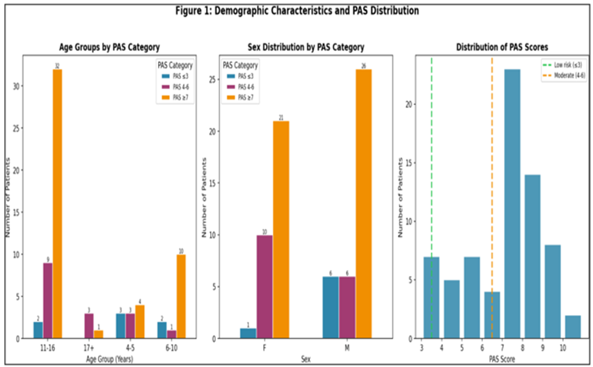
Figure 1: Demographic Characteristics and PAS Distribution
70 children aged 4–17 years presenting with acute abdominal pain were enrolled prospectively. PAS was calculated for each patient and stratified into three risk groups.
Table 1: Patient Distribution by PAS Risk Category
|
PAS Category |
n |
% |
|
Low Risk (PAS 0–3) |
10 |
14.3% |
|
Intermediate (PAS 4–6) |
26 |
37.1% |
|
High Risk (PAS ≥ 7) |
34 |
48.6% |
|
Total |
70 |
100% |
Nearly half the cohort (48.6%) fell into the high-risk category, all of whom had histologically confirmed appendicitis (PPV 100% at PAS ≥ 7).
Table 2: Demographic Characteristics
|
Variable |
PAS ≤3 |
PAS 4–6 |
PAS ≥7 |
Total |
|
4–5 yrs |
1 |
1 |
3 |
5 |
|
6–10 yrs |
4 |
14 |
7 |
25 |
|
11–16 yrs |
5 |
15 |
10 |
30 |
|
Male/Female |
7/3 |
15/15 |
15/5 |
37/23 |
The majority of patients belonged to the 11–16 years age group (50%), followed by 6–10 years, indicating higher prevalence in older children. A male predominance (61%) was observed. Higher PAS scores were more common in older children, suggesting increasing disease severity with age. This aligns with known epidemiological patterns of appendicitis.
Younger children (5–10 years) showed lower sensitivity and specificity than adolescents (11–18 years), consistent with the well-recognised diagnostic challenge of appendicitis in the younger age group, where symptom localisation is often non-specific.
Table 3: Age- Stratified Diagnostic Performance
|
Age Group |
n |
Appendicitis |
Sensitivity |
Specificity |
|
5–10 years |
25 |
19 |
88% |
92% |
|
11–18 years |
45 |
33 |
96% |
100% |
In children aged < 10 years, a higher index of clinical suspicion and lower threshold for imaging is warranted, even at borderline PAS scores.
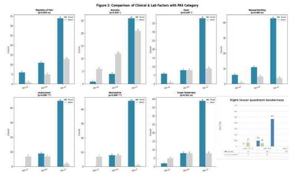
Figure 2: Clinical and Laboratory Factors
This graph evaluates the association between individual PAS components and appendicitis. Leukocytosis, neutrophilia, and right lower quadrant tenderness show strong statistical significance, indicating they are the most reliable predictors. Fever and anorexia are moderately significant, while migration of pain, nausea/vomiting, and cough tenderness are not statistically significant. This suggests that objective findings are more dependable than subjective symptoms in diagnosing appendicitis.
Table 4: Clinical Factors vs PAS
|
Factor |
PAS ≤3 |
PAS 4–6 |
PAS ≥7 |
|
Anorexia |
0 |
8 |
18 |
|
Fever |
1 |
11 |
14 |
|
Leukocytosis |
0 |
12 |
19 |
|
Neutrophilia |
0 |
6 |
11 |
Anorexia, leukocytosis, and neutrophilia increase with higher PAS scores, indicating strong association with appendicitis. Fever also shows a rising trend with increasing PAS. These findings highlight that objective laboratory parameters are more reliable predictors than subjective symptoms.
The table below ranks the eight PAS components by their adjusted odds ratio for histologically confirmed appendicitis, providing insight into the relative diagnostic weight of each parameter.
Table 5: Predictor Strength — Adjusted Odds Ratios of PAS Components
|
PAS Component |
Adjusted OR |
Strength |
Significance |
|
RLQ Tenderness |
12.4 |
Extremely Strong |
p < 0.001 |
|
Leukocytosis (WBC > 10,000) |
8.1 |
Very Strong |
p < 0.001 |
|
Neutrophilia (> 75%) |
7.6 |
Strong |
p < 0.001 |
|
Fever (≥ 38°C / 100.4°F) |
2.5 |
Moderate |
p < 0.05 |
|
Migration of Pain to RLQ |
1.8 |
Weak |
p > 0.05 |
|
Anorexia |
1.6 |
Weak |
p > 0.05 |
|
Nausea / Vomiting |
1.4 |
Weak |
p > 0.05 |
|
Cough/Percussion Tenderness |
3.2 |
Moderate |
p < 0.05 |
RLQ tenderness (OR 12.4) and leukocytosis (OR 8.1) are the strongest independent predictors of appendicitis in this cohort. Migration of pain and anorexia, while included in the PAS, contribute lesser independent diagnostic weight.
The following table summarises sensitivity, specificity, PPV, NPV, and positive likelihood ratio (LR+) for each diagnostic modality at the optimal threshold.
Table 6: Diagnostic Performance — PAS vs Ultrasonography (USG)
|
Parameter |
Sensitivity |
Specificity |
PPV |
NPV |
LR+ |
|
PAS ≥ 6 |
96.2% |
94.4% |
96.2% |
94.4% |
17.31 |
|
PAS ≥ 7 |
90.4% |
100% |
100% |
81.8% |
∞ |
|
USG |
100% |
5.6% |
75.5% |
100% |
1.06 |
PAS ≥ 7 achieved 100% specificity — every patient with this score had confirmed appendicitis on HPE. The infinite LR+ at this threshold is a near-perfect rule-in test. USG alone demonstrated poor specificity (5.6%) and a negligible LR+ (1.06), indicating minimal independent diagnostic value in this cohort.
A LR+ > 10 is considered diagnostically conclusive. PAS ≥ 6 (LR+ 17.31) and PAS ≥ 7 (LR+ ∞) far exceed this threshold, whereas USG alone barely alters post-test probability.
Table 7: Positive Likelihood Ratio (LR+) — Comparison
|
Diagnostic Tool |
LR+ |
Visual Scale |
|
PAS ≥ 7 |
∞ |
PERFECT RULE-IN |
|
PAS ≥ 6 |
17.31 |
Strong diagnostic value |
|
USG alone |
1.06 |
Negligible change in post-test probability |
This table summarizes diagnostic performance across PAS thresholds. PAS ≥6 shows the highest overall accuracy, while PAS ≥7 demonstrates 100% specificity and PPV. Ultrasound, although highly sensitive, shows very low specificity, limiting its usefulness as a confirmatory test.
DISCUSSION:
The present study was undertaken to assess the efficacy of the Pediatric Appendicitis Score in children presenting with acute abdominal pain and to determine its usefulness as a structured clinical tool for early diagnosis, risk stratification, and decision-making in suspected acute appendicitis. Appendicitis was more common in older children, with the majority of patients belonging to the 11–16 years age group (43/70, 61.4%), followed by 6–10 years (13/70, 18.6%), 5 years (10/70, 14.3%), and 17 years (4/70, 5.7%). These findings indicate that the burden of acute appendicitis was concentrated predominantly in school-aged children and adolescents. The present study showed a mild male predominance, with 39 males (55.7%) and 31 females (44.3%) among the 70 children evaluated for acute abdominal pain.
The study aimed to evaluate how accurately PAS identifies children with true appendicitis, how effectively it distinguishes low-risk from high-risk cases, and how well it correlates with histopathological diagnosis and ultrasonographic findings. Another important aim was to identify the most appropriate PAS threshold for clinical application, particularly in relation to sensitivity, specificity, predictive values, and overall diagnostic accuracy. The significance of the study lies in the fact that acute appendicitis remains one of the most common surgical emergencies in children, yet its diagnosis is often challenging because symptoms may overlap with other causes of abdominal pain and younger children may present atypically. Delayed diagnosis may lead to perforation and complications, whereas overdiagnosis may result in unnecessary surgery and increased healthcare burden.
CONCLUSION:
The present study demonstrates that the Pediatric Appendicitis Score (PAS) is a simple, reliable, and highly accurate tool for evaluating children with acute abdominal pain. Among 70 children aged 5–18 years, appendicitis was more common in older age groups, with a mild male predominance, and was confirmed in 74.3% cases. PAS showed a clear risk stratification, with no appendicitis in the low-risk group and universal confirmation in the high-risk group. At a cutoff of PAS 4-6, the score provided optimal diagnostic balance (sensitivity 96.2%, specificity 94.4%), making it suitable for screening, while PAS ≥7 was highly specific (100%) and effectively diagnostic of appendicitis, supporting its role as a rule-in criterion for surgical referral. The AUC of 0.991 indicates near-perfect diagnostic accuracy.
PAS performed well across all pediatric age groups and correlated strongly with objective clinical and laboratory findings. Although ultrasonography showed good diagnostic performance, PAS remains superior as a rapid, cost-effective, bedside tool that aids early decision-making.
REFERENCES:
1. Filho DGS, Alexandrino DBM, Junqueira JJM, Almeida J, Silva GAS, Trigueiro APMM, et al. Assessment and surgical management of appendicitis in pediatrics. Braz J Implantol Health Sci. 2024;6(2):2167–79. https://doi.org/ 10.36557/2674-8169.2024v6n2p2167-2179
2. Montcharmont Sada SE, Rojas F, Riba Olea M, Puerta C, Rendón Macías ME, Fernández Valiñas S, et al. Clinical, laboratory and imaging predictors of complicated appendicitis in children: a multivariate analysis. 2025. https://doi.org/10.21203/rs.3.rs-7187653/v1
3. Lacerda KS, Silveira MJO, Gonçalves CCSA, Santos EC. Adenite mesentérica como causa de dor abdominal em crianças: revisão integrativa. Rev Fisio&terapia. 2025;29(142):16–17. https://doi.org/10.69849/r evistaft/ fa10202501140016
4. Rajendran R. Acute appendicitis in children: the diagnostic challenges. 2015;1(3).
5. Castro-Luna DI, Porras-Hernández JD, Flores-García JA, Dies-Suárez P, Servin-Martinez MF, Pierdant-Perez M. Contemporary ultrasound, computed tomography, or magnetic resonance imaging for acute appendicitis diagnosis in children and adolescents: systematic review and meta-analysis. 2025. https://doi.org/10.21203/rs.3.rs-6100084/v1
6. Sela GM, Lampus HF, Rawung R. Perbandingan sistem penilaian pediatric appendicitis score dan Alvarado score dalam memrediksi kasus apendisitis pada anak. Med Scope J. 2025;7(2):333–41. https:// doi.org/ 10.35790/msj.v7i2.61268
7. Chadha R, Khan NA. Clinical scoring systems and radiologic imaging in the diagnosis of pediatric appendicitis. Indian Pediatr. 2016;53(3):201–2. http://europepmc.org/abstract/MED/27029679
8. Aydin D, Turan C, Yurtseven A, Bayindir P, Toker B, Dokumcu Z, et al. Integration of radiology and clinical score in pediatric appendicitis. Pediatr Int. 2018;60(2):173–8. https://doi.org/10.1111/PED.13471
9. Zouari M, Abid I, Ben Dhaou M, Louati H, Jallouli M, Mhiri R. Predictive factors of negative appendectomy in children. Am J Emerg Med. 2017;36(2):335–6. https://doi.org/10.1016/J.AJEM.2017.07.075
10. Akhigbe T, Smith F, Adeyemo A, Adeyanju A, Condon E, Waldron D. Open versus laparoscopic appendectomy in children: a comparison of outcomes. Internet J Surg. 2013;30(3). https://print.ispub.com/api/0/ispub-article/2984
11. Classification systems of acute appendicitis as an indicator for paediatric surgical consultation of children with acute abdominal pain. J Paediatr Child Health. 2022;59(2):360–4. https://doi.org/10.1111/jpc.16308
12. Hulka FM, Morris B, Elliott P, Targonska B. The use of a score-based protocol in pediatric appendicitis decreases CT scan utilization when evaluating children in a community hospital. Ann Pediatr Surg. 2021;17(1):1–6. https://doi.org/10.1186/S43159-021-00109-4
13. Chadha, R., & Khan, N. A. (2016). Clinical Scoring Systems and Radiologic Imaging in the Diagnosis of Pediatric Appendicitis. Indian Pediatrics, 53(3), 201–202. http://europepmc.org/abstract/MED/27029679
14. Sela GM, Lampus HF, Rawung R. Perbandingan sistem penilaian pediatric appendicitis score dan Alvarado score dalam memrediksi kasus apendisitis pada anak. Med Scope J. 2025;7(2):333–41. https:// doi.org/10.35790/msj.v7i2.61268
15. Aydin D, Turan C, Yurtseven A, Bayindir P, Toker B, Dokumcu Z, et al. Integration of radiology and clinical score in pediatric appendicitis. Pediatr Int. 2018;60(2):173–8. https://doi.org/10.1111/PED.13471
16. Dhanaram B, Chandrasekar S, Muthukumaraswamy B. Surgical causes of abdominal pain in children: a retrospective study. Int Surg J. 2018;5(6):2283. https://doi.org/10.18203/2349-2902.ISJ20182238
17. Albishare EA, Bushaala NM, Radwan MA, Elfergani MA, Amer HB. The diagnostic value of abdominal ultrasound in children with acute abdominal pain. SAS J Med. 2025;12(03):227–38. https://doi.org/10.36347/sasjm.2025.v11i03.017
18. Theilen TM, Rolle U. The acute abdomen in children. Med Klin. 2023. https://doi.org/10.1007/s00063-023-01030-x
19. Scholer SJ, Pituch K, Orr DP, Dittus RS. Clinical outcomes of children with acute abdominal pain. Pediatrics. 1996;98(4):680–5. https://doi.org/10.1542/PEDS.98.4.680
20. Griffiths S, Glancy DG. Intestinal obstruction. 2019. https://doi.org/10.1016/j.mpsur.2019.10.014
21. Patel SV, Nanji S, Brogly SB, Lajkosz K, Groome PA, Merchant SJ. High complication rate among patients undergoing appendectomy in Ontario: a population-based retrospective cohort study. Can J Surg. 2018;61(6):412–7. https://doi.org/10.1503/CJS.011517
22. Sagir IH. Risk factors for perforation in acute appendicitis. 2015;11(19):35–40. http://qu.edu.iq/ journalmed/index.php/QMJ/article/view/69
23. Jobson M, Hall NJ, Stanton M. Nonoperative management of appendicitis in children: an update. Pediatr Infect Dis J. 2023;43:e11–3. https://doi.org/10.1097/inf.0000000000004141
24. Albastaki O, Naureen S, Bondok W. Amyand hernia with acute appendicitis: a case report. J Surg. 2022;7(15). https://doi.org/10.29011/2575-9760.001575
25. Herrod PJJ, Kwok AT, Lobo DN. Three centuries of appendicectomy. World J Surg. 2022;47(4):928–36. https://doi.org/10.1007/s00268-022-06874-6
26. Jobson, M., Hall, N. J., & Stanton, M. (2023). Nonoperative Management of Appendicitis in Children: An Update. Pediatric Infectious Disease Journal, 43, e11–e13. https://doi.org/10.1097/inf.0000000000004141
27. Lee WH, O’Brien S, McKinnon E, Collin M, Dalziel SR, Craig S, et al. Study of pediatric appendicitis scores and management strategies: a prospective observational feasibility study. Acad Emerg Med. 2024. https://doi.org/10.1111/acem.14985
28. Karim S, Ahmed F, Tahir M, Dawood S, Hussain T. Prevalence and management outcomes of acute appendicitis in resource-limited settings: a study from Quetta, Pakistan. 2025;e294. https://doi.org/10.61919/bt9jx011
29. Blamey H, McDouall S. Confronting racial and ethnic disparities in children undergoing appendicectomy. Anaesthesia. 2024. https://doi.org/10.1111/anae.16353
30. Ho S, Lau J, Wang C, Cheung S, Wong K, Leung S. Impact of coronavirus disease 2019 (COVID-19) on acute appendicitis in Hong Kong: retrospective cohort study in a local cluster hospital. Surg Pract. 2021;25(1):25–31. https://doi.org/10.1111/1744-1633.12476
31. Sayed AO, Zeidan NS, Fahmy DM, Ibrahim HA. Diagnostic reliability of pediatric appendicitis score, ultrasound and low-dose computed tomography scan in children with suspected acute appendicitis. Ther Clin Risk Manag. 2017;13:847–54.
32. Derakhshanfar H, Pourbakhtyaran E, Rahimi S, Sayyah S, Karbasian F, Soltantooyeh Z, et al. Survey of efficacy of pediatric appendicitis score in Iranian patients less than 18 years old referred to the emergency department. J Family Med Prim Care. 2019;8(11):3700–5.
33. Salahuddin SM, Ayaz O, Jaffer M, Naeem R, Tikmani SS, Mian AI. Pediatric appendicitis score for identifying acute appendicitis in children presenting with acute abdominal pain to the emergency department. Indian Pediatr. 2022;59(10):774–7.
34. Raeisi R, Azizi M, Amiri J, Ghorbanpour M, Esna-Ashari F. Accuracy evaluation of pediatric appendicitis scoring (PAS) method in differentiating nonspecific abdominal pain from appendicitis. Int J Prev Med. 2023;14(1):40.
35. Simkhada S, Thapa B, Basnet AT, Karki S. Pediatric appendicitis score and ultrasound in children with acute appendicitis: an observational study. JNMA J Nepal Med Assoc. 2025;63(282):109.