Agreement Between Preoperative High-Resolution Computed Tomography and Intraoperative Findings in Squamous-Type Chronic Otitis Media: A Cohen’s Kappa Analysis
- Dr. Akhila Albert , Senior Resident Department of Otorhinolaryngology, Government Medical College, Kozhikode, Kerala, India
- Dr. Sreejith M K , Associate Professor Department of Otorhinolaryngology, Government Medical College, Kozhikode, Kerala, India
- Dr. Swathilal S A , 3Professor, Department of Otorhinolaryngology, Government Medical College, Kozhikode, Kerala, India
- Dr. Sagesh M , Associate Professor Department of Otorhinolaryngology, Government Medical College, Kozhikode, Kerala, India
Article Information:
Abstract:
Background: High-resolution computed tomography (HRCT) is routinely requested before surgery for squamous-type chronic otitis media (COM), yet published agreement with operative findings varies between series and between anatomical structures. Objective: To measure how closely preoperative HRCT findings match what is found at surgery in squamous-type COM. Methods: Preoperative HRCT findings of 100 patients with squamous-type COM admitted for tympanomastoid surgery were studied and were compared with intraoperative findings recorded on a structured proforma: soft-tissue opacification at five middle-ear and mastoid subsites; erosion of the malleus, incus, stapes, scutum, tegmen tympani, bony labyrinth, facial canal and sigmoid sinus plate; lateral semicircular canal fistula; tympanosclerosis; and mastoid pneumatisation. Agreement was measured with Cohen’s kappa (κ), and sensitivity and specificity were derived from the cross-tabulations. Results: Agreement was perfect (κ = 1.00) for soft tissue in the mastoid antrum, bony labyrinth erosion, facial canal erosion, sigmoid sinus plate erosion, lateral semicircular canal fistula and mastoid pneumatisation. All other findings showed almost perfect agreement (κ = 0.87–0.98); scutum erosion was lowest (κ = 0.87; 95% CI 0.76–0.98). Of 1500 paired binary assessments, 21 (1.4%) disagreed, and 20 of those were findings that HRCT had missed. Specificity was at least 98.6% throughout. Conclusion: In this series HRCT matched intraoperative findings almost perfectly, which supports its use in surgical planning for squamous-type COM. Estimates for very rare findings (sigmoid sinus plate erosion and lateral semicircular canal fistula, one case each) are imprecise, and a normal scan does not rule out erosion of small structures such as the stapes and scutum.
Keywords:
Article :
INTRODUCTION:
Chronic otitis media (COM) is a long-standing or recurrent middle-ear infection marked by persistent inflammation, repeated purulent discharge and hearing loss, and it remains a substantial public health problem [1]. In the squamous (unsafe, cholesteatomatous) type, keratinising squamous epithelium builds up in the middle ear and mastoid and gradually destroys the surrounding bone [2,3]. Apart from deafness and discharge, extensive disease can cause intracranial abscess, meningitis or sigmoid sinus thrombosis [1,4].
The treatment is surgical, and planning it safely requires knowing how far the disease extends and whether the ossicles, scutum, tegmen tympani, facial canal, bony labyrinth and sigmoid sinus plate are intact. HRCT shows fine bony detail and is the imaging test most often used to answer these questions [5]; it may also influence how extensive an operation is chosen. Whether every patient needs a scan is still debated. Leighton et al. suggested reserving it for particular groups, namely children, patients with a single hearing ear, previous mastoid surgery, or suspected intratemporal or intracranial complications [6].
Many studies have set preoperative CT against operative findings [7–19]. Jackler et al. found good correlation for bony erosion and disease extent, but CT alone could not reliably characterise soft-tissue masses [7]. Later series disagree: one found good correlation for cholesteatoma, ossicular erosion and semicircular canal dehiscence but weak specificity for facial canal dehiscence and dural plate erosion [20], while another concluded that CT could not reliably tell cholesteatoma from chronic mucosal disease [21]. To add to this evidence, we compared a structured set of preoperative HRCT findings with the operative findings for the same structures in consecutive patients with squamous-type COM, using Cohen’s kappa to correct for chance agreement.
MATERIALS AND METHODS:
Study design, setting and ethics
This descriptive single-centre study took place in the Department of Otorhinolaryngology, Government Medical College, Kozhikode, Kerala, India, over two years. The Institutional Ethics Committee approved the protocol (Ref. No. GMCKKD/RP 2023/IEC/07; 9 January 2023), and all participants gave written informed consent, in English or Malayalam.
Participants
We enrolled consecutive patients with squamous-type COM, active or inactive, who were evaluated and admitted for tympanomastoid surgery. We excluded patients with congenital ear disease, those admitted for revision surgery and those with known temporal bone neoplastic or granulomatous disease.
We calculated the sample size for a kappa statistic as n = [4z²(1 − κ)/d²] × [(1 − κ)(1 − 2κ) + κ(2 − κ)/(2pq)], with z = 1.96, expected κ = 0.8, precision d = 0.3 and expected proportion p = 0.2 (q = 1 − p). This gives a minimum of 98 patients, and we enrolled 100.
Clinical evaluation
Every patient had a history taken, a clinical examination, otoendoscopy and pure-tone audiometry, recorded on a structured proforma.
HRCT
Every patient had a non-contrast HRCT of the temporal bones before surgery, acquired as thin-collimation volumetric data with axial and coronal bone-algorithm reformats [22] Images were viewed on DICOM software. For each scan we recorded whether the following were present: soft-tissue opacification in the attic, aditus, mastoid antrum, posterior mesotympanum and anterior epitympanum; erosion of the scutum, malleus, incus, stapes, tegmen tympani, bony labyrinth, facial canal and sigmoid sinus plate; lateral semicircular canal (LSCC) fistula; and tympanosclerosis. Mastoid pneumatisation was graded as sclerotic, diploic or pneumatised.
Intraoperative findings
During mastoid exploration the operating surgeon noted the same items on the proforma .HRCT opacification at a subsite was compared with cholesteatoma or disease tissue found at that subsite, and the intraoperative findings served as the reference standard.
Statistical analysis
Data were entered in Microsoft Excel and analysed in SPSS version 20.0. We report categorical variables as counts and percentages and quantitative variables as mean ± standard deviation. Agreement between HRCT and surgery was measured with Cohen’s κ and interpreted using the Landis and Koch bands [23]: <0.00 poor, 0.00–0.20 slight, 0.21–0.40 fair, 0.41–0.60 moderate, 0.61–0.80 substantial and 0.81–1.00 almost perfect. We call a κ of 1.00 perfect agreement. A p value <0.05 was taken as significant. From the reported 2 × 2 tables we also derived 95% confidence intervals for κ (asymptotic standard error) and the sensitivity and specificity of HRCT, with Wilson score intervals.
RESULTS:
Participants
The study included 100 patients (Table 1). Mean age was 33.9 ± 12.7 years, and 36% were aged 20–29 years. There were 52 men (52%) and 48 women (48%). Disease was unilateral in 88 patients (right 60, left 28) and bilateral in 12. Fifty-five patients had active and 45 inactive squamous disease.
Table 1. Characteristics of the 100 study participants.
|
Characteristic |
n |
% |
|
Age (years), mean ± SD: 33.9 ± 12.7 |
||
|
<20 |
9 |
9.0 |
|
20–29 |
36 |
36.0 |
|
30–39 |
20 |
20.0 |
|
40–49 |
18 |
18.0 |
|
≥50 |
17 |
17.0 |
|
Sex |
||
|
Male |
52 |
52.0 |
|
Female |
48 |
48.0 |
|
Side and disease activity |
||
|
Right, active |
39 |
39.0 |
|
Right, inactive |
21 |
21.0 |
|
Left, active |
12 |
12.0 |
|
Left, inactive |
16 |
16.0 |
|
Bilateral, active |
4 |
4.0 |
|
Bilateral, inactive |
8 |
8.0 |
Prevalence of findings
On HRCT, soft-tissue opacification was commonest in the mastoid antrum (71%), anterior epitympanum (67%) and attic (66%). Incus (61%) and malleus (49%) erosion were the most frequent bony findings. Tegmen tympani, scutum and facial canal erosion were seen in 27%, 25% and 13% of patients; bony labyrinth erosion (5%), sigmoid sinus plate erosion (1%) and LSCC fistula (1%) were uncommon. The mastoid was sclerotic in 92%, diploic in 7% and pneumatised in 1%. Frequencies at surgery were the same or slightly higher (Table 2).
Table 2. Frequency of findings on preoperative HRCT and at surgery (n = 100).
|
Finding |
HRCT, n (%) |
Surgery, n (%) |
|
Soft-tissue opacification / disease |
||
|
Attic |
66 (66%) |
68 (68%) |
|
Aditus |
57 (57%) |
59 (59%) |
|
Posterior mesotympanum |
35 (35%) |
37 (37%) |
|
Anterior epitympanum |
67 (67%) |
70 (70%) |
|
Mastoid antrum |
71 (71%) |
71 (71%) |
|
Bony erosion and other findings |
||
|
Malleus erosion |
49 (49%) |
50 (50%) |
|
Incus erosion |
61 (61%) |
62 (62%) |
|
Stapes erosion |
31 (31%) |
34 (34%) |
|
Tegmen tympani erosion |
27 (27%) |
28 (28%) |
|
Scutum erosion |
25 (25%) |
28 (28%) |
|
Bony labyrinth erosion |
5 (5%) |
5 (5%) |
|
Facial canal erosion |
13 (13%) |
13 (13%) |
|
Sigmoid sinus plate erosion |
1 (1%) |
1 (1%) |
|
Lateral semicircular canal fistula |
1 (1%) |
1 (1%) |
|
Tympanosclerosis |
8 (8%) |
9 (9%) |
|
Mastoid pneumatisation |
||
|
Sclerotic |
92 (92%) |
92 (92%) |
|
Diploic |
7 (7%) |
7 (7%) |
|
Pneumatised |
1 (1%) |
1 (1%) |
Agreement between HRCT and intraoperative findings
Every finding showed almost perfect or perfect agreement, with κ from 0.87 to 1.00 (all p < 0.01; Table 3, Figure 1). For soft-tissue disease, κ was 0.95 in the attic, 0.96 in the aditus, 0.96 in the posterior mesotympanum, 0.93 in the anterior epitympanum and 1.00 in the mastoid antrum. Ossicular erosion gave κ = 0.98 for the malleus, 0.98 for the incus and 0.93 for the stapes. Tegmen tympani erosion scored 0.97 and tympanosclerosis 0.94. Scutum erosion had the lowest agreement, κ = 0.87 (95% CI 0.76–0.98). Agreement was perfect (κ = 1.00) for bony labyrinth erosion, facial canal erosion, sigmoid sinus plate erosion, LSCC fistula and mastoid pneumatisation; for pneumatisation, all 100 patients were classified identically (92 sclerotic, 7 diploic, 1 pneumatised).
Of 1500 paired binary assessments (15 findings × 100 patients), 21 (1.4%) disagreed. Twenty were false negatives, where HRCT was normal but the finding was present at surgery, and one was a false positive (scutum erosion). Sensitivity ranged from 85.7% (scutum erosion) to 100%, and specificity was 100% for every finding except scutum erosion (98.6%). Three missed stapes erosions, four missed scutum erosions and three missed cases of anterior epitympanic soft tissue made up about half of all disagreements. Findings that occurred in very few patients had wide confidence intervals for sensitivity, for example 21–100% for sigmoid sinus plate erosion and LSCC fistula, one case each.
Table 3. Agreement between preoperative HRCT and intraoperative findings (n = 100).
|
Finding |
TP |
FP |
FN |
TN |
κ (95% CI) |
Sensitivity, % (95% CI) |
Specificity, % (95% CI) |
|
Attic |
66 |
0 |
2 |
32 |
0.95 (0.89–1.00) |
97.1 (89.9–99.2) |
100.0 (89.3–100.0) |
|
Aditus |
57 |
0 |
2 |
41 |
0.96 (0.90–1.00) |
96.6 (88.5–99.1) |
100.0 (91.4–100.0) |
|
Posterior mesotympanum |
35 |
0 |
2 |
63 |
0.96 (0.90–1.00) |
94.6 (82.3–98.5) |
100.0 (94.3–100.0) |
|
Anterior epitympanum |
67 |
0 |
3 |
30 |
0.93 (0.85–1.00) |
95.7 (88.1–98.5) |
100.0 (88.6–100.0) |
|
Mastoid antrum |
71 |
0 |
0 |
29 |
1.00 (–) |
100.0 (94.9–100.0) |
100.0 (88.3–100.0) |
|
Malleus erosion |
49 |
0 |
1 |
50 |
0.98 (0.94–1.00) |
98.0 (89.5–99.6) |
100.0 (92.9–100.0) |
|
Incus erosion |
61 |
0 |
1 |
38 |
0.98 (0.94–1.00) |
98.4 (91.4–99.7) |
100.0 (90.8–100.0) |
|
Stapes erosion |
31 |
0 |
3 |
66 |
0.93 (0.86–1.00) |
91.2 (77.0–97.0) |
100.0 (94.5–100.0) |
|
Tegmen tympani erosion |
27 |
0 |
1 |
72 |
0.97 (0.93–1.00) |
96.4 (82.3–99.4) |
100.0 (94.9–100.0) |
|
Scutum erosion |
24 |
1 |
4 |
71 |
0.87 (0.76–0.98) |
85.7 (68.5–94.3) |
98.6 (92.5–99.8) |
|
Bony labyrinth erosion |
5 |
0 |
0 |
95 |
1.00 (–) |
100.0 (56.6–100.0) |
100.0 (96.1–100.0) |
|
Tympanosclerosis |
8 |
0 |
1 |
91 |
0.94 (0.81–1.00) |
88.9 (56.5–98.0) |
100.0 (95.9–100.0) |
|
Facial canal erosion |
13 |
0 |
0 |
87 |
1.00 (–) |
100.0 (77.2–100.0) |
100.0 (95.8–100.0) |
|
Sigmoid sinus plate erosion |
1 |
0 |
0 |
99 |
1.00 (–) |
100.0 (20.7–100.0) |
100.0 (96.3–100.0) |
|
Lateral SCC fistula |
1 |
0 |
0 |
99 |
1.00 (–) |
100.0 (20.7–100.0) |
100.0 (96.3–100.0) |
|
Mastoid pneumatisation† |
– |
– |
– |
– |
1.00 (–) |
– |
– |
TP, HRCT-positive and surgery-positive; FP, HRCT-positive and surgery-negative; FN, HRCT-negative and surgery-positive; TN, HRCT-negative and surgery-negative. Surgery was the reference standard. κ: all p < 0.01; upper confidence limits truncated at 1.00; (–) confidence interval not estimable when observed agreement was 100%. Sensitivity and specificity intervals: Wilson score. †Three-category variable; all 100 patients concordant.
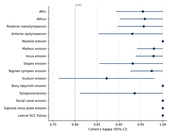
Figure 1. Cohen’s κ (95% CI) for agreement between preoperative HRCT and intraoperative findings. The dashed line marks κ = 0.80, the lower bound of “almost perfect” agreement. Points without bars have κ = 1.00 and no estimable confidence interval. Mastoid pneumatisation (κ = 1.00, three categories) is not shown.
DISCUSSION:
In these 100 consecutive patients operated on for squamous-type COM, preoperative HRCT agreed almost perfectly or perfectly with surgery (κ 0.87–1.00) for all 16 parameters. Disagreements were rare and nearly all went one way: 20 of the 21 were structures involved at surgery that the scan had not shown. Apart from one case of scutum erosion, every positive HRCT finding was confirmed in the operating theatre.
Agreement for soft-tissue disease was consistently high across subsites (κ 0.93–1.00), in line with Walshe et al. [14]. Two caveats apply. Opacification on HRCT is non-specific and cannot on its own separate cholesteatoma from granulation tissue, effusion or mucosal disease [7,21]. And our reference standard was the surgeon’s own assessment, without histology, in a surgical population with a high pre-test probability of extensive disease, where soft tissue in the attic and antrum is likely to be cholesteatoma.
Agreement for malleus and incus erosion (κ 0.98 each) is consistent with earlier reports [15,17]. The stapes scored slightly lower (κ 0.93), with three false negatives; its small size, and the difficulty of resolving the crura when they are surrounded by soft-tissue opacity, may explain this. Tegmen tympani erosion (κ 0.97) matched surgery, as Gerami et al. also found [8]. Others have likewise described perfect agreement for LSCC fistula [15,24–26], and similar agreement for the facial canal [17] and sigmoid sinus plate [15]. Scutum erosion had the lowest agreement (κ 0.87), with four false negatives and one false positive. Early blunting of the scutum is a subtle sign, and judging it at surgery is itself partly subjective. Jadia et al. reported perfect agreement for this finding [16].
Our results for the critical structures are more favourable than the weak specificity for facial canal dehiscence and dural plate erosion reported by Yildirim-Baylan et al. [20]. We cannot say why from these data; scanner technology, the criteria used to call erosion, case mix and reader blinding could all contribute. Mastoid pneumatisation agreed completely, as others have reported [7,13,15], but 92% of mastoids were sclerotic, so there was little variation against which to test HRCT.
Clinically, these results support using HRCT to map the extent of disease and to anticipate dehiscence of the tegmen, facial canal, labyrinth and sigmoid plate before operating, which can help with counselling and with choosing the approach. A normal scan should not replace careful inspection of small structures such as the stapes and scutum at surgery, since seven of the 20 missed findings involved them.
Strengths and limitations
The study has several strengths: consecutive enrolment, a structured proforma used for both HRCT and surgery, a calculated sample size, and agreement reported structure by structure with a chance-corrected statistic.
The study also has limitations. It was carried out at one centre and was restricted to squamous-type COM without revision surgery, so the results may not apply to mucosal disease or revision cases. Intraoperative findings rested on surgical inspection without histopathology. The interval between HRCT and surgery leaves room for interval progression of disease. Cohen’s κ also depends on prevalence. For findings present in very few patients (sigmoid sinus plate erosion and LSCC fistula, one case each; bony labyrinth erosion, five cases) and for the skewed distribution of mastoid pneumatisation, κ = 1.00 is imprecise, as the wide confidence intervals for sensitivity show. Finally, we compared HRCT soft-tissue opacity with intraoperative cholesteatoma or disease tissue, so high agreement for soft tissue shows that the two assessments coincided in this population, not that HRCT can distinguish cholesteatoma from other soft tissue. Larger multicentre studies with blinded, independent readers and histopathological correlation are needed.
CONCLUSION:
Preoperative HRCT of the temporal bone agreed almost perfectly or perfectly with intraoperative findings in squamous-type COM (κ 0.87–1.00), with high specificity and rare, mostly false-negative, disagreement. It is a reliable aid to surgical planning in this setting, although estimates for uncommon findings are imprecise and small structures such as the stapes and scutum can be missed.
REFERENCES:
1. Khairkar M, Deshmukh P, Maity H, Deotale V. Chronic suppurative otitis media: a comprehensive review of epidemiology, pathogenesis, microbiology, and complications. Cureus. 2023 Aug;15(8).
2. Castle JT. Cholesteatoma pearls: practical points and update. Head Neck Pathol. 2018 Sep;12:419-29.
3. Olszewska E, Rutkowska J, Özgirgin N. Consensus-based recommendations on the definition and classification of cholesteatoma. J Int Adv Otol. 2015;11(1):81-7.
4. Sharma N, Jaiswal AA, Banerjee PK, Garg AK. Complications of chronic suppurative otitis media and their management: a single institution 12 years experience. Indian J Otolaryngol Head Neck Surg. 2015 Dec;67(4):353-60.
5. Juliano AF, Ginat DT, Moonis G. Imaging review of the temporal bone: part I. Anatomy and inflammatory and neoplastic processes. Radiology. 2013 Oct;269(1):17-33.
6. Leighton SE, Robson AK, Anslow P, Milford CA. The role of CT imaging in the management of chronic suppurative otitis media. Clin Otolaryngol Allied Sci. 1993 Feb;18(1):23-9.
7. Jackler RK, Dillon WP, Schindler RA. Computed tomography in suppurative ear disease: a correlation of surgical and radiographic findings. Laryngoscope. 1984 Jun;94(6):746-52.
8. Gerami H, Naghavi E, Wahabi-Moghadam M, Forghanparast K, Akbar MH. Comparison of preoperative computerized tomography scan imaging of temporal bone with the intra-operative findings in patients undergoing mastoidectomy. Saudi Med J. 2009 Jan;30(1):104-8.
9. Keskin S, Çetin H, Töre HG. The correlation of temporal bone CT with surgery findings in evaluation of chronic inflammatory diseases of the middle ear. Eur J Gen Med. 2011;8(1):24-30.
10. Chatterjee P, Khanna S, Talukdar R. Role of high resolution computed tomography of mastoids in planning surgery for chronic suppurative otitis media. Indian J Otolaryngol Head Neck Surg. 2015 Sep;67(3):275-80.
11. Payal G, Pranjal K, Gul M, Mittal MK, Rai AK. Computed tomography in chronic suppurative otitis media: value in surgical planning. Indian J Otolaryngol Head Neck Surg. 2012 Sep;64:225-9.
12. Badran K, Ansari S, Al Sam R, Al Husami Y, Iyer A. Interpreting pre-operative mastoid computed tomography images: comparison between operating surgeon, radiologist and operative findings. J Laryngol Otol. 2016 Jan;130(1):32-7.
13. Vlastarakos PV, Kiprouli C, Pappas S, Xenelis J, Maragoudakis P, Troupis G, et al. CT scan versus surgery: how reliable is the preoperative radiological assessment in patients with chronic otitis media? Eur Arch Otorhinolaryngol. 2012 Jan;269(1):81-6.
14. Walshe P, McConn Walsh R, Brennan P, Walsh M. The role of computerized tomography in the preoperative assessment of chronic suppurative otitis media. Clin Otolaryngol Allied Sci. 2002 Apr;27(2):95-7.
15. Datta G, Mohan C, Mahajan M, Mendiratta V. Correlation of preoperative HRCT findings with surgical findings in unsafe CSOM. J Dent Med Sci. 2014;13(1):120-5.
16. Jadia S, Qureshi S, Sharma S, Mishra K. Correlation of preoperative 'HRCT temporal bone' findings with 'surgical findings' in unsafe CSOM. Indian J Otolaryngol Head Neck Surg. 2021 Mar;73(1):33-40.
17. Rai T. Radiological study of the temporal bone in chronic otitis media: prospective study of 50 cases. Indian J Otol. 2014;20(2):48-55.
18. Aljehani M, Alhussini R. The correlation between preoperative findings of high-resolution computed tomography (HRCT) and intraoperative findings of chronic otitis media (COM). Clin Med Insights Ear Nose Throat. 2019 Aug;12:1179550619870471.
19. Singh J, Bhardwaj B. How efficacious is HRCT temporal bone in determining the ossicular erosion in cases of safe and limited squamous type CSOM? Indian J Otolaryngol Head Neck Surg. 2019 Nov;71:1179-82.
20. Yildirim-Baylan M, Ozmen CA, Gun R, Yorgancilar E, Akkus Z, Topcu I. An evaluation of preoperative computed tomography on patients with chronic otitis media. Indian J Otolaryngol Head Neck Surg. 2012;64:67-70.
21. Alzoubi FQ, Odat HA, Al-Balas HA, Saeed SR. The role of preoperative CT scan in patients with chronic otitis media. Eur Arch Otorhinolaryngol. 2009 Jun;266(6):807-9.
22. Bhalla AS, Jana M. Clinico Radiological Series: Temporal Bone Imaging. Jaypee Brothers Medical Publishers; 2020.
23. Landis JR, Koch GG. The measurement of observer agreement for categorical data. Biometrics. 1977 Mar;33(1):159-74.
24. Chee NW, Tan TY. The value of pre-operative high resolution CT scans in cholesteatoma surgery. Singapore Med J. 2001 Apr;42(4):155-9.
25. Mafee MF, Levin BC, Applebaum EL, Campos M, James CF. Cholesteatoma of the middle ear and mastoid: a comparison of CT scan and operative findings. Otolaryngol Clin North Am. 1988 May;21(2):265-93.
26. 26.Rocher P, Carlier R, Attal P, Doyon D, Bobin S. Contribution and role of the scanner in the preoperative evaluation of chronic otitis. Radiosurgical correlation apropos of 85 cases. Ann Otolaryngol Chir Cervicofac. 1995;112(7):317-23.