Analyse Platelet Morphology And Platelet Indices (Mpv And Pdw) In Acs Patients, Comparing These Values With Age- And Sex-Matched Controls.
- Dr. Bhavini Harsiyani , Junior Resident, Department of Pathology, D. Y. Patil Medical College, Kolhapur.
- Dr. Vinayak Deshmukh , Department of General Medicine, Assistant Professor, Government Medical College, Goa.
- Dr. Aishwarya Darak , Junior Resident, Department of Pathology, D. Y. Patil Medical College, Kolhapur.
- Dr. Suchita V Deshmukh , HOD and Professor, Department of Pathology, D. Y. Patil Medical College, Kolhapur.
Article Information:
Abstract:
Background: Acute coronary syndrome (ACS) is associated with platelet activation and thrombus formation. Platelet indices such as platelet count (PC), mean platelet volume (MPV), platelet distribution width (PDW), and plateletcrit (PCT) are inexpensive, routinely available parameters that may reflect platelet activation. Aim: To assess platelet morphology and platelet indices in patients with ACS and compare them with age- and sex-matched healthy controls, and to evaluate differences among ACS subtypes. Methods: A prospective case-control study was conducted at the Central Clinical Laboratory, D.Y. Patil Medical College, Hospital and Research Institute, Kolhapur, over a two-year period. The study included 100 patients with clinically diagnosed ACS and 100 age- and sex-matched healthy controls. Blood samples were collected within six hours of symptom onset. Platelet indices were measured using an automated hematology analyzer, and peripheral blood smears were examined for platelet morphology, size variation, clumping, and distribution. Results: The mean age was 58.40 ± 10.60 years in ACS patients and 54.20 ± 9.80 years in controls, with no significant difference (p=0.547). Hypertension and diabetes mellitus were significantly more frequent among ACS patients (p<0.001). Platelet count was significantly higher in ACS patients than controls (p=0.047). MPV was significantly increased in ACS patients compared with controls (9.24 ± 1.25 vs. 8.31 ± 0.82 fL, p<0.001), as was PDW (14.93 ± 3.36% vs. 12.41 ± 1.96%, p<0.001). PCT was also significantly elevated in ACS (p<0.001). Large platelets were observed in 36% of ACS patients but were absent in controls. Among ACS subtypes, MPV and PDW showed significant differences, whereas PC and PCT did not. Conclusion: ACS was associated with significant alterations in platelet indices and morphology, particularly increased MPV, PDW, PCT, platelet count, and the presence of large platelets. MPV and PDW also differed significantly among ACS subtypes and may serve as simple, cost-effective adjunctive markers for assessment and risk stratification in ACS.
Keywords:
Article :
INTRODUCTION:
The Global Health Observatory (GHO) has brought attention to a silent epidemic—non-communicable diseases (NCDs), which have developed as the primary causes of mortality worldwide. These include major health disorders such as heart disease, stroke, diabetes, cancer, and chronic respiratory illnesses. According to the World Health Report (2017), ischemic heart disease (IHD) ranked as the foremost cause of mortality globally, accountable for a projected 9.5 million mortalities in 2016. [1]
Coronary artery disease (CAD) symbolizes the most prevalent type of cardiovascular disease (CVD). In 2010, an estimated 47 million individuals in India were affected by CAD. That same year, the disease accounted for around 2.3 million deaths in India, in contrast to 404,000 deaths showen in United States. Throughout the last four decades, the incidence of CAD in India has risen by more than 300% and continues to grow annually at a rate of 5 to 6%. Consequently, CAD persists as a major determinant of morbidity and death in the country. [2]
CAD is a blood vessel problem that mainly targets the coronary arteries, and it’s part of a bigger group of diseases called atherosclerosis. This process includes the gradual appearance of atheromas that narrow the arteries and reduce blood circulation, which results in critical lack of oxygen and, in some cases, necrosis of portions of cardiac muscles. Patients who suffer from coronary artery disease usually do not experience any symptoms. For example, patients may suffer from chronic stable angina that is characterized by chest pain. However, the situation becomes even more complicated if blood flow remains at the mentioned low level for extended periods of time without proper medical intervention; in such cases, Acute Coronary Syndrome may appear as a result of the condition. It is necessary to note that ACS embraces two conditions that include unstable angina (UA) and various forms of heart attack, including ST-segment elevation myocardial infarction (STEMI) and non-ST-segment elevation myocardial infarction (NSTEMI). [3] Unstable angina is the first symptom of CAD, and it may become a frequent occurrence for patients. If chest pain intensifies, urgent assistance will be required. During STEMI, there is a whole blockage of one heart's artery, which means that an extensive portion of cardiac muscles is endangered. In case of NSTEMI, blockage is partial, and a smaller portion of muscle tissue is affected. [4,5]
Platelets are very important in the progression of ACS. In the event of an atherosclerotic plaque, platelet activation takes place, resulting in the formation of a thrombus and leading to occlusion of the coronary arteries. The activated platelets tend to be larger in size, evidenced by their higher mean platelet volume (MPV), with higher size variation in platelets referred to as platelet distribution width (PDW). [6,7] Bigger platelets are also more metabolically active, exhibit stronger adhesive properties, and have a greater tendency to aggregate. Consequently, they contribute to the formation of intraluminal thrombi, starting the sequence of clinical proceedings related to ACS. Elevated platelet aggregation is commonly observed in cases of MI and UA. [8] Platelet indices such as PDW, MPV, and plateletcrit (PCT) are simple, cost-effective parameters that can usually be evaluated through a complete blood count (CBC). [9]
Based on the aforementioned context, the current research aimed to determine the predictable significance of platelet indices (MPV, PDW, PCT) in patients with ACS as compared to those presenting with non-cardiac chest pain in a tertiary care setting. Measurements of these platelet indices, including MPV, PDW, and P-LCR, along with platelet count (PC) could be useful in identifying patients who are more susceptible to acute coronary events.
METHODOLOGY:
A prospective case-control study was conducted at the Central Clinical Laboratory of the Department of Pathology, D.Y. Patil Medical College, Hospital and Research Institute, Kolhapur, between a period of two years. A total of 100 cases meeting the inclusion criteria, along with 100 age- and sex-matched healthy controls, were considered for this study.
Inclusion criteria
All clinically diagnosed patients of acute coronary syndrome including myocardial infarction and unstable angina, Age and sex matched healthy volunteers with normal ECG and without any history of ACS in previous two years as controls.
Exclusion criteria
Those with known platelet disorders (thrombocytopenia or thrombocytosis) or bleeding or clotting disorder, Patients on fibrinolytics or anti-cancer therapy or antiplatelet therapy, Critically ill patients.
Methodology
All patients who met the inclusion criteria provided written informed consent. After receiving approval from the institutional ethics committee, the study was carried out. Blood samples from the cases were collected aseptically in plain and EDTA vacutainers within six hours of symptom onset and were received at the central clinical laboratory of the Department of Pathology, D.Y. Patil Medical College, Hospital and Research Institute, Kolhapur.
These samples were tested using Horiba Yumizen H550 5 part, automated hematology analyzer, during the study period.
Similarly, blood samples from 100 controls were collected aseptically in plain and EDTA vacutainers.
Platelet parameters of both cases and controls were recorded immediately after sample processing to avoid pre-analytical variations. In addition, peripheral blood smears were prepared from EDTA blood samples, stained using Leishman stain, and examined microscopically for assessment of platelet morphology, platelet clumping, platelet size variation, and adequacy of platelet distribution. Detailed observations were documented in the proforma for comprehensive analysis.
A comparative analysis of platelet parameters between cases of Acute Coronary Syndrome (ACS) and controls was performed to evaluate the association and correlation of platelet indices with ACS.
RESULTS:
Age
The mean age was 58.40 ± 10.60 years in the case group and 54.20 ± 9.80 years in the control group. Although the case group had a higher mean age, the difference was not statistically significant (P = 0.547) (Table 1).
Table 1. Comparison of the age between groups
|
Groups |
Age (years) |
P-value |
|
|
Mean |
SD |
||
|
Case |
58.40 |
10.60 |
0.547 |
|
Control |
54.20 |
9.80 |
|
Gender
Each group comprised 100 participants. In the case group, there were 68 males and 32 females, whereas the control group included 52 males and 48 females. Although males were more prevalent in the case group, the difference in gender distribution between the groups was not statistically significant (P = 0.105) (Table 2).
Table 2. Distribution of Gender
|
Gender |
Groups (n) |
P-value |
|
|
Case |
Control |
||
|
Male |
68 |
52 |
0.105 |
|
Female |
32 |
48 |
|
|
Total |
100 |
100 |
|
Alcohol intake
Among the study participants, 36 cases reported alcohol intake compared to 28 control group, while 64 cases and 72 controls reported no alcohol consumption. Although alcohol intake was more common in the case group, the variance between the two groups was not statistically significant (P = 0.226) (Table 3).
Table 3. Distribution of alcohol intake
|
Alcohol intake |
Groups (n) |
P-value |
|
|
Case |
Control |
||
|
Yes |
36 |
28 |
0.226 |
|
No |
64 |
72 |
|
|
Total |
100 |
100 |
|
Smoking status
In the case group, 42 participants were smokers, and 58 were non-smokers, whereas in the control group, 33 participants were smokers and 67 were non-smokers. Although smoking was more common in the case group than in the control group, the variance was not statistically significant (P = 0.189) (Table 4 and Figure 9).
Table 4. Distribution of smoking status
|
Smoking status |
Groups (n) |
P-value |
|
|
Case |
Control |
||
|
Yes |
42 |
33 |
0.189 |
|
No |
58 |
67 |
|
|
Total |
100 |
100 |
|
Comorbidities
Hypertension was noted in 56 cases compared to 20 controls, while diabetes was present in 44 cases and 10 controls. Both hypertension and diabetes were more frequent in the case group compared to the control group, and these differences were observed to be highly statistically significant (P < 0.001 for both) (Table 5 and Figure 1).
Table 5. Distribution of comorbidities
|
Comorbidities |
Groups (n) |
P-value |
|
|
Case |
Control |
||
|
Hypertension |
56 |
20 |
<0.001 |
|
Diabetes |
44 |
10 |
<0.001 |
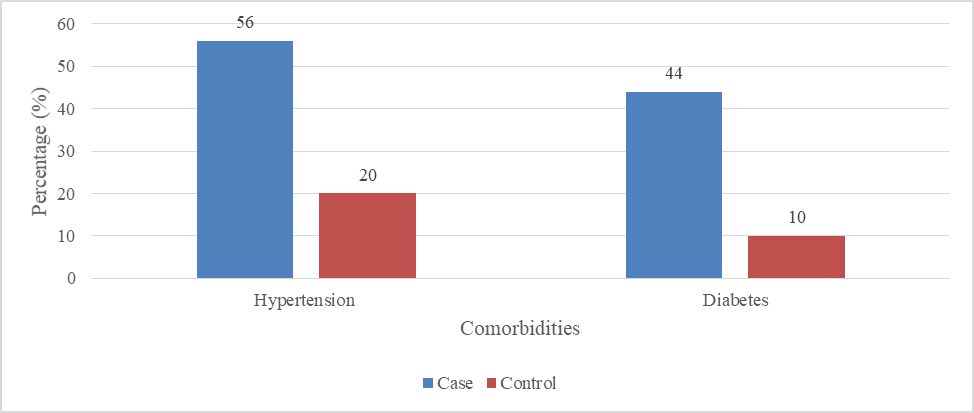
Figure 1. Distribution of comorbidities
Type of ACS
Among the study participants, STEMI was the most common type of ACS, observed in 66 cases. This was followed by NSTEMI in 23 cases and UA in 11 cases (Table 6 and Figure 2).
Table 6. Distribution of the type of ACS
|
Type of ACS |
Frequency (n) |
Percentage (%) |
|
NSTEMI |
23 |
23 |
|
STEMI |
66 |
66 |
|
UA |
11 |
11 |
|
Total |
100 |
100 |
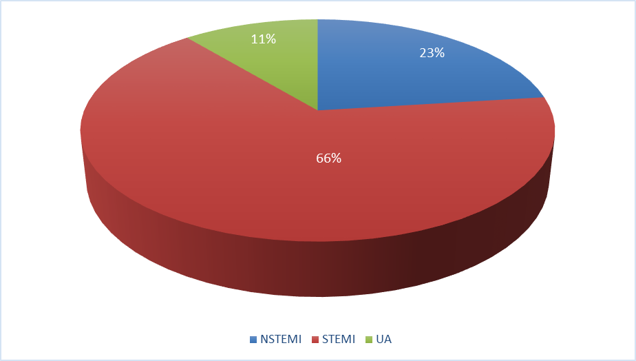
Figure 2. Distribution of the type of ACS
Hb
The mean Hb level in the case group was 11.84 ± 2.52 g/dL, whereas in the control group it was 13.21 ± 3.09 g/dL. Although the mean Hb was lower in the case group compared to the control group, the difference was not statistically significant (P = 0.149) (Table 7 and Figure 3).
Table 7. Comparison of Hb between groups
|
Groups |
Hb (g/dl) |
P- value |
|
|
Mean |
SD |
||
|
Case |
11.84 |
2.52 |
0.149 |
|
Control |
13.21 |
3.09 |
|

Figure no 3. Comparison of Hb between groups
Total WBC count
The mean total WBC count was observed to be higher in the case group (12397.29 ± 6192.55) compared to the control group (8463.44 ± 4097.40). This difference was observed to be statistically highly significant (p < 0.001), indicating elevated leukocyte counts in the case group (Table 8 and Figure 4).
Table 8. Comparison of total WBC count between groups
|
Groups |
Total WBC count |
P-value |
|
|
Mean |
SD |
||
|
Case |
12397.29 |
6192.55 |
<0.001 |
|
Control |
8463.44 |
4097.40 |
|
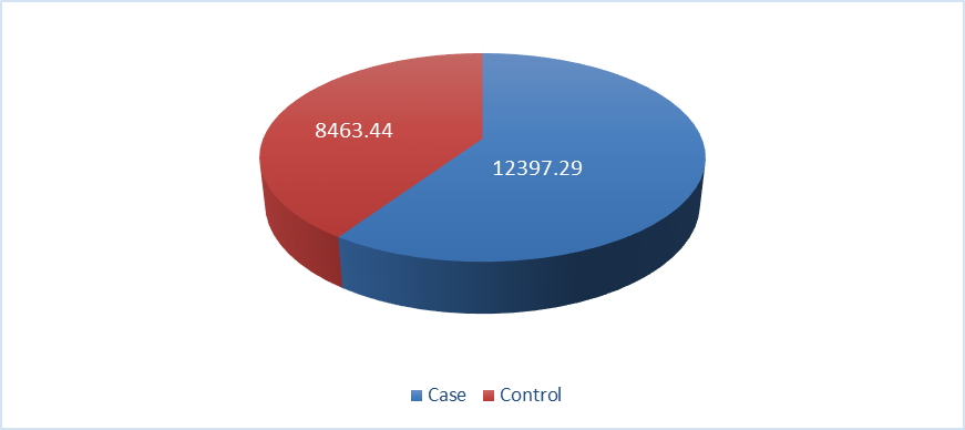
Figure 4. Comparison of total WBC count between groups PC
The mean PC was slightly higher in the case group (244.53 ± 92.23 ×10³/µL) compared to the control group (221.30 ± 71.40 ×10³/µL). This difference was noted to be statistically significant (p = 0.047), representing a modest but significant elevation in PC in the case group (Table 9 and Figure 5).
Table no 9. Comparison of between groups PC
|
Groups |
PC (x103/µL) |
P-value |
|
|
Mean |
SD |
||
|
Case |
244.53 |
92.23 |
0.047 |
|
Control |
221.30 |
71.40 |
|
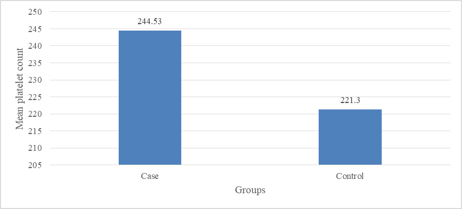
Figure no 5 Comparison of between groups PC
MPV
The MPV was significantly higher in the case group (9.24 ± 1.25 fL) compared to the control group (8.31 ± 0.82 fL). This change was observed to be statistically highly significant (p < 0.001), suggesting increased platelet activation in the case group (Table 10 and Figure 6).
Table 10. Comparison of MPV between groups
|
Groups |
MPV (fL) |
P-value |
|
|
Mean |
SD |
||
|
Case |
9.24 |
1.25 |
<0.001 |
|
Control |
8.31 |
0.82 |
|
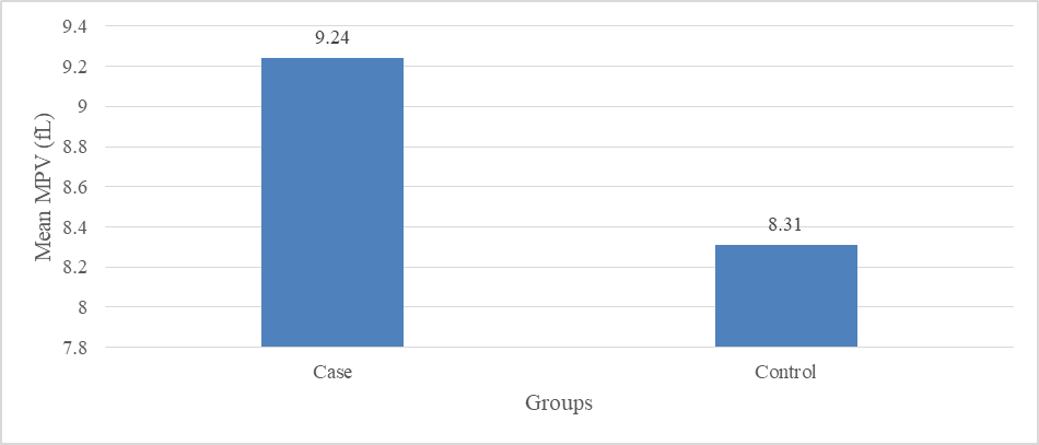
Figure 6. Comparison of MPV between groups
PDW
The mean PDW was significantly greater in the case group (14.93 ± 3.36%) compared to the control group (12.41 ± 1.96%). This difference was noted to be statistically highly significant (p < 0.001), indicating greater variation in platelet size among cases (Table 11 and Figure 7).
Table 11. Comparison of PDW between groups
|
Groups |
PDW (%) |
P-value |
|
|
Mean |
SD |
||
|
Case |
14.93 |
3.36 |
<0.001 |
|
Control |
12.41 |
1.96 |
|
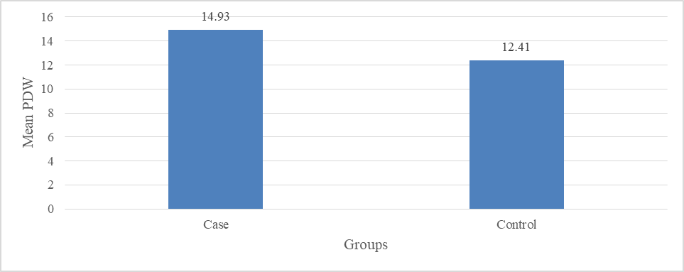
Figure 7. Comparison of PDW between groups
PCT
The mean PCT was slightly greater in the case group (0.22 ± 0.08%) compared to the control group (0.19 ± 0.06%). This difference was found to be statistically highly significant (p < 0.001), indicating an increased total platelet mass in the case group (Table 12 and Figure 8).
Table 12. Comparison of PCT between groups
|
Groups |
PCT (%) |
P-value |
|
|
Mean |
SD |
||
|
Case |
0.22 |
0.08 |
<0.001 |
|
Control |
0.19 |
0.06 |
|
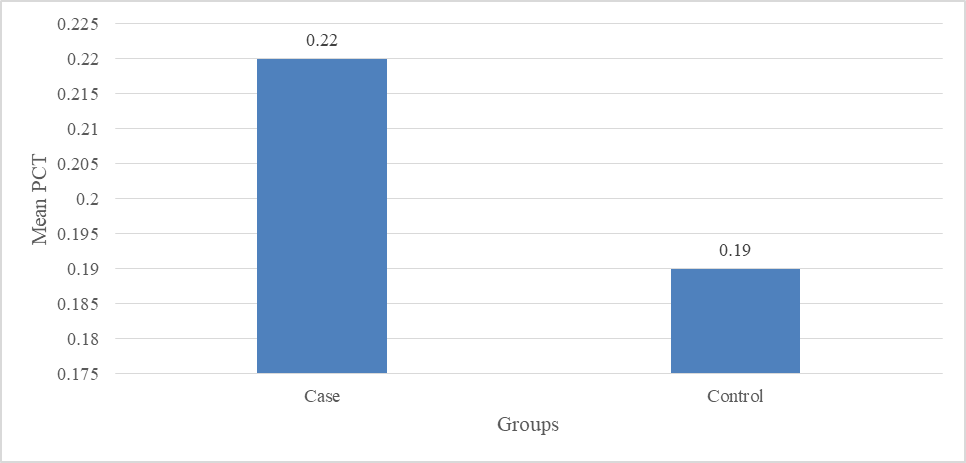
Figure 8. Comparison of PCT between groups
Platelet morphology
The distribution of platelet morphology presented a statistically highly significant variance between the case and control groups (p < 0.001). In the case group, the majority of patients had adequate platelets (n = 62), followed by large platelets (n = 36), with a small number showing decreased platelets (n = 2). In contrast, the control group predominantly demonstrated platelets of normal size with adequate platelet count (n = 89), while a few cases showed decreased platelets (n = 5), increased platelets (n = 5), and slightly reduced platelet count (n = 1). Notably, large platelets were not observed in the control group (Table 13 and Figure 9).
Table 13. Distribution of platelet morphology
|
Platelet morphology |
Groups (n) |
P-value |
|
|
Case |
Control |
||
|
Adequate |
62 |
89 |
<0.001 |
|
Decreased |
2 |
5 |
|
|
Increased |
0 |
5 |
|
|
Large platelet |
36 |
0 |
|
|
Slightly reduced |
0 |
1 |
|
|
Total |
100 |
100 |
|
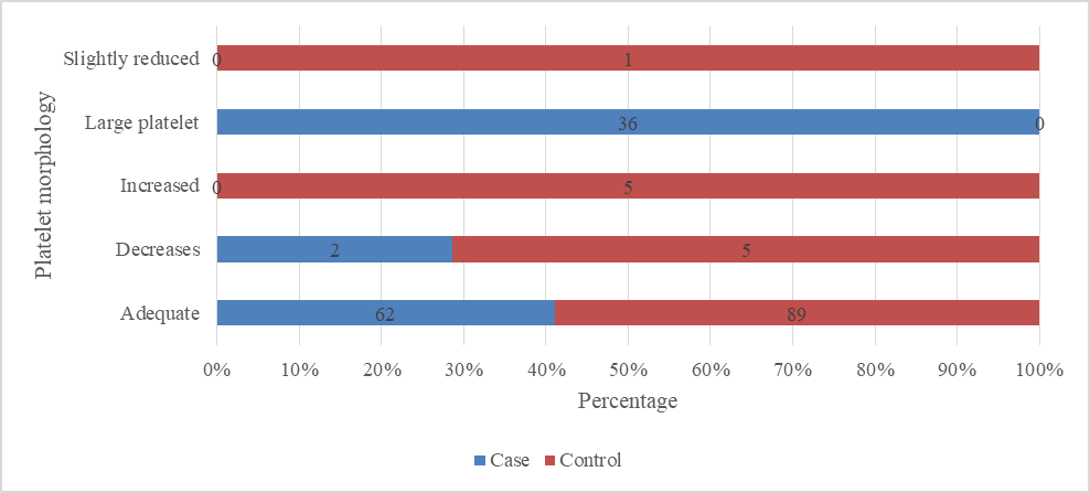
Figure 9. Distribution of platelet morphology.
Comparison of platelet indices according to ACS
The comparison of platelet indices among different types of ACS showed varying patterns. The mean PC was comparable across NSTEMI (236 ± 84), STEMI (252 ± 96), and UA (221 ± 72), with no statistically significant variance (p = 0.231).
In contrast, MPV exhibited a statistically highly significant difference among the groups (p < 0.001), with the highest values observed in STEMI (9.68 ± 1.32 fL), followed by NSTEMI (9.01 ± 1.08 fL), and the lowest in UA (8.54 ± 0.92 fL).
Similarly, PDW values were highest in STEMI (15.84 ± 3.48%), followed by NSTEMI (14.12 ± 2.91%) and UA (13.10 ± 2.44%), showing a statistically highly significant difference among the groups (p < 0.001).
PCT showed comparable values across NSTEMI (0.214 ± 0.076%), STEMI (0.228 ± 0.091%), and UA (0.201 ± 0.063%), with no statistically significant difference (p = 0.162). (Table 14).
Table 14. Comparison of platelet indices according to ACS
|
Parameters |
Type of ACS |
P-value |
||
|
STEMI |
NSTEMI |
UA |
||
|
PC |
252 ± 96 |
236 ± 84 |
221 ± 72 |
0.231 |
|
MPV |
9.68 ± 1.32 |
9.01 ± 1.08 |
8.54 ± 0.92 |
< 0.001 |
|
PDW |
15.84 ± 3.48 |
14.12 ± 2.91 |
13.10 ± 2.44 |
< 0.001 |
|
PCT |
0.228 ± 0.091 |
0.214 ± 0.076 |
0.201 ± 0.063 |
0.162 |
Large platelet
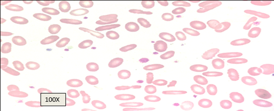
Figure 10 – Peripheral smear from ACS case showing Large Platelet (100x oil immersion)
DISCUSSION:
ACS is a spectrum of clinical conditions resulting from acute myocardial ischemia, most commonly due to atherosclerotic plaque rupture and subsequent thrombosis. Platelets are pivotal in the pathogenesis of ACS through activation, aggregation, and thrombus formation. In recent years, platelet indices such as MPV and PDW have emerged as inexpensive and readily available biomarkers reflecting platelet activation and reactivity. The present study assessed platelet morphology and platelet indices in ACS subjects and compared them with age- and sex-matched controls.
Demographic and clinical profile
In the present study, the mean age was greater in the case group (58.40 ± 10.60 years) compared to controls (54.20 ± 9.80 years), although this variance was not statistically significant (p = 0.547). Similarly, males were more common among cases (68%) compared to controls (52%), but without statistical significance. This male preponderance and higher age distribution among ACS cases are consistent with Assiri AS et al [10] and Mir AA et al [11], indicating increased cardiovascular risk with advancing age and in males, largely attributed to hormonal, metabolic, and behavioural risk profiles. Comparable observations have been reported in large population-based studies such as the Framingham Heart Study, which established increasing prevalence of CAD with age and greater prevalence in males. [12]
In the present study smoking and alcohol intake were more frequent in cases compared to controls, though differences were not statistically significant. However, hypertension and diabetes mellitus were significantly more prevalent in ACS patients (p < 0.001). These results are consistent with Chowdekar VS and Peddi N [13], identifying hypertension and diabetes as major independent risk features for CAD. Chronic hyperglycemia and hypertension contribute to oxidative stress, endothelial dysfunction, and accelerated atherosclerosis, thereby increasing thrombotic risk. [86, 14]
The present study demonstrated a significantly elevated total leukocyte count in ACS patients compared to controls (p < 0.001). Leukocytosis as an inflammatory process in ACS indicates the rupture of plaque with subsequent myocardial ischemia and necrosis. Cytokines, released by inflammatory cells, may lead to instability of plaque and cause proteolysis. Grewal T et al [15] made similar conclusions: the authors noted that elevated WBC values could be an indicator of poor heart prognosis in itself. In the current study, the level of Hb was reduced in cases compared to the control group, though statistically the difference wasn't very significant. Anemia in ACS is an indicator of unfavorable prognosis, because it implies a reduction of oxygenation in the myocardium. However, its predictive value is controversial. [10, 11]
PC and indices
The current study demonstrated a significant increase of platelets count in patients with ACS compared to control group participants (p = 0.047). The platelets count is a poor indicator of their activation; nevertheless, elevation of platelet count may suggest an increased activity and/or synthesis of platelets possibly due to inflammation or thrombogenic stimuli. The fact is that all platelet indices, calculated in the current research, showed significant differences between two groups; those differences can hardly be random.
MPV was significantly higher in ACS patients (9.24 ± 1.25 fL vs 8.31 ± 0.82 fL, p < 0.001).
PDW was also significantly elevated in cases (14.93 ± 3.36% vs 12.41 ± 1.96%, p < 0.001).
PCT was significantly greater in cases (p < 0.001).
MPV reflects the average platelet volume and serves as an indicator of their activity. Larger platelets are more metabolically active and produce more thromboxane A2, thus making platelets more prone to aggregation. Patients with ACS in our study demonstrated increased MPV levels, similar to previously obtained results in research conducted by Chowdekar VS and Peddi N [86], Grewal T et al. [15], and Yadav S, Yadav R. [16]. These researchers also reported that patients with MI and UA have elevated MPV levels compared to patients without heart disease.
Meanwhile, PDW indicates the variability in platelet volume. This parameter indirectly reflects all kinds of morphological abnormalities occurring as a result of uneven platelet activity. Significantly increased PDW levels were detected in ACS patients, indicating platelet activation and accelerated production. Previous research proves that the increase in PDW levels reflects high thrombotic activity. [89] Also, an extremely high level of PCT, reflecting total platelet mass, was observed in our group of patients with ACS. An increase in PCT is attributed either to the presence of an increased number of platelets or their enlarged volume, leading to increased clot formation. [84]
One of the findings from the present research is that of large platelets, which were mostly found in patients with ACS (36%) but were absent in healthy individuals. Large platelets have been identified as platelets that have higher reactivity, pro-coagulant potential, granule content, and adhesion capability. The absence of large platelets in controls further supports their association with pathological platelet activation in ACS. These morphological differences reinforce the functional alterations occurring in platelet biology during acute coronary events. [13]
When platelet indices were compared among different types of ACS, platelet count and plateletcrit did not show any statistically significant difference between STEMI, NSTEMI, and unstable angina cases. This suggests that these parameters may not be very useful in distinguishing the different ACS subtypes.
However, MPV showed a statistically highly significant difference among the groups. Higher MPV in the more severe forms of ACS may be due to increased platelet activation, as larger platelets are more active and have greater tendency to form thrombus. This supports the role of platelet activation in the development and progression of acute coronary events.
Similarly, PDW also showed a statistically highly significant difference among the ACS subtypes. Increased PDW reflects variation in platelet size, which occurs due to the release of larger and more reactive platelets during acute thrombotic events. The higher MPV and PDW observed in severe ACS cases indicate increased platelet activity and thrombotic burden.
Similar findings were reported by Mir AA et al. [11], who also observed significantly elevated MPV and PDW values in severe forms of ACS.
Overall, the findings of the present study strongly support the role of platelet activation in the pathophysiology of ACS. Elevated MPV, PDW, and platelet morphological abnormalities in ACS patients indicate increased platelet reactivity and heterogeneity, contributing to thrombus formation and coronary occlusion. These indices, being inexpensive and routinely available, may serve as adjunctive biomarkers for risk stratification in ACS.
Higher MPV and PDW in STEMI compared to NSTEMI and UA may suggest greater platelet activation and size variability in more severe forms of ACS. However, literature on subtype-specific platelet indices remains inconsistent, with some studies reporting higher values in STEMI due to more extensive thrombotic burden, while others show no significant differences.
CONCLUSION:
The current study demonstrated significant differences in platelet morphology and indices between patients with acute coronary syndrome (ACS) and age- and sex-matched controls. Platelet count (PC), mean platelet volume (MPV), platelet distribution width (PDW), and plateletcrit (PCT) were significantly elevated in ACS, suggesting increased platelet activation and a prothrombotic state. Among ACS subtypes (STEMI, NSTEMI, and UA), only MPV and PDW showed significant differences, whereas PC and PCT did not, indicating that MPV and PDW may be more useful for differentiating ACS severity.
The presence of large platelets on peripheral smear further supports platelet activation in acute coronary events. Overall, MPV and PDW appear to be simple, cost-effective adjunctive markers for ACS assessment and risk stratification. Larger multicentric studies with advanced platelet analysis and long-term follow-up are needed to establish their prognostic utility.
REFERENCES:
1. Abu-Assi E, López-López A, González-Salvado V, Redondo-Diéguez A, Pena-Gil C, Bouzas-Cruz N, Raposeiras-Roubín S, Abumuaileq RR, García-Acuña JM, González-Juanatey JR. The risk of cardiovascular events after an acute coronary event remains high, especially during the first year, despite revascularization. Revista Española de Cardiología (English Edition). 2016 Jan 1;69(1):11-8.
2. Al-Hassan M., Sagr L. Stress and stressors of myocardial infarction patients in the early period after discharge. J Adv Nurs. 2002;40(2):181-8.
3. Falastine R. Hamdan F.R., Shishani K., Nabolsi M., Froelicher E.S. (2010). Delay Times and Decision Making Processes in Acute Myocardial Infarction: A Qualitative Literature Review. J Med J. Vol. 44. Issue 4. Pages 447-455.
4. French DP, Marteau TM, Senior V, Weinman JA. Eliciting causal beliefs about heart attacks: A comparison of implicit and explicit methods. Journal of Health Psychology. 2002 Jul;7(4):433-44.
5. Frohmader TJ, Lin F, Chaboyer W. Patient perceptions of nurse mentors facilitating the Aussie Heart Guide: A home‐based cardiac rehabilitation programme for rural patients. Nursing Open. 2016 Jan;3(1):41-50.
6. Endler G, Klimesch A, Sunder‐Plassmann H, Schillinger M, Exner M, Mannhalter C, Jordanova N, Christ G, Thalhammer R, Huber K, Sunder‐Plassmann R. Mean platelet volume is an independent risk factor for myocardial infarction but not for coronary artery disease. Br J Haematol 2002 May;117(2)399-404.
7. Schoene NW. Design criteria: tests used to assess platelet function. Am J Clin Nutr 1997 May 1;65(5)1665S-8S.
8. Ahamed H, Henry RA, Pai R. Association of mean platelet volume and acute coronary syndrome. Int J Res Med Sci 2017 Mar 28;5(4)1217-20.
9. Abass AE, Yassir T, Saeed Y, Taha A, Basheer A, Mubarak E, Zonoon F, Faroog G, Mohammad N. Investigation of platelets count and indices in coronary artery diseases among Sudanese patients. J Dent Med Sc Jan 2016 Jan;15(1)35-8.
10. Assiri AS, Jamil AM, Mahfouz AA, Mahmoud ZS, Ghallab M. Diagnostic importance of platelet parameters in patients with acute coronary syndrome admitted to a tertiary care hospital in southwest region, Saudi Arabia. Journal of the Saudi Heart Association. 2012 Jan 1;24(1):17-21.
11. Mir AA, Khan AA, Sheikh JM, Dar MI, Rather HA, Lone AA, Hafeez I, Rashid A. Analysis of Platelet Volume Indices and their utility in Acute Coronary Syndrome. Heart Views. 2024 Oct 1;25(4):229-35.
12. Chen G, Levy D. Contributions of the Framingham heart study to the epidemiology of coronary heart disease. JAMA cardiology. 2016 Oct 1;1(7):825.
13. Chowdekar VS, Peddi N. Evaluation of platelet indices in patients with acute coronary syndrome and chronic stable angina. International Journal of Research in Medical Sciences. 2020 Nov;8(11):3886-90.
14. Care D. Cardiovascular disease and risk management. Diabetes Care. 2017 Jan 1;40(1):S75-87.
15. Grewal T, Gunasekaran A, Kapoor M. Study of the Diagnostic Importance of Platelet Distribution Width in Acute Coronary Syndrome. Acta Scientific Cardiovascular System. 2021;1(1):01-06.
16. Yadav S, Yadav R. Study comparing platelet indices between stable angina, acute coronary syndrome with age sex matched normal controls. International Journal of Medical and Pharmaceutical Research. 2025 Dec 31;6:2156-60.