Management Of Immature Permanent Teeth Using Regenerative Endodontic Therapy, Traditional Apexification And Mta Apical Barrier Approach: An 1-Year Follow-Up Case Report.
- Dr. Shreyasi Chakraborty , Associate Professor, Department Of Paediatric and Preventive Dentistry, Dr R Ahmed Dental College and Hospital, Kolkata.
- Dr. Sadique Mohammad , BDS, Mds, Consultant Dental Practitioner, Apollo Dental, Camac Street, Kolkata.
- Dr. Monalisa Das , Associate Professor and Hod, Department Of Dentistry, Raiganj Medical College and Hospital, Raiganj.
Article Information:
Abstract:
Background: Traumatic dental injuries frequently resulting in pulp necrosis in children arrest root development of immature permanent incisors. Case presentation: An 8.5-year-old girl presented one month after a sports injury with Ellis class III fractures of the maxillary right central incisor (11), left central incisor (21) and left lateral incisor (22). All three teeth were nonresponsive to electric pulp testing, tender to percussion, showed Grade II mobility and had open apices (Cvek stage IV), thickening of the lamina dura, and a diagnosis of symptomatic apical periodontitis was made. Because remaining crown structure differed between the teeth, three apical management strategies were used: regenerative endodontic therapy with injectable platelet-rich fibrin (i-PRF) in 11, calcium hydroxide apexification in 21 and an MTA apical barrier in 22. Behaviour management included video modelling, a take-home schematic chart and the tell-show-do technique, and informed parental consent was obtained. After canal disinfection, each tooth was treated according to its protocol and restored with a post, core and polycarbonate crown where required. Follow-up was clinical and radiographic, with crowns placed on all three teeth and review over two years. Conclusion: Treatment selection for immature, traumatised incisors should be individualised according to remaining tooth structure, restorative needs and patient cooperation. Regenerative endodontics with i-PRF, apexification and MTA barriers can each manage the open apex, but regeneration alone offers the prospect of continued root development.
Keywords:
Article :
INTRODUCTION:
Traumatic dental injuries are common in school-age children, and the maxillary central incisors are the teeth most often affected, particularly during sports and play.1 When trauma causes pulp necrosis before root formation is complete, the tooth is left with thin, fragile dentinal walls, a short root and a wide open apical foramen. These features make conventional root canal obturation difficult and increase the risk of cervical root fracture,2 which is why immature non-vital teeth have long been considered a clinical challenge.
Traditionally, apexification with long-term calcium hydroxide has been used to induce an apical calcific barrier against which the root filling can be condensed.3 Although effective, it requires multiple visits over several months, depends heavily on patient compliance, and prolonged exposure to calcium hydroxide has been reported to weaken root dentine and increase the susceptibility to fracture.4 Mineral trioxide aggregate (MTA) was introduced as an apical plug material because of its sealing ability, biocompatibility and capacity to be placed as an immediate barrier in a single visit, reducing the treatment time and the dependence on recall.5,6 Neither technique, however, promotes further root maturation, so root length and wall thickness remain as they were at the time of treatment.
Regenerative endodontic therapy offers a biologically based alternative. After the report of continued root development in an immature tooth with apical periodontitis,7 Banchs and Trope proposed a revascularisation protocol based on minimal instrumentation, copious irrigation, intracanal disinfection with antibiotic medicament and evoked bleeding to create a blood-clot scaffold.8 The current approach relies on disinfection of the canal, delivery of stem cells from the apical papilla by evoked bleeding, a suitable scaffold, and a coronal seal.9 Platelet concentrates, particularly platelet-rich fibrin (PRF), are attractive scaffolds because they are autologous, easy to prepare and release growth factors over several days. A systematic review reported similar resolution of the signs and symptoms of infection with all approaches, while regenerative procedures generally showed further root maturation, although the results were variable.10
The case described here is unusual because three adjacent traumatised incisors in the same child, with the same diagnosis, were managed with three different apical strategies chosen on the basis of the amount of remaining coronal structure. The aim of this report is to describe the clinical decision-making and protocol for each approach and to compare the findings with those of previous studies.
CASE PRESENTATION:
An eight-and-a-half-year-old girl attended the outpatient clinic of the Department of Paediatric and Preventive Dentistry with a chief complaint of fractured upper front teeth after a sports injury sustained one month earlier. The medical history was non-contributory, and she was classified as ASA I according to the American Society of Anaesthesiologists Physical Status Classification System.
Extraoral examination showed no regional lymphadenopathy. Intraoral examination revealed complicated crown fractures (Ellis class III) of the maxillary right central incisor (11), the maxillary left central incisor (21) and the maxillary left lateral incisor (22), with more than two-thirds of the crowns of 21 and 22 lost (Figure 1A). Palpation of the alveolar mucosa over the three teeth elicited tenderness, and the teeth were also tender to percussion. Moreover, the teeth exhibited Grade II mobility according to Miller's classification. Electric pulp testing produced no response in any of the three teeth, denoting loss of pulp sensitivity.
An intraoral periapical radiograph of 11, 21 and 22 demonstrated widening of the periodontal ligament space and periapical radiolucency. The roots were of nearly complete length, but the apices were wide open with thin dentinal walls, corresponding to Cvek stage IV of root development (Figure 1B). A final diagnosis of pulp necrosis with symptomatic apical periodontitis was made for 11, 21 and 22.
Tooth 11 retained sufficient coronal structure and did not require a post and core, so regenerative endodontic therapy using injectable PRF (i-PRF) was planned to give it the chance of continued root maturation. Teeth 21 and 22 had lost more than two-thirds of the crown and required a post, core and provisional crown. A conventional calcium hydroxide apexification was therefore planned for 21, and a single-visit MTA apical barrier was planned for 22. The options, their advantages, limitations and expected recall schedule were explained to the parents, and written informed consent was obtained. The child was given a schematic diagram and a chart to study at home, and video modelling and the tell-show-do technique were used to obtain her assent and cooperation.
Treatment began with regenerative endodontic therapy for tooth 11. Local anaesthesia was administered with 2% lignocaine with 1:200,000 epinephrine. Under rubber dam isolation, access cavities were prepared in the involved teeth. Minimal instrumentation was performed, and the canals were irrigated with 20 mL of 2.5% sodium hypochlorite, followed by 5 mL of sterile saline and a final rinse with 20 mL of 17% EDTA (Pyrax, Roorkee, India). The canals were dried with sterile paper points, calcium hydroxide paste (UltraCal XS, 35%, Ultradent, Utah, USA) was placed as an intracanal medicament and the access was sealed with Cavit (ESPE, Cergy-Pontoise, France).
At the second visit three weeks later, tooth 11 was asymptomatic and no longer tender to palpation or percussion. Anaesthesia was given with 2% lignocaine without epinephrine, a rubber dam was placed, and the temporary seal was removed. The working length was estimated with an apex locator (Canal Pro, Coltene Whaledent) and confirmed radiographically. The canal was irrigated with 20 mL of 17% EDTA for 5 minutes to remove the calcium hydroxide, followed by sterile saline to reduce the cytotoxic effect of EDTA, and thereafter was dried with sterile paper points.
For preparation of i-PRF, 10 mL of venous blood was drawn from the median cubital vein into a plain tube without anticoagulant (Figure 1C) and centrifuged at 700 rpm for 3 minutes. The upper yellow liquid layer (i-PRF) was collected as close as possible to the red cell layer (Figure 1D). A K-file was introduced 2 to 3 mm beyond the wide apical foramen to provoke bleeding. When blood filled the apical third of the canal, i-PRF was injected to a level approximately 3 mm below the canal orifice (Figure 1E). After an interval of 10 to 15 minutes to allow for coagulation, a 3-mm layer of MTA putty (Kids-E-Dental, Mumbai, India) was placed over the PRF, and the access cavity was closed with Type II glass ionomer cement (Fuji, Japan).
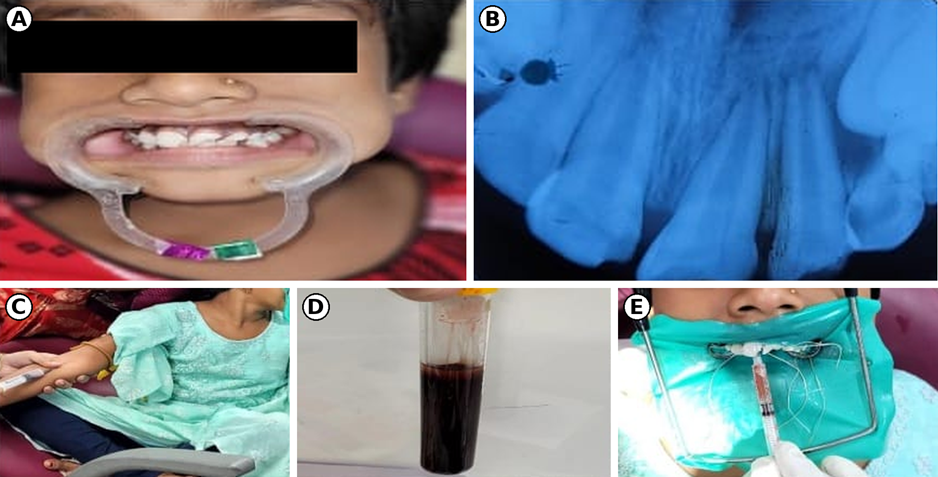
Figure 1. Regenerative endodontic therapy using injectable platelet-rich fibrin (i-PRF) in tooth 11. (A) Pre-operative intraoral photograph showing fractured maxillary incisors 11, 21 and 22. (B) Pre-operative periapical radiograph showing open apices and incomplete root development of 11, 21 and 22. (C) Venous blood being drawn from the median cubital vein. (D) Blood centrifuged without anticoagulant, with the upper yellow i-PRF layer visible. (E) i-PRF being injected into the canal under rubber dam isolation.
Tooth 21 was disinfected during the first visit in the same manner as the other teeth intracanal calcium hydroxide was placed as medicament. At the recall visit six weeks after the first appointment, the tooth was isolated with a rubber dam, and the calcium hydroxide was removed by irrigation with 2.5% sodium hypochlorite. A size 15 paper point was inserted to the apex and, when its butt end splayed, a firm apical barrier formation was confirmed clinically and radiographically. The barrier had formed about 2 mm short of the radiographic apex. The canal was obturated with gutta-percha, and the access was sealed with Type II glass ionomer cement.
Four weeks after the first appointment, tooth 22 was asymptomatic and non-tender on percussion and palpation. Under local anaesthesia and rubber dam isolation, the canal was irrigated copiously with 2.5% sodium hypochlorite, and the working length was determined with the apex locator and by radiograph. MTA putty was introduced to a thickness of 4 mm from the apex. A moist cotton pellet was placed in the canal to aid in setting, and the access was closed with Cavit. The next day, the dressing was removed and the MTA was found to have set into a hard barrier. Tooth 22 was then restored with a fibre post and dual-cure core build-up material (Fluorocore 2+, Dentsply, USA). The teeth were restored provisionally with Polycarbonate crowns as the occlusion was in a mixed dentition stage. [Figure 2B].
The patient was reviewed clinically and radiographically at regular intervals for one year, and an uneventful healing was observed. Tooth 11, treated with regenerative endodontic therapy, remained unresponsive to electric pulp testing at the 3-, 9-, and 12-month follow-ups. However, the one-year radiograph (Figure 1B and 2A) showed continued root lengthening, increased root wall thickness, and apical closure.
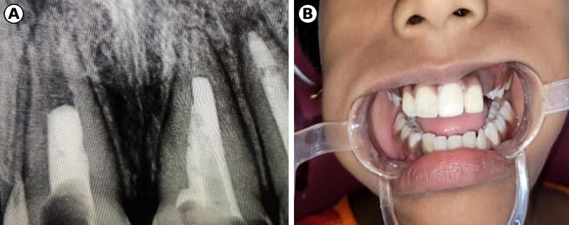
Figure 2. One-year follow-up. (A) Follow-up periapical radiograph of 11, 21 and 22 after treatment. (B) Intraoral photograph after placement of polycarbonate crowns on 11, 21 and 22.
DISCUSSION:
Immature necrotic permanent incisors can be managed by apexification, an MTA apical plug or regenerative endodontic therapy, and none of these has been shown to be superior in every situation. In the present case, the choice of treatment for each tooth was determined by the restorative needs rather than by the diagnosis, which was identical for all three teeth. This is consistent with the view that the remaining coronal structure, the need for a post and the anticipated compliance of the patient should be weighed alongside the biological aim of the treatment.
Apexification with calcium hydroxide has a long record of clinical success, but it requires a prolonged period of treatment and repeated visits, which may be difficult in a young child. The long-term use of the medicament has also been linked to a reduction in the fracture resistance of immature roots.4 Witherspoon and colleagues, in a retrospective analysis of open-apex teeth obturated with MTA, reported healing in more than 90% of the teeth recalled at one year or longer when MTA was placed in open-apex teeth with no apical barrier,6 and this supports the use of MTA in tooth 22, in which a post and core were required and a rapid, predictable apical seal was desirable. Our finding of a hard-set barrier on the day after placement agrees with the known setting behaviour and sealing ability of the material.5
The regenerative approach used in tooth 11 aims to go beyond apical closure and promote angiogenesis, reinnervation and continued root formation16. The protocol followed the principles of disinfection with calcium hydroxide, irrigation with EDTA to release growth factors from dentine, evoked bleeding to introduce apical papilla stem cells, and a coronal MTA seal.9 The use of i-PRF as the scaffold differs from the classic blood clot in that it is an autologous, fibrin-rich preparation obtained at a low centrifugation force, which, according to its developers, retains more leucocytes, platelets and growth factors than earlier protocols.11 Bakhtiar and colleagues described the use of PRF as a scaffold in a case series of regenerative endodontic treatment,12 and the course of tooth 11 in the present case supports the feasibility of platelet concentrates as a scaffold in young patients.
Several comparative studies have examined how regeneration performs against apexification, with differing conclusions. In the Mahidol retrospective study of 61 teeth, Jeeruphan and colleagues found that revascularisation produced significantly greater increases in root length and thickness than either calcium hydroxide or MTA apexification, and survival was 100% for revascularisation, 95% for MTA and 77.2% for calcium hydroxide.13 In contrast, Alobaid and colleagues, in a pilot retrospective cohort of 31 teeth, found that revascularisation was not superior to apexification in clinical or radiographic outcomes.14 In a prospective randomised controlled trial of 118 teeth followed for 12 months, Lin and colleagues reported that regeneration and apexification achieved comparable resolution of symptoms and apical healing, whereas regeneration produced a significantly greater increase in root length and thickness on cone-beam imaging.15
The review by Kahler and colleagues similarly concluded that clinical outcomes in terms of resolution of infection were similar for all approaches, and that continued root maturation after regeneration was generally seen but variable.10 Our case is consistent with these reports in that all three teeth were treated successfully with respect to the resolution of symptoms, although the three techniques cannot be compared directly because each tooth differed in structure and treatment. According to Zbanska et al17. the key factor establishing the success of Regenerative Endodontic therapy is continued growth of the root in length, thickness and closure of the root apex as noticed in a radiograph. In our case the teeth did not give positive response to electric pulp testing, but it showed an increase in root length, thickness and root closure in the one-year follow -up radiograph, pointing toward a successful Regenerative Endodontic therapy.
CONCLUSION:
Open-apex traumatised incisors can be managed successfully with regenerative endodontic therapy, apexification or an MTA apical barrier. The selection of technique should be individualised according to the remaining tooth structure, restorative requirements, the need for biological root maturation and the cooperation of the child and parents. Long-term clinical and radiographic follow-up remains essential.
REFERENCES:
1. Glendor U. Epidemiology of traumatic dental injuries - a 12-year review of the literature. Dent Traumatol. 2008;24(6):603-611.
2. Cvek M. Prognosis of luxated non-vital maxillary incisors treated with calcium hydroxide and filled with gutta-percha. A retrospective clinical study. Endod Dent Traumatol. 1992;8(2):45-55.
3. Rafter M. Apexification: a review. Dent Traumatol. 2005;21(1):1-8.
4. Andreasen JO, Farik B, Munksgaard EC. Long-term calcium hydroxide as a root canal dressing may increase risk of root fracture. Dent Traumatol. 2002;18(3):134-137.
5. Torabinejad M, Chivian N. Clinical applications of mineral trioxide aggregate. J Endod. 1999;25(3):197-205.
6. Witherspoon DE, Small JC, Regan JD, Nunn M. Retrospective analysis of open apex teeth obturated with mineral trioxide aggregate. J Endod. 2008;34(10):1171-1176.
7. Iwaya SI, Ikawa M, Kubota M. Revascularization of an immature permanent tooth with apical periodontitis and sinus tract. Dent Traumatol. 2001;17(4):185-187.
8. Banchs F, Trope M. Revascularization of immature permanent teeth with apical periodontitis: new treatment protocol? J Endod. 2004;30(4):196-200.
9. Hargreaves KM, Diogenes A, Teixeira FB. Treatment options: biological basis of regenerative endodontic procedures. J Endod. 2013;39(3 Suppl):S30-S43.
10. Kahler B, Rossi-Fedele G, Chugal N, Lin LM. An evidence-based review of the efficacy of treatment approaches for immature permanent teeth with pulp necrosis. J Endod. 2017;43(7):1052-1057.
11. Choukroun J, Ghanaati S. Reduction of relative centrifugation force within injectable platelet-rich-fibrin (PRF) concentrates advances patients' own inflammatory cells, platelets and growth factors: the first introduction to the low speed centrifugation concept. Eur J Trauma Emerg Surg. 2018;44(1):87-95.
12. Bakhtiar H, Esmaeili S, Fakhr Tabatabayi S, Ellini MR, Nekoofar MH, Dummer PMH. Second-generation platelet concentrate (platelet-rich fibrin) as a scaffold in regenerative endodontics: a case series. J Endod. 2017;43(3):401-408.
13. Jeeruphan T, Jantarat J, Yanpiset K, Suwannapan L, Khewsawai P, Hargreaves KM. Mahidol study 1: comparison of radiographic and survival outcomes of immature teeth treated with either regenerative endodontic or apexification methods: a retrospective study. J Endod. 2012;38(10):1330-1336.
14. Alobaid AS, Cortes LM, Lo J, Nguyen TT, Albert J, Abu-Melha AS, et al. Radiographic and clinical outcomes of the treatment of immature permanent teeth by revascularization or apexification: a pilot retrospective cohort study. J Endod. 2014;40(8):1063-1070.
15. Lin J, Zeng Q, Wei X, Zhao W, Cui M, Gu J, et al. Regenerative endodontics versus apexification in immature permanent teeth with apical periodontitis: a prospective randomized controlled study. J Endod. 2017;43(11):1821-1827.
16. Murray, P.E.; Garcia-Godoy, F.; Hargreaves, K.M. Regenerative Endodontics: A Review of Current Status and a Call for Action. J. Endod. 2007;33, 377–390.
17. Zbańska, J.; Herman, K.; Kuropka, P.; Dobrzyński, M. Regenerative Endodontics as the Future Treatment of Immature Permanent Teeth. Appl. Sci. 2021, 11, 6211.