Congenital Cholesteatoma in an Adolescent - A Rare, Delayed Presentation in a 16 Year Old Boy.
- Dr. Alagammai Sivaraman , Assistant Professor, Department of ENT, Dhanalakshmi Srinivasan Medical College and Hospital, Siruvachur, Perambalur, Tamil Nadu, India
- Dr. Laavanya Selvaraju , Assistant Professor, Department of ENT, Dhanalakshmi Srinivasan Medical College and Hospital, Siruvachur, Perambalur, Tamil Nadu, India
- Dr. Ravindran Ambika , Professor and Head of the Department, Department of ENT, Dhanalakshmi Srinivasan Medical College and Hospital, Siruvachur, Perambalur, Tamil Nadu, India
- Dr. Geetha J , Professor, Department of Anaesthesia, Dhanalakshmi Srinivasan Medical College and Hospital, Siruvachur, Perambalur, Tamil Nadu, India
- Dr. Natasha Renchi Mathew , Postgraduate, Department of ENT, Dhanalakshmi Srinivasan Medical College and Hospital, Siruvachur, Perambalur, Tamil Nadu, India
- Dr. Sindhuja B , Postgraduate, Department of ENT, Dhanalakshmi Srinivasan Medical College and Hospital, Siruvachur, Perambalur, Tamil Nadu, India.
Article Information:
Abstract:
Background: Congenital cholesteatoma is a rare lesion arising behind an intact tympanic membrane. We report a 16-year-old boy with progressive unilateral hearing loss. Otoscopy revealed a pearly white middle-ear mass, and imaging confirmed congenital cholesteatoma. Surgical excision was performed successfully. This case highlights the rare adolescent presentation of congenital cholesteatoma.
Keywords:
Article :
INTRODUCTION:
Congenital cholesteatoma is a rare lesion accounting for 1–5% of all cholesteatomas.[1] It is characterized by keratinizing squamous epithelium within the middle ear behind an intact tympanic membrane in the absence of previous otorrhea, tympanic membrane perforation, or ear surgery. Most cases are diagnosed in early childhood with mean age being 5.6 +/- 2.8 years.[2] Adolescent presentation is uncommon and may indicate delayed diagnosis or slow disease progression. We report a rare case of congenital cholesteatoma in a 16-year-old boy presenting with long-standing unilateral hearing loss.
CASE REPORT:
A 16-year-old boy presented to the Department of Otorhinolaryngology at Dhanalakshmi Srinivasan Medical College at Perambalur with complaints of progressive hearing loss in the left ear for 10 years. There was no history of ear discharge, otalgia, tinnitus, vertigo, facial weakness, trauma, previous ear surgery, or recurrent episodes of otitis media.
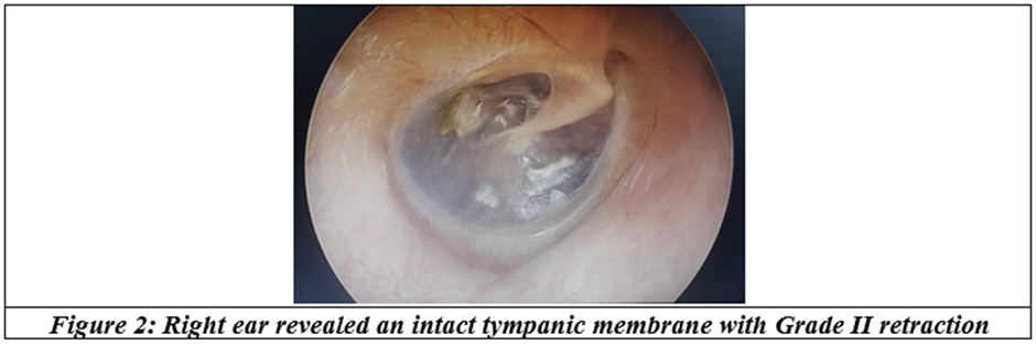
General physical examination was unremarkable. Otoscopic examination of the left ear (Fig. 1) revealed an intact tympanic membrane with a pearly white mass involving all 4 quadrants of the middle ear. The external auditory canal was normal. Examination of the right ear (Fig. 2) revealed Grade II retraction of the tympanic membrane.
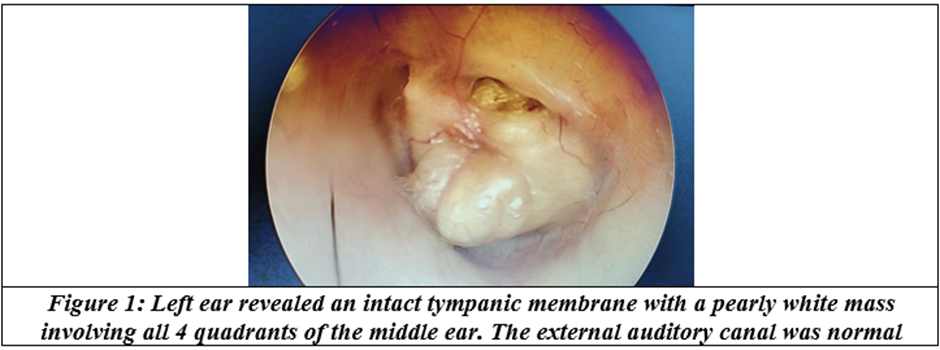

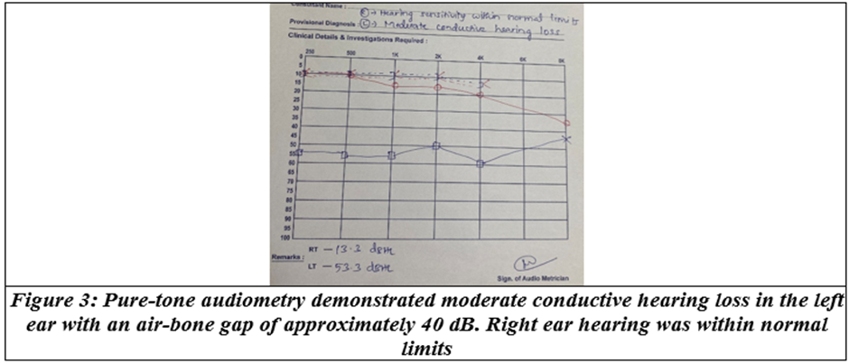
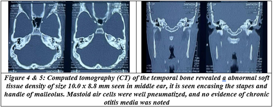
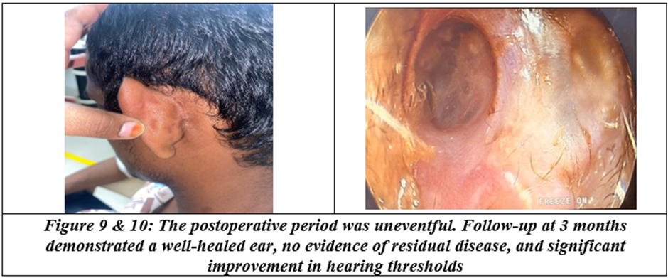
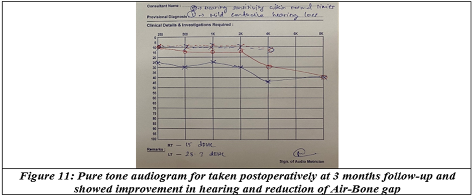
Based on the clinical and radiological findings, a diagnosis of congenital cholesteatoma was made. The lesion was classified as Stage III according to the Potsic classification due to ossicular involvement.
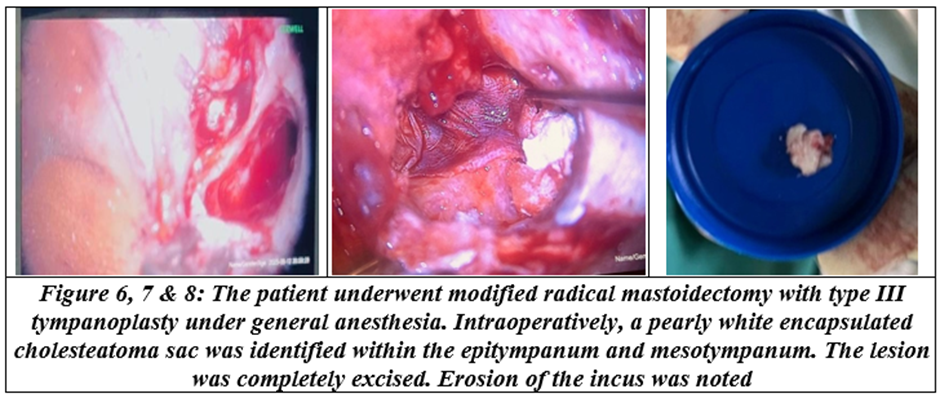
Histopathological examination confirmed the diagnosis of cholesteatoma, showing a cyst lined by keratinizing stratified squamous epithelium containing laminated keratin debris.
DISCUSSION:
Congenital cholesteatoma is believed to arise from embryonic epithelial cell rests that persist within the middle ear cavity.[3] Several theories have been proposed regarding its origin, including the epidermoid formation theory, amniotic fluid contamination theory, and epithelial metaplasia theory. Levenson et al. defined congenital cholesteatoma as a white mass medial to an intact tympanic membrane in the absence of previous otorrhea, perforation, or ear surgery.[4] Most cases are diagnosed before the age of ten years. Presentation in adolescence is unusual and may indicate slow-growing disease or delayed diagnosis. Progressive unilateral conductive hearing impairment is the symptom most frequently reported at presentation. Otoscopic examination typically reveals a white, pearly lesion behind an intact tympanic membrane, most frequently in the anterosuperior quadrant.
The Potsic staging system[5] is widely used:
• Stage I: Single quadrant involvement
• Stage II: Multiple quadrants without ossicular or mastoid involvement
• Stage III: Ossicular involvement
• Stage IV: Mastoid extension
Surgical excision remains the definitive treatment.[6] The choice of procedure depends on disease extent and surgeon preference. Early diagnosis and complete removal are essential to prevent recurrence and preserve hearing.[7,8]
This case is noteworthy because of the unusually late presentation at 16 years of age despite the congenital origin of the lesion. Awareness of this possibility can facilitate timely diagnosis and management.
CONCLUSION:
Congenital cholesteatoma should be considered in adolescents presenting with unilateral conductive hearing loss and an intact tympanic membrane. Careful otoscopic examination and appropriate imaging are crucial for diagnosis. Surgical excision provides excellent outcomes when performed before the development of extensive complications.
REFERENCES:
1. Solanki KJ, Nakum NN, Mehta MV. Congenital cholesteatoma of middle ear: clinical features and surgical outcomes. International Journal of Research in Medical Sciences 2026;14(3):1162-6.
2. Nelson M, Roger G, Koltai PJ, et al. Congenital cholesteatoma: classification, management, and outcome. Arch Otolaryngol Head Neck Surg 2002;128(7):810-4.
3. Levenson MJ, Michaels L, Parisier SC. Congenital cholesteatomas in children: an embryologic correlation. Laryngoscope 1988;98(9):949-55.
4. Levenson MJ, Michaels L, Parisier SC. Congenital cholesteatomas of the middle ear in children: origin and management. Otolaryngologic Clinics of North America 1989;22(5):941-54.
5. Potsic WP, Korman SB, Samadi DS, et al. Congenital cholesteatoma: 20 years’ experience at The Children’s Hospital of Philadelphia. Otolaryngol Head Neck Surg 2002;126(4):409-14.
6. Hasan B, Hamdan Z, Al Baker R. Early diagnosis and complete excision of congenital cholesteatoma in a 4-year-old boy: a case report. Int J Surg Case Rep 2025;134:111820.
7. McGill TJ, Merchant S, Healy GB, et al. Congenital cholesteatoma of the middle ear in children: a clinical and histopathological report. Laryngoscope 1991;101(6):606-13.
8. Kazahaya K, Potsic WP. Congenital cholesteatoma. Curr Opin Otolaryngol Head Neck Surg 2004;12(5):398-403.