Anatomical Variations and Extralaryngeal Branching of the Recurrent Laryngeal Nerve in Relation to the Inferior Thyroid Artery.
- Satishkumar M. Patel , Assistant professor, Department of Anatomy, GMERS Medical College Valsad, Gujarat, India.
- Rakesh K. Vora , Professor, Department of Anatomy, Dr. M. K. Shah Medical College & Research Centre, Ahmedabad, Gujarat, India.
Article Information:
Abstract:
Introduction: Variations in the relationship between the recurrent laryngeal nerve (RLN) and inferior thyroid artery (ITA), including extralaryngeal branching, are relevant to thyroid surgery. This study aimed to describe these relationships, branching patterns and bilateral concordance. Materials & Methods: A descriptive cadaveric study was planned in the Department of Anatomy at a medical college in Gujarat, India, involving 30 cadavers and 60 heminecks. RLN–ITA relationships were classified as anterior, posterior or between arterial branches. Extralaryngeal branching was categorized as single trunk, bifurcation, trifurcation or multiple branching. Frequencies, percentages and bilateral concordance were summarized descriptively. Results: The RLN was posterior to the ITA in 38 heminecks (63.3%), anterior in 15 (25.0%) and between its branches in seven (11.7%). Posterior relationships occurred in 17 right (56.7%) and 21 left heminecks (70.0%). A single trunk occurred in 45 heminecks (75.0%), bifurcation in 12 (20.0%) and trifurcation in three (5.0%). No multiple branching was represented. Bilateral concordance occurred in 19 cadavers (63.3%); 11 (36.7%) were discordant. Conclusion: The RLN predominantly coursed posterior to the ITA as a single trunk. Anatomical variations emphasize the importance of identifying the nerve and its branches on each side during thyroid surgery.
Keywords:
Article :
INTRODUCTION:
The recurrent laryngeal nerve (RLN) is an important anatomical structure encountered during thyroid and parathyroid surgery. Its close relationship with the thyroid gland and surrounding vessels makes it vulnerable during dissection, vascular ligation and gland mobilization. Injury may cause vocal fold dysfunction, affecting phonation, swallowing and airway protection. Consequently, knowledge of the nerve’s course and anatomical variations is essential for surgical identification and preservation. Indian surgical studies have emphasized the relevance of the inferior thyroid artery (ITA), tracheoesophageal groove and other adjacent landmarks in locating the RLN. [1] [2]
The cervical course of the RLN differs between sides. The right nerve loops around the subclavian artery, whereas the left loops around the aortic arch before ascending towards the larynx. Near the thyroid gland, the nerve may pass anterior to the ITA, posterior to it, or between its branches. Although the artery provides a useful landmark, its relationship with the nerve is variable. Recognizing this variability is particularly important when either structure divides before their crossing point. [2] [3]
Extralaryngeal branching introduces additional anatomical complexity. The nerve may remain undivided or separate into two, three or more terminal branches before entering the larynx. Identification of the main trunk alone may therefore be insufficient to characterize the complete terminal anatomy. Recent Indian studies have documented diverse branching patterns, supporting careful tracing of branches towards their laryngeal entry. [3] [4]
Available Indian evidence includes both surgical observations and cadaveric investigations. Samorekar reported side-specific RLN–ITA relationships during thyroid surgery, while Dadwani and colleagues investigated branching and nerve–artery relationships in Gujarat cadaveric and autopsy specimens. A subsequent Telangana cadaveric study by Aenumulapalli and colleagues also demonstrated variation in arterial relationships and terminal branching. [5] [6] [7] Differences in specimen selection, pathological distortion, dissection technique and classification limit direct comparison between studies.
Bilateral examination permits assessment of whether the configuration observed on one side is reproduced contralaterally. Documenting concordance alongside side-specific frequencies may strengthen the anatomical description and discourage assumptions of symmetry. Accordingly, the proposed study framework examines RLN–ITA relationships, extralaryngeal terminal branching and bilateral concordance in 30 adult cadavers comprising 60 heminecks at a medical college in Gujarat. Its intended contribution is descriptive anatomical evidence relevant to surgical training and nerve identification; it does not directly evaluate postoperative outcomes.
MATERIALS AND METHODS:
Study design and setting: A descriptive cross-sectional cadaveric study was conducted in the Department of Anatomy, Medical College, Gujarat, India, during 2023 to 2025 year.
Study sample and selection: Thirty adult human cadavers were included, providing 60 heminecks for bilateral examination. Cadavers were selected by availability-based convenience sampling from specimens available in the department during the study period. Cadavers with adequately preserved cervical anatomy permitting identification of the RLN and ITA were eligible. Specimens with previous cervical surgery, extensive trauma, gross pathological distortion, or dissection damage preventing reliable assessment were excluded from the relevant analysis. Any exclusion and its reason were documented separately for each side.
Dissection procedure: Cadavers were positioned supine with the neck extended sufficiently to expose the anterior cervical region. A midline skin incision was made from the lower border of the mandible to the suprasternal notch, supplemented by transverse incisions along the lower border of the mandible and the clavicles. The skin, superficial fascia and platysma were reflected laterally. The infrahyoid muscles were exposed and retracted to identify the thyroid gland and adjacent structures. Careful dissection preserved the nerves and vessels and minimized displacement of their anatomical relationships. The ITA was identified and traced towards the thyroid gland. The RLN was identified in the lower cervical region and followed superiorly to its laryngeal entry. The nerve–artery relationship was examined before dividing or mobilizing either structure. The same assessment procedure was followed on both sides.
Anatomical observations: The RLN–ITA relationship was classified as anterior to the artery, posterior to the artery, or between its branches at the crossing region adjacent to the thyroid gland. When the RLN divided before crossing the artery, the relationship of each terminal branch was recorded individually. Configurations in which all nerve branches shared the same relationship were assigned to the corresponding category; mixed or unclassifiable configurations were documented separately. Extralaryngeal terminal branching was categorized as an undivided single trunk, bifurcation, trifurcation, or multiple branching comprising four or more terminal branches. Only branches traced into the larynx were counted. Small branches supplying the trachea, oesophagus or adjacent structures were excluded from this classification. Bilateral concordance was defined as an identical RLN–ITA relationship on both sides of a cadaver; differing relationships were classified as discordant. Findings were entered on a standardized recording sheet using a unique cadaver identifier and separate right- and left-sided fields.
Statistical analysis: Data were entered and analysed using Microsoft Excel 2024 (Microsoft Corporation, Redmond, WA, USA). Categorical variables were summarized as frequencies and percentages, using evaluable nerves as the denominator. Bilateral concordance was calculated among cadavers with complete paired observations. Analysis was descriptive; no inferential statistical tests were performed.
RESULTS:
A total of 30 cadavers, comprising 60 heminecks equally distributed between the right and left sides, were represented in the dataset. The recurrent laryngeal nerve (RLN) was posterior to the inferior thyroid artery (ITA) in 38 heminecks (63.3%), anterior in 15 (25.0%), and between its branches in seven (11.7%). On the right side, the nerve was posterior to the ITA in 17 heminecks (56.7%), anterior in nine (30.0%), and between its branches in four (13.3%). Corresponding findings on the left side were 21 (70.0%), six (20.0%), and three (10.0%), respectively (Table 1; Figure 1).
Table 1. Relationship of RLN to ITA
|
Relationship |
Right (N = 30), n (%) |
Left (N = 30), n (%) |
Total (N = 60), n (%) |
|
Anterior to ITA |
9 (30.0) |
6 (20.0) |
15 (25.0) |
|
Posterior to ITA |
17 (56.7) |
21 (70.0) |
38 (63.3) |
|
Between branches of ITA |
4 (13.3) |
3 (10.0) |
7 (11.7) |
|
Total |
30 (100.0) |
30 (100.0) |
60 (100.0) |
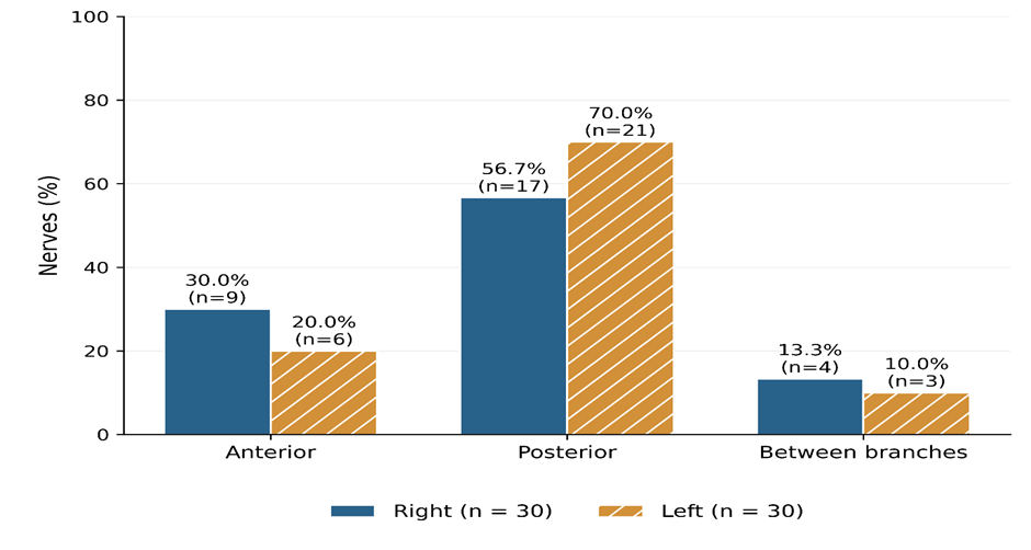
Figure 1. RLN relationship to ITA
A single RLN trunk without extralaryngeal division was recorded in 45 heminecks (75.0%). Extralaryngeal branching was present in 15 heminecks (25.0%), comprising bifurcation in 12 (20.0%) and trifurcation in three (5.0%). On the right side, 21 nerves (70.0%) remained undivided, seven (23.3%) bifurcated, and two (6.7%) trifurcated. On the left side, 24 nerves (80.0%) remained undivided, five (16.7%) bifurcated, and one (3.3%) trifurcated. No nerve with four or more terminal branches was recorded (Table 2; Figure 2).
Table 2. Extralaryngeal branching patterns
|
Branching pattern |
Right (N = 30), n (%) |
Left (N = 30), n (%) |
Total (N = 60), n (%) |
|
Single trunk |
21 (70.0) |
24 (80.0) |
45 (75.0) |
|
Bifurcation |
7 (23.3) |
5 (16.7) |
12 (20.0) |
|
Trifurcation |
2 (6.7) |
1 (3.3) |
3 (5.0) |
|
Multiple branches (≥4) |
0 (0.0) |
0 (0.0) |
0 (0.0) |
|
Total |
30 (100.0) |
30 (100.0) |
60 (100.0) |
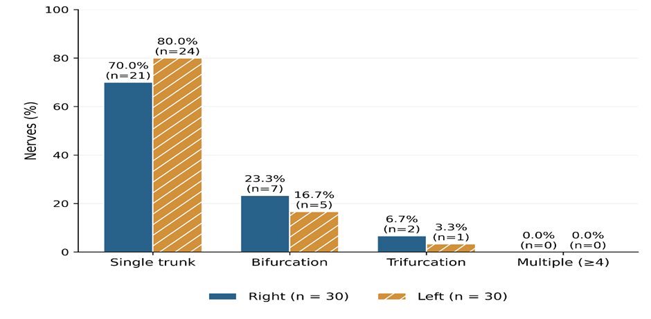
Figure 2. Extralaryngeal branching
The RLN–ITA relationship was bilaterally concordant in 19 cadavers (63.3%) and discordant in 11 (36.7%). Bilateral posterior positioning was the most frequent concordant configuration, occurring in 14 cadavers (46.7%), followed by bilateral anterior positioning in four (13.3%) and positioning between the arterial branches on both sides in one (3.3%). Among discordant configurations, an anterior relationship on the right and posterior relationship on the left occurred in four cadavers, while a between-branch relationship on the right and posterior relationship on the left occurred in three. A posterior relationship on the right and anterior relationship on the left occurred in two cadavers. The remaining two configurations—anterior on the right with between-branch positioning on the left, and posterior on the right with between-branch positioning on the left—occurred in one cadaver each (Table 3).
Table 3. Simulated bilateral concordance of RLN–ITA relationships
|
Right-side relationship ↓ / Left-side relationship → |
Anterior |
Posterior |
Between branches |
Total |
|
Anterior |
4 |
4 |
1 |
9 |
|
Posterior |
2 |
14 |
1 |
17 |
|
Between branches |
0 |
3 |
1 |
4 |
|
Total |
6 |
21 |
3 |
30 cadavers |
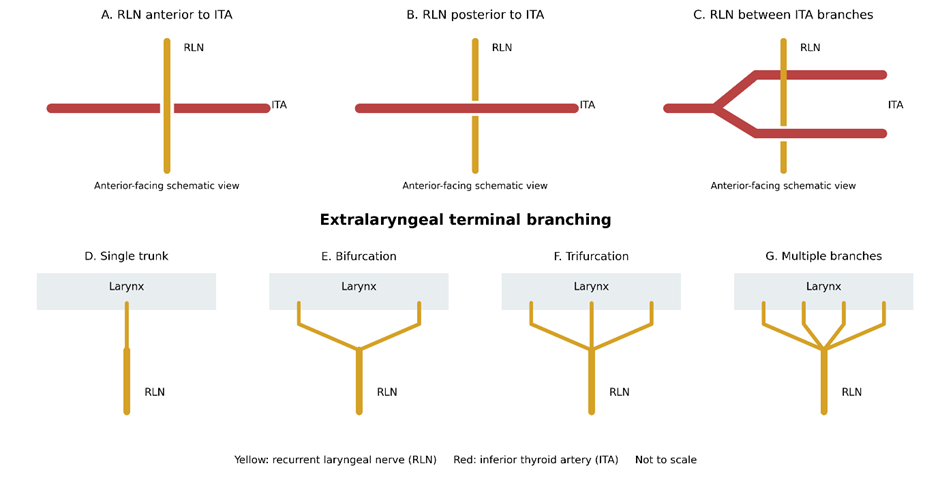
Figure 3: Anatomical schematic: recurrent laryngeal nerve variations
DISCUSSION:
This study demonstrates a predominantly posterior RLN–ITA relationship, observed in 63.3% of heminecks, followed by anterior positioning in 25.0% and passage between arterial branches in 11.7%. Posterior positioning was more frequent on the left than on the right, at 70.0% and 56.7%, respectively. These distributions provide an example of how anatomical findings can be compared with published studies. However, they cannot establish population frequencies until verified against actual specimen records.
The overall posterior proportion is numerically close to Samorekar’s Indian surgical series, which identified 43 posterior relationships among 69 nerves, corresponding to 62.3%. Anterior and between-branch relationships occurred in 33.3% and approximately 4.3%, respectively. Side-specific posterior proportions were 59.5% on the right and 65.6% on the left. [5]. Aenumulapalli and colleagues examined 40 heminecks and reported posterior, anterior and between-branch relationships in 57.5%, 25.0% and 17.5%, respectively. [7] Erfan and colleagues reported posterior positioning in 72% of their surgical observations, whereas Kumar and colleagues reported approximately 82%. [3] [2]
Saldanha and colleagues found the RLN deep to the ITA in 125 of 140 identified nerves, approximately 89.3%. [1] Their description of the artery as superficial to the nerve must be translated consistently when comparing nerve-centred classifications. Gujrathi and colleagues reported posterior relationships in 65.38% of right-sided nerves; on the left, posterior and anterior relationships were equally frequent at 45.45%. Their anatomical observations concerned the nerve-identification group, rather than every patient enrolled. [8]
Table 4. Comparison of present study findings with other studies
|
Study |
RLN anterior to ITA (%) |
RLN posterior/deep to ITA (%) |
RLN between ITA branches (%) |
Extralaryngeal branching (%) |
|
Present study |
25.0 |
63.3 |
11.7 |
25.0 |
|
Saldanha et al., 2019 [1] |
10.7 |
89.3 |
0 |
1.4† |
|
Kumar et al., 2023 [2] |
17.9 |
82.1 |
0 |
10.7 |
|
Erfan et al., 2024 [3] |
28.0 |
72.0 |
0‡ |
28.0 |
|
Samorekar, 2023 [5] |
33.3 |
62.3 |
4.3 |
NR |
|
Aenumulapalli et al., 2025 [7] |
25.0 |
57.5 |
17.5 |
12.5 |
|
Gujrathi et al., 2021 [8] |
33.3† |
56.3† |
10.4† |
NR |
Dadwani’s Gujarat study is geographically relevant but requires particular caution. Its RLN–ITA categories total 79 observations despite a stated sample of 73 nerves, and the corresponding percentages exceed 100%. Branching counts also fail to reconcile with the stated side-specific denominators. [6] Consequently, those proportions should not be treated as precise quantitative benchmarks without clarification. Similarly, the laryngeal-entry findings in Aenumulapalli’s paper differ between the narrative and table, although its RLN–ITA counts reconcile. [7]. Extralaryngeal branching occurs in 25% of nerves, comprising bifurcation in 20% and trifurcation in 5%. Erfan reported bifurcation in 26% and trifurcation in 2%, whereas Kumar reported extralaryngeal branching in 10.7%. [3] [2] Aenumulapalli reported branching in 12.5% of heminecks. [7] Kale documented substantially more frequent branching, but its side-specific nerve observations and patient-level summary use different denominators, limiting straightforward comparison. [4] Consistent definitions distinguishing terminal laryngeal branches from small adjacent-organ branches are therefore necessary. Bilateral concordance is 63.3%, with discordance in 36.7%. This demonstrates why observations on one side cannot invariably predict contralateral anatomy. However, bilateral concordance concerns paired cadaver observations, whereas pooled relationship frequencies concern individual heminecks. These denominators must remain distinct, and descriptive percentage differences should not be labelled statistically significant.
CONCLUSION:
The recurrent laryngeal nerve predominantly coursed posterior to the inferior thyroid artery and remained an undivided single trunk. Variations in nerve–artery relationships, extralaryngeal branching and bilateral concordance highlight the importance of identifying the nerve and its branches independently on each side during thyroid surgery. Larger studies incorporating standardized morphometric measurements are needed to further characterize these anatomical variations.
REFERENCES:
1. Saldanha M, Jayaramaiah SK, Aroor R, Bhat VS, Varghese S. Relationship of recurrent laryngeal nerve with inferior thyroid artery. Int J Otorhinolaryngol Clin. 2019;11(2):27–29.
2. Kumar P, Chatterjee M, Gupta A. Variations in recurrent laryngeal nerve in thyroidectomy. Indian J Otolaryngol Head Neck Surg. 2023;75:2856–2861.
3. Erfan S, Saha S, Guha R, Sen I, Kulkarni S. Varying course of external branch of superior laryngeal nerve (EBSLN) and recurrent laryngeal nerve (RLN) in thyroidectomy—an observational study. Indian J Otolaryngol Head Neck Surg. 2024;76:1959–1964.
4. Kale VD, Prajosh A, Chavan SS, Patra A. A prospective observational study on the anatomical variations of recurrent laryngeal nerve and its application in the prevention of injury during thyroid surgery in a tertiary care centre. Egypt J Otolaryngol. 2024;40:132.
5. Samorekar AV. Anatomical variations of recurrent laryngeal nerve (RLN) in relation with inferior thyroid artery (ITA) in the patients undergoing thyroid surgeries: a descriptive study. Int J Pharm Clin Res. 2023;15(12):272–279.
6. relationship between the recurrent laryngeal nerve and the inferior thyroid artery: insights from cadaveric dissections. Indian J Clin Anat Physiol. 2024;11(2):71–74.
7. Aenumulapalli A, Repalle S, Vazir D. Topographic anatomy of the thyroid gland and recurrent laryngeal nerve in cadavers: a descriptive cross-sectional cadaveric study with surgical relevance. Stud J Health Res Afr. 2025;6(6).
8. Gujrathi AB, Kurande HS, Gadpayale N, Paikrao Y. Identification of recurrent laryngeal nerve: a dilemma in thyroid surgery. Int J Otorhinolaryngol Head Neck Surg. 2021;7(9):1450–1454.