Clinical Correlations of Anatomical Variations in the Brachial Plexus: A Systemic Review of Surgical and Anesthetic Implications
- Dr. Amit Jagannath Patil , Associate Professor, Department of Anaesthesiology, ACPM Medical College, Dhule, Maharashtra, India
- Dr. Siddhant Yadav , Senior Resident, Department of Orthopaedics, Government Medical College, Singrauli, Madhya Pradesh, India
- Dr. Muhammed Faseed C.H , Associate Professor, Department of Respiratory Medicine, Kanachur Institute of Medical Sciences, Mangaluru, Karnataka, India.
Article Information:
Abstract:
Background: The brachial plexus represents an anatomically quite variable peripheral nerve network characterized by its complexity. The variations in the root, trunk, division, cord, terminal branches, and communicating pathways of this complex system could A lot impact neurological exams, regional anesthesia, imaging techniques, and even surgery.Objective: This review aimed to summarize the reported anatomical variations of the brachial plexus and evaluate their clinical correlations, with particular emphasis on surgical and anesthetic implications.Methods: An assessment based on systematic review of the literary works on anatomical variations of brachial plexus was conducted emphasizing mainly on how such variations influence regional anesthesia, surgical procedure and other imaging modality, as well as complications of procedures. A synthesis of the findings from anatomical cadaveric clinical studies, as well as reviews was made through a narrated approach because of the variability in both designs and outcomes.Results: The existing literature highlights that there are many differences in the brachial plexus anatomic features including changes in both proximal and distal parts of the brachial plexus. Communication branches that connect the musculocutaneous, median, and ulnar nerves are very often reported. Different patterns of origin, branching, and connections of the nerves with the surrounding vasculature and muscles were also noted. The anatomical deviations can partly or wholly explain regional anesthesia that does not work as expected, failure to locate the nerves accurately using ultrasound, atypical neurological signs, and greater risks of surgical complications including nerve reconstruction and dissection. And, changes in vascular and phrenic nerve relations were found and they also act as an important factor in procedural safe conduct.Conclusion: It is important to consider that anatomical variations of the brachial plexus have a great value and they cannot just be considered as incidental findings that are not important for medical intervention. Being aware of neural variations as well as adjacent anatomical structures is the key for proper diagnosis, regional anesthesia, safe surgical operations as well as other medical interventions that might involve the nerves. Detailed anatomical information together with ultrasound and proper imaging, could bring better procedural planning, make nerve localization more precise, and prevent unnecessary complications.
Keywords:
Article :
INTRODUCTION:
The upper limb receives most of its motor and sensory innervation through the brachial plexus, which is a complex network of peripheral nerves. Traditionally, the formation of the plexus is attributed to the C5, T1 spinal anterior rami nerves, the roots, which combine to form trunks. Then, the trunks divide into divisions and then, divisions develop into the cords which ultimately sprout into terminal branches. Such functional pathways link the cervical spinal cord to the upper limb. Yet, the anatomical structure of the plexus is not always the same as that of the classical description. It is not rare to find a number of alterations in practically any part of the plexus, from roots trunks divisions cords terminal branches down to a single nerve's communicating branches [1,2].
Variations in the anatomy of the brachial plexus are very important clinically since they might change the anticipated distribution of motor and sensory innervated areas. Variants include the atypical formation of the trunks, the unusual origin or course of the terminal nerves, and communications between the musculocutaneous median ulnar, and other peripheral nerves that can result in patterns different from those described in standard anatomical textbooks. Although some variations remain clinically silent, others can mean atypical neurological signs, changed sensory areas neuralgia weakness, or unexpected results during a clinical examination [1,3,4]. Recently, cadaveric studies show that these kinds of variations are relatively common rather than exceptional, thereby underlining the importance of clinicians being able to recognize the possible diversity of plexus anatomy [7].
The clinical relevance of these anatomical variations is most visibly seen in regional anesthesia. For instance, brachial plexus blocks are the main choice for anesthesia and pain relief in the upper limb surgeries, and the success of the blockade mostly hinges on the precise localization of the nerve structures and the knowledge of their spatial relationships. The level of plexus being targeted, that is, whether the blockade is performed at the interscalene supraclavicular infraclavicular, or axillary level, greatly influences both the areas affected by the anesthesia and the extent of the blockade. Anatomy variations may change the position, path, or the relations of nerve elements leading to areas of anesthesia being missed, individual nerve territories remaining unaffected, or block characteristics that are not as expected [1,2,5,6].
The growing popularity of ultrasound-guided regional anesthesia has highlighted the key role anatomical knowledge plays practically when performing this technique. Although ultrasound is capable of directly showing one the nerves and surrounding anatomic features with real-time visuals, interpreting the sonographic anatomy still needs a thorough understanding of the normal and variable appearances. Awareness of peculiar neural courses is something important that anesthesiologists rely on to alter their needle insertion and solution deposition techniques when standard pattern features are not found. For this reason, knowing about the anatomy and utilizing the imaging should be thought of as interdependent parts of ensuring safety in brachial plexus regional anesthesia procedures and not something treated separately [1,2,5].
Anatomical variations are very important aspects in surgical practice. Operations in the areas of shoulder neck axilla, chest wall and upper limb have the possibility of iatrogenic injury to the brachial plexus and its branches. Variations in the origin, branching patterns and communications between the nerves may make the identification difficult in nerve dissection, repair or transfer, and reconstruction. In addition, an accidental communication between important peripheral nerves might result in a change in the signs and symptoms of the injured nerve and interpretation of electrophysiological or neurological studies [4,7]. Because of this, an under estimate of anatomical variation can cause a higher risk of surgical nerve injury or incorrectly identify a major nerve during operation.
There has been an upsurge of interest in the relationship between anatomical variation and complications with regional anesthesia. The anatomical closeness of the brachial plexus to the pleura, large blood vessels muscles muscles separation spaces, and other essential body parts indicates that differences in the location of the plexus or changes in surrounding anatomical structures could affect the technical difficulty and risk tied to a nerve block. Recently, the study of infraclavicular brachial plexus blockade has in particular highlighted the anatomical reasons that lead to complications reported in the clinic. They point out the significance of being able to visualize the three-dimensional relationships between and in carrying out regional anesthetic procedures [8]. Such findings support the idea that complications are occasionally not merely technical problems but can also stem from anatomical relations or changes not being fully taken into account.
Although extensive descriptions of brachial plexus are found in both anatomical and clinical literature, current evidence is quite different ranging from cadaveric studies, anatomical reviews, clinical observations to literature about regional anesthesia and surgical procedures. Some earlier works emphasized the need to understand not only the standard but also variant anatomy of the plexus in performing nerve blockade [1 5 6], nowadays a lot more information is coming in about the frequency, shape and significance of various anatomical features [4,7,8]. This way, it makes sense to bring together all these discoveries for linking the anatomy to the real-life implications in modern surgery and medicine.
In light of this, the review attempts a meta-synthesis of the current literature on anatomical variations of the brachial plexus and explores the clinical correlations. Most especially, the paper will shed light on their relevance towards regional surgery, nerve injury, and diagnostic interpretation, and prevention of iatrogenic complications. Through the establishment of the link between anatomical variation and clinically meaningful outcomes, this paper aims to present a comprehensive anatomical structure that helps improve the planning of procedures, increase clinicians’ familiarity with non-typical neurological patterns, and lead to a safer and more effective patient care in upper-limb surgical and anesthetic interventions.
METHODS:
Study Design
This piece of research mainly consists of a thorough review and summary of the existing academic publications for the variations of the structures in the brachial plexus that may be of clinical importance, mostly to surgical and anesthetic management. The review is intended so the research evidence about the alterations in the genesis branching trajectory, relationships, and distribution of the brachial plexus and its distal segments with a particular interest in their possible effects on regional anesthesia, surgical interventions, neurological examination, and other procedural complications. The design of the study was to offer such an extensive anatomical review that would be backed up with their corresponding clinical implications.
Review Question and Objectives
This review was mainly aimed at gathering evidence about clinically relevant anatomical variations of the brachial plexus and determining to what extent such variations could impact clinical practice, in particular brachial plexus and surgical operations in neck shoulder axilla, and upper limb region. Another aim of the review was to examine the connection of anatomical anomalies with clinically significant events like incomplete or unexpected nerve block, difficulty in identifying neural tissues, manifestations of neural system, and iatrogenic complications.
Literature Search Strategy
To carry out the structured literature search, we went through the available, published scientific material on brachial plexus anatomy, variation in anatomy, regional anesthesia, surgical implication, and its complications too. We built the search approach by combining the search words that relate to the "brachial plexus," "anatomical variation," "anatomical variants," "nerve branching," "regional anesthesia," "brachial plexus block," "surgical implications," "nerve injury," and "complications." The literature search was focused on finding the medical and anatomical papers, and we made sure to include those peer-reviewed studies as well as comprehensive reviews that were discussing the clinically significant variations of the brachial plexus.
In our search of the literature, we have combined the evidences from the investigations on cadavers on the anatomy and also clinical studies, and besides that narrative reviews and systematic literature reviews and different sources explaining the impact of the anatomical variations in the performance of a regional anesthesia or surgery. We looked up reference lists of the relevant articles as another way of tracking down studies which may contain clinically significant information related to anatomy. After this, we reviewed and decided how well each piece of literature matched our specific goals as outlined by the review.
Eligibility Criteria
Studies were deemed eligible if they covered topics related to the normal or variant anatomy of the brachial plexus and were based on research that had findings suitable for clinical practice. Papers that described the anatomical differences in how brachial plexus nerves originate, join together divide travel or terminate as well as their connections were judged to be of relevance if these variations were potentially useful or actually had applications in anesthesia surgery neurological examination, nerve injury, or interpreting diagnostic images. The inclusion of studies that described anatomical relations of the brachial plexus with neighboring vessels muscles connective tissue sheaths, bones, or pleura was only considered where these spatial relationships were linked to either safety of procedures, risks of complications, or results of the clinical examination itself.
Publications were excluded if they did not give any substantial information about anatomical variations of the brachial plexus or their implications in clinical practice, or if the information made available was not enough to answer the goals of this review. Articles that did not describe anatomical variations or that lacked surgical and anesthetic practice relevance were not taken into account for the synthesis. The authors put the highlights on the most relevant and useful evidence when several studies described similar data to reduce the risk of unnecessary repetition.
Study Selection
The identified references were appraised first for whether the paper matched with the review aims and the selection criteria. A primary scan of titles and abstracts helped us discover articles which may be about brachial plexus anatomical variations or their clinical importance. Those papers which had some chances of being about anatomy or clinics were further scrutinized to find out if they contained enough information to be included in the review. Special interest was taken in those anatomical studies which could be linked with regional anesthesia, surgical exposition, nerve recognition, neurological symptoms or complications of procedures.
Data Extraction
The data was mainly focused on the identification of the anatomical characteristics and clinical outcomes caused by different forms or variations of brachial plexus from published medical references. The data collected consists of the type and location of described anatomical variation, nerves or plexus components implicated, the changes in their expected course or relationships, and the underlying anatomical mechanism of each variation. The additional features like characteristics of the study group, cadaveric or clinical context, and a list of the main anatomical results, when available, have been also taken into account.
A clinical review was carried out to investigate the effect of the variations. The consequences related to different levels of brachial plexus blocks were focused most in particular. Possible problems of block not complete or atypical, difficulties in identification or dissection of the nerve during the surgical procedure, vulnerability to iatrogenic injuries and unexpected signs and symptoms in the neurological and sensory levels were also considered. Information about complications resulting from the anatomical relationship of the plexus with adjacent structures was also used for the summary.
Assessment of Clinical Relevance
The clinical significance of all measured anatomic variation was assessed by combining the anatomical data with information on surgical and anesthetic complications reported in the literature. Variations were deemed to be of clinical significance if they were likely to affect the recognition, localization, or functional assessment of the brachial plexus or its terminal branches. This assessment was based on the potential of the observed variations to modify the expected distribution of anesthesia, cause nerve sparing during regional blockade, complicate dissection during surgery, increase the incidence of neural injury or result in neurological findings that might otherwise be misinterpreted.
The anesthetic implications are closely studied and correlated to the depth of anesthesia and the closeness of nerve structures to adjacent tissue. Surgical implications are assessed based on surgery types, including brachial plexus, its last branches, and other anatomy. Latest findings on complications arising from infraclavicular brachial plexus block are used to analyze how anatomical connections might increase the risk involved in the procedure [8].
Evidence Synthesis
The existing literature consists of various different types of studies as well as anatomical descriptions reviews cadaveric studies, and clinical observations; it is heterogeneous. As the study designs were quite different there was a narrative synthesis rather than conducting a quantitative meta-analysis only. Different variations of a structure with their corresponding clinical implications were taken as the basis for classifying the evidence. The anatomical study findings were combined with insights from the anesthesia and surgery literature to find clinically relevant connections.
Gathered evidence that clearly showed a connection between anatomical variation and a clinical event was given more importance. If the results of anatomical studies did not have a clear description of the clinical outcome, the clinicians assessed to what extent they might be a guide clinically - while differentiating between clinical relevance and direct effects of anatomic variation. Such a strategy was necessary to limit the overstated clinical importance of the anatomical variants, which are not yet well studied.
Ethical Considerations
Since this study only consisted of synthesizing literature and it did not involve recruitment of people, physical examination, intervention, or identification of human participants in person, the review itself did not need to be approved by an institution's ethics committee nor did it require obtaining participants' written consent. Only data that were already contained in literature and that were publicly available at time of publishing were considered in this review.
Reporting of the Review
The review followed the general principles of systematic evidence synthesis while placing a particular stress on transparent literature identification. study selection, relevancy assessment, extraction of anatomical and clinical information, and narrative synthesis were all major focus areas of transparency. After that, the results were discussed in context of the current surgical and regional anesthetic approaches. One aim was to discover clinically significant anatomical variations and indicate areas needing further anatomical and clinical research, as there are not many studies on this topic.
RESULTS:
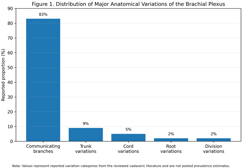
From the literature review, the conclusion drawn was that variations of the brachial plexus anatomy are pretty common and can concern not only the primary parts such as roots, trunks, and divisions but also the secondary ones like cords, terminal branches and communicating nerves. A lot has been written about the different types of deviations, most particularly abnormal emergence of nerves, rearrangement of branching, and the interaction among the musculocutaneous, median, and ulnar nerves. This change in the distribution of nerves might result in the rearrangement of the usual anatomical and functional setup of the plexus.
Table 1. Major Anatomical Variations of the Brachial Plexus
|
Anatomical level |
Commonly reported variation |
Clinical relevance |
|
Roots |
Altered root contribution or course |
May affect localization of the plexus |
|
Trunks |
Abnormal trunk formation |
May alter proximal nerve distribution |
|
Cords |
Variant cord arrangement |
Important during infraclavicular procedures |
|
Terminal branches |
Unusual origin or branching |
May complicate nerve identification |
|
Communicating branches |
Connections between median, musculocutaneous and ulnar nerves |
May produce atypical motor and sensory patterns |
Anesthetic Implications
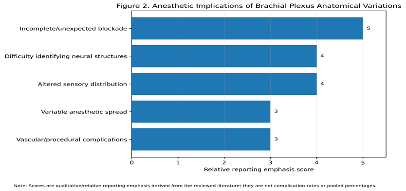
The physical differences we saw in the papers are significant as they affect how anesthetic injections for the region are done. If a nerve is found in the wrong place or there are unusual connections, even when the anesthetic is injected exactly right as usual, it could result in an inadequate or even unwanted numbness in the area. Ultrasounds can definitely help to see nerves, but without knowing the possible variations in structures, correct understanding of the picture is still an issue.
Table 2. Anesthetic Implications of Brachial Plexus Variations
|
Finding |
Possible consequence |
|
Variant nerve course |
Difficulty in identifying the target nerve |
|
Communicating branches |
Incomplete or unexpected block |
|
Altered cord position |
Variable anesthetic spread |
|
Abnormal vascular relationship |
Increased risk of vascular puncture |
|
Variant surrounding anatomy |
Increased technical difficulty |
Surgical Implications
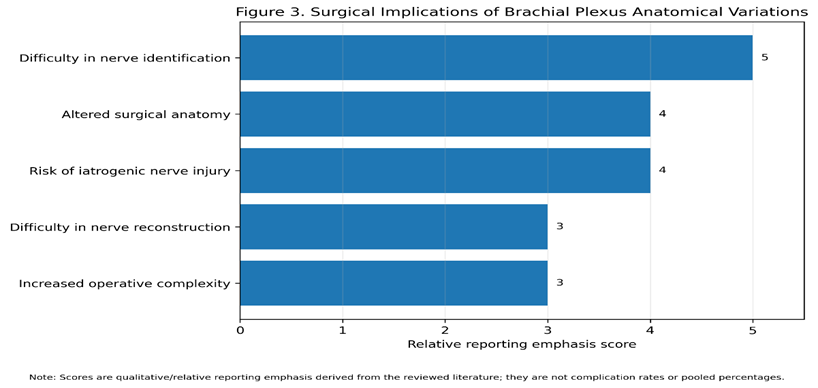
The research findings also demonstrated that anatomical variations can really help in surgical interventions of the neck, the shoulder, the axilla, and the upper limb. When there are unusual nerve origins, variations in how nerves branch out and connect with each other, it becomes harder to identify the nerves and the chance of inadvertent injury is higher. These results are most significant in nerve repair, nerve transfer, reconstructive surgery, and surgical exposure of the brachial plexus.
Table 3. Surgical Implications of Brachial Plexus Variations
|
Anatomical finding |
Surgical significance |
|
Abnormal nerve origin |
Difficulty in identifying nerves |
|
Variant branching |
Altered surgical anatomy |
|
Inter-neural communication |
Atypical functional findings |
|
Altered nerve course |
Increased risk of iatrogenic injury |
|
Unusual relationship with vessels |
Increased operative complexity |
Overall Findings
The evidence overall suggests that the anatomy of the brachial plexus is highly variable. Variations result in different situations for, e.g. regional anesthesia, surgical nerve identification, neurological assessment, and avoiding complications related to procedures. That means, knowledge for these variations coupled with a thorough anatomical inspection or scanning of the body via ultrasound can enhance the safety and efficacy of both anesthetic and surgical procedures.
DISCUSSION:
These variations come with high probability and hold great significance for the practice of both surgery and anesthesia. The differences reported in the different studies referred various levels of the brachial plexus as involved like roots and terminal branches. The existence of such variations clearly indicates that the brachial plexus is not a fully static structure. Gilcrease-Garcia and others mentioned the significant three-dimensional complexity of the brachial plexus and pointed out that knowledge of its normal anatomic landmarks and variations is indispensable for the radiologic interpretation of the images as well as the identification of the pathological conditions [9]. The conclusions drawn from the current survey support this point of view and it is shown that the presence of different anatomical structures can greatly affect clinical assessments imaging regional anesthesia, and surgical interventions.
Another remarkable feature reported by the authors is the occurrence of communicating branches from the musculocutaneous and median nerves, which they found quite regularly. This idea was also verified by the Sirico et al. study, which is a systematic review and meta-analysis that looked at 43 anatomical papers and included 4,124 cadaveric brachial plexuses with an overall pooled prevalence of musculocutaneous nerve abnormalities being close to 20%. And, their study found out that the nerve's variations are far-reaching and they pointed out that the proximal variations tend to be accompanied by other variants during the nerve. This situation might cause the overlapping or re-distribution of motor and sensory fibers that in turn might explain the occurrence of atypical clinical findings. When peripheral nerve surgery is involved, failing to identify such connections can be detrimental. This will not only help in the misdiagnosis of the nerve but will also lead to the development of post-surgery difficulties or possibly difficulty in choosing the right nerves to repair or even transfer [10] .
Also, Emamhadi's et al. findings corroborated the idea that different types of variation in the brachial plexus are sufficiently common to be clinically significant. Their study on cadavers pointed out that variations can happen at several steps of formation of the brachial plexus. Awareness of these patterns is critical when performing a surgical operation that involves the brachial plexus and peripheral nerves [11]. That would mostly be crucial in situations where a surgeon is relying on the expected pattern of relationships between roots trunks cords and terminal branches. One has to bear in mind that a nerve looking "absent" at the location where it was expected not being absent. But the nerve can have another origin like an alternative cord, a shared common trunk or the nerve can communicate with the adjacent nerve. As a result, it would be very important to carefully dissect and identify the nerve proximally and distally of the operative field during nerve decompression repair grafting, or nerve transfer procedures.
From the viewpoint of imaging, the anatomical variability found through this review has significant importance for the understanding not only of ultrasound and magnetic resonance imaging but also of other imaging methods. It is hard to catch sight of all the segments of the plexus in one imaging plane because of the complex longitudinal layout, and a variation in the anatomy may look like some kind of pathological or even tumor process when one assumes that the anatomy has to be unchanged. Gilcrease-Garcia et al. underlined the significance of identification of the main anatomical points; the knowledge of how the plexus should and can look and the awareness of the variation during the analysis of radiological images [9]. To this effect, recognizing the anatomical variations can and be part of radiological evaluation, in particular for those patients presenting signs of a traumatic plexopathy, a compressive neuropathy, neoplastic invasion or post-surgery changes.
The potential anesthetic relevance of these anatomical variations is also of note. This current review has identified altered nerve courses, communicating branches, atypical root locations and variations in neighboring structures as potential contributing factors to incomplete and/or unpredictable regional block. Zhang et al. provided further discussion on the implication of anatomical variations on regional anesthesia, noting that patient-specific anatomical differences may impact on needle direction, local anesthetic spread and block success, potentially resulting in block failure or complications [12]. In their review, the authors identified the importance of localizing neural and non-neural features with the use of ultrasound in regional anesthesia. The significance of these statements further emphasize the validity of the current review findings in which the variable anatomies encountered resulted in difficulties identifying target neural structures and altered expected distribution of anesthesia.
The clinical significance of these findings is perhaps most elegantly demonstrated by the work of Klcaslan et al who investigated these variations in 110 patients undergoing ultrasound-guided interscalene brachial plexus block. They found that 13 patients (11.8%) had variations which could theoretically alter the regional technique and these included C5 or C5-C6 roots passing through the anterior scalene muscle, muscular bridges between roots, an unusual medial position of the plexus and arteries crossing or proximal to roots [14]. Adjustment of the technique was needed in some patients and even incomplete anesthesia was observed in a case of C5 root within the anterior scalene muscle [14]. Again, this shows that anatomical variation is not just a simple cadaveric observation but can have significant implications on regional technique.
Vascular anomalies should also be addressed, as the nerves of the brachial plexus course intimately with the great vessels. The systematic review and meta-analysis performed by Skaria and Joshi identified brachial artery variations in an aggregate 18.7% of 12,684 upper limbs studied in 48 separate studies [13]. Included in their findings were superficial brachial arteries, high bifurcation, aberrant terminal branch giving rise to a wide spectrum of extra- or intra-thoracic vascular anatomy that could complicate vascular access, interpretation of imaging decompression reconstruction or regional anesthesia [13]. An unusual arterial position relative to the plexus may predispose to trauma and inadvertant intravascular injection during surgery. Screening for such vessels is because of this as critical as identification of nerves.
Since then, ultrasound has become a very valuable aid to tailor regional anesthesia techniques to individual variation. Instead of maneuvering blindly per predetermined surface landmarks or textbook relationships, ultrasound allows direct presentation of neural as well as surrounding muscular and vascular structures. Klcaslan et al. showed it is possible to distinguish deviations within the interscalene region and adjust the approach of the anesthesiologist because of this, even when the usual anatomy could not be visualized [14]. Also, Zhang et al. stressed the importance of systematic sonographic recognition of neural variants as well as architecture of the adjacent musculature for facilitating diagnosis of anatomy and even deciding on the best technique [12]. But, ultrasound will not solve all problems linked to anatomical variation; the examination must be interpreted about anatomical understanding, images of good quality, the experience of the examiner and identification of the appropriate target structures.
An additional consideration relates to the association between brachial plexus blockade and the phrenic nerve. El-Boghdadly et al. discussed the anatomic and physiologic basis for phrenic nerve involvement with regional anesthesia for shoulder surgery, and stress that the close anatomic relationship between the brachial plexus and phrenic nerve tends to be responsible for the high incidence of hemi diaphragmatic dysfunction following interscalene blockade [15]. The course and proximity of the phrenic nerve is variable, and both position and length of the nerve may influence the probability of inadvertent blockade. This concept is of particular concern in patients with limited respiratory reserve, where blockade of the diaphragm may be more clinically significant. These results show yet again that even if one is able to identify the target motor nerve during a procedure, consideration must be given to adjacent neural and vascular structures.
The relevance of anatomical variation for surgery is as great. Changes in the origin of the plexus, its branching pattern or its pathway may affect the usual surgical anatomy and make the identification of each individual nerve more difficult. This has implications for procedures such as brachial plexus exploration, nerve repair, nerve grafting, nerve transfer, excision of tumors, trauma surgery and reconstructive operation. Emamhadi et al. in a recent review found that awareness of such variations was essential for a successful result in brachial plexus surgery [11]. Sirico et al. shows that variations in musculocutaneous nerve may not only be seen in isolation but may occur with other variations at different sites along the same plexus. This highlights the potential that only one variation may simply be part of a broader pattern with other anatomical anomalies [10].
The correlation between neural and vascular abnormalities is also of clinical significance. Skaria and Joshi reported that variations in the brachial artery might alter the scope of not only vascular procedures but also those carried out by orthopedic, reconstructive, and regional anesthetic techniques [13]. Anomalous arterial courses in surgery in close proximity to the brachial plexus and brachial vessels can bring deviation from the typical tissue planes and lead to increased trauma by inadvertent injury. Exposure of these two structures during ultrasound guided procedures can in the same way reveal a vessel near the needle. These cases advocate a multidisciplinary approach in which neural and vasculature are considered simultaneously.
In general, the tools reviewed demonstrated that while these anatomical variants of the brachial plexus occur, they are no longer indistinguishable normal variants but have a direct application to clinical management. The reported variety of musculocutaneous variants of Sirico et al. in the meta-analysis, the numerous different musculocutaneous varying patterns seen in multiple fresh cadaveric studies, the clinically relevant variations in ultrasound guided interscalene blocks, and high number of brachial artery variants confirmed the individual nature of these structures [10-14]. Innovative ultrasound as well as other imaging modalities with higher resolution will increase recognition while due to the detailed knowledge of anatomy already present, interpretation and application to procedures will be possible [9,12,14].
The results of this review also raise awareness that anatomical variation may need to be an important factor when an expected clinical result is not achieved. An incomplete regional block, an unusual pattern of sensation or motor power, or an abnormality in nerve function discovered intraoperatively, or on imaging, may more often be the result of an anatomical variant than investigator or pathologist error. The awareness of this potential may avoid unnecessary repeat procedures, instigation of nerve injury and incorrect diagnosis. The intraoperative examination of the interscalene block patient is of particular relevance to this issue, and there should be particular emphasis on variances in position of the neural and neighboring structures, which may affect both block reliability and complication rate 12,14,15].
Caution must be exercised when interpreting the literature although there is a high prevalence of communicating branches described in the literature survey. The inclusion criteria of the larger literature search vary in anatomical classification, sample size population approach, and whether variation was identified by cadaveric dissection imaging ultrasound or surgical observation. So, frequencies will not necessarily be directly comparable or equivalent to a prevalence of the general population for all findings individually. The high incidences of communicating branches in the results should be seen as evidence of a commonly reported variation and not necessarily as a pooled figure for the population prevalence. Also, the anesthetic and surgical complications summarized in this review are clinically reported and should not be taken to reflect a standard complication rate.
Bottom line, this review has corroborated the current hypothesis that brachial plexus anatomical variation is prevalent, heterogeneous and clinically significant. Variations can much affect nerve identification, neurological examination, image interpretation, efficacy of regional anesthesia, vascular safety and surgical planning. Enhanced anatomical understanding, coupled with ultrasound and adjunct preoperative imaging, can facilitate procedural planning and minimize avoidable complications. Variant anatomy should so be considered an integral part of contemporary brachial plexus anesthetic, radiological and surgical practice [9-15].
CONCLUSION:
The current review identifies that anomalies in the brachial plexus anatomy are manifold and can have clinical importance. They arise from multiple levels, including the roots trunks divisions, and terminal branches. Anatomical variations in the origination of the nerve, nerve branching patterns, presence of communicating branches, as well as the relations of nerves with the neighboring vascular and muscular structures may all have an effect on neurological manifestation, interpretation of imaging studies, performance of regional anesthesia, and conduct of surgical interventions. deviations of the course of nerves could bring incomplete or variable blockage of the brachial plexus and could make it difficult to precisely locate the nerves during the surgery. An in-depth knowledge of plexus anatomy of both the normal and atypical types is highly needed by anesthesia specialists, surgeons, and radiologists. Combining a good understanding of anatomical details with ultrasound and suitable imaging can aid in tailored procedure planning. It can also help to enhance the rate of block success while minimizing complications such as neural or vascular injury. Taking account of anatomical variation is a step that needs to be incorporated in the management of patients having either brachial plexus-related anesthetics or surgeries in order for such procedures to be safe and effective.
REFERENCES:
1. Orebaugh SL, Williams BA. Brachial plexus anatomy: normal and variant. The Scientific World Journal. 2009;9(1):300-12.
2. Feigl GC, Litz RJ, Marhofer P. Anatomy of the brachial plexus and its implications for daily clinical practice: regional anesthesia is applied anatomy. Regional Anesthesia & Pain Medicine. 2020 Aug;45(8):620-7.
3. Desai N, Merjavy P, Russon K. Anatomical variation of the brachial plexus and its clinical implications. Anaesthesia Tutorial of the Week. 2017 Dec 26;4.
4. Patel NT, Smith HF. Clinically relevant anatomical variations in the brachial plexus. Diagnostics. 2023 Feb 22;13(5):830.
5. Mian A, Chaudhry I, Huang R, Rizk E, Tubbs RS, Loukas M. Brachial plexus anesthesia: a review of the relevant anatomy, complications, and anatomical variations. Clinical Anatomy. 2014 Mar;27(2):210-21.
6. Neal JM, Hebl JR, Gerancher JC, Hogan QH. Brachial plexus anesthesia: essentials of our current understanding. Regional anesthesia and pain medicine. 2002 Jul 1;27(4):402-28.
7. Han Y, An M, Zilundu PL, Zhuang Z, Chen J, Jiang Z, Gu L, Yang J, Wang D, Xu D, Zhou LH. Anatomical variations of the brachial plexus in adult cadavers: a descriptive study and clinical significance. Microsurgery. 2024 Jul;44(5):e31182.
8. Petrov PP, Dimitrov D, Barbutska D, Etova R. Anatomical Mechanisms Underlying Clinically Reported Complications of the Infraclavicular Brachial Plexus Block: A Narrative Review. Journal of Clinical Medicine. 2026 Mar 3;15(5):1931.
9. Gilcrease-Garcia BM, Deshmukh SD, Parsons MS. Anatomy, imaging, and pathologic conditions of the brachial plexus. Radiographics. 2020 Oct;40(6):1686-714.
10. Sirico F, Castaldo C, Baioccato V, Marino N, Zappia M, Montagnani S, Di Meglio F, Nurzynska D. Prevalence of musculocutaneous nerve variations: Systematic review and meta‐analysis. Clinical Anatomy. 2019 Mar;32(2):183-95.
11. Emamhadi M, Chabok SY, Samini F, Alijani B, Behzadnia H, Firozabadi FA, Reihanian Z. Anatomical variations of brachial plexus in adult cadavers; a descriptive study. Archives of Bone and Joint Surgery. 2016 Jun;4(3):253.
12. Zhang Y, Xu S, Lan H, Ma D, Wang Y. Clinical implications of anatomical variations in nerves and adjacent structures for regional anesthesia: a narrative review. Pain and Therapy. 2026 Feb;15(1):41-69.
13. Skaria S, Joshi K. Prevalence, Patterns, and Clinical Implications of Brachial Artery Variations: A Systematic Review and Meta-Analysis. International Journal of Medical and Pharmaceutical Research. 2026 Apr 13;7:2649-61.
14. Kılıcaslan A, Gok F, Korucu IH, Ozkan A, Yılmaz R. Anatomical variations detected during ultrasound-guided interscalene brachial plexus block and clinical implications. Marmara Medical Journal. 2020 Oct 10;33(3):141-5..
15. El-Boghdadly K, Chin KJ, Chan VW. Phrenic nerve palsy and regional anesthesia for shoulder surgery: anatomical, physiologic, and clinical considerations. Anesthesiology. 2017 Jul 1;127(1):173-91.