Clinical Profile And Prognosis In Lens Induced Glaucoma In A Tertiary Care Centre: A Prospective Observational Study.
- DR. SANJAY CHOUDHARY , Postgraduate Resident, Department Of Ophthalmology, Chhattisgarh Institute Of Medical Sciences (Cims), Bilaspur, Chhattisgarh, India.
- DR. PRABHA SONWANI , Associate Professor, Department Of Ophthalmology, Chhattisgarh Institute Of Medical Sciences (Cims), Bilaspur, Chhattisgarh, India.
- DR. SUCHITA SINGH , Professor And Head Of Department, Department Of Ophthalmology, Chhattisgarh Institute Of Medical Sciences (Cims), Bilaspur, Chhattisgarh, India.
Article Information:
Abstract:
Background: Lens-induced glaucoma (LIG) is a preventable cause of visual impairment associated with advanced cataract and delayed treatment. This study evaluated the clinical profile and visual outcomes of patients with LIG. Materials and Methods: This prospective observational study included 40 patients aged ≥40 years with LIG at a tertiary care center in Chhattisgarh. Clinical presentation, cataract morphology, intraocular pressure (IOP), type of LIG, visual acuity, fundus findings, and postoperative outcomes up to day 30 were assessed. Results: The mean age was 59.3 ± 11.2 years; 52.5% were males and 90% were from rural areas. Diminution of vision (100%), pain (95%), and redness (90%) were the common symptoms. Phacomorphic glaucoma accounted for 80% and phacolytic glaucoma for 20% of cases. Mean preoperative IOP was 42.85 mmHg, with 60% having IOP >40 mmHg. Preoperatively, 92.5% had visual acuity between <1/60 and perception of light. By day 30, 12.5% achieved 6/6–6/12 and 45% achieved 6/18–6/60 vision. Glaucomatous changes were present in 30% of patients. Conclusion: LIG commonly presented with advanced cataract, severe visual impairment, and markedly elevated IOP. Early diagnosis and timely cataract surgery are essential to improve visual outcomes and prevent irreversible glaucomatous optic nerve damage.
Keywords:
Article :
INTRODUCTION:
Lens-induced glaucoma (LIG) is an important cause of secondary glaucoma and preventable visual morbidity, particularly in developing countries where delayed cataract surgery allows mature and hypermature cataracts to persist.[1,2] Cataract remains a major cause of avoidable blindness worldwide, and disparities in access to timely cataract treatment continue to affect rural and socioeconomically disadvantaged populations.[3,4]
LIG develops due to changes in the crystalline lens that result in elevated intraocular pressure (IOP). The two predominant forms are phacomorphic and phacolytic glaucoma.[5,6] Phacomorphic glaucoma results from an intumescent lens causing pupillary block and secondary angle closure, whereas phacolytic glaucoma occurs when proteins from a mature or hypermature cataract obstruct aqueous outflow through the trabecular meshwork.[7–9]
Patients commonly present with severe diminution of vision, ocular pain, redness, corneal edema, inflammation, and markedly elevated IOP.[10–12] Visual prognosis depends largely on the duration of symptoms, severity and duration of IOP elevation, optic nerve damage, and delay in treatment.[13] Definitive management consists of cataract extraction after initial control of IOP and inflammation. Manual small-incision cataract surgery is particularly useful for advanced cataracts, although surgery may be challenging because of corneal edema, shallow anterior chamber, inflammation, and zonular weakness.[14,15]
LIG is largely preventable through early detection and timely cataract surgery. Delayed presentation due to poor awareness, fear of surgery, financial constraints, and limited access to ophthalmic services may result in irreversible glaucomatous optic nerve damage and poor visual recovery.[4,13] Therefore, the present prospective observational study was undertaken to evaluate the demographic and clinical profile and visual outcomes of patients with lens-induced glaucoma presenting to a tertiary care centre.
MATERIALS AND METHODS:
This prospective observational study was conducted in the Department of Ophthalmology, Chhattisgarh Institute of Medical Sciences (CIMS), Bilaspur, Chhattisgarh, over one year. Patients presenting to the ophthalmology OPD or emergency services with lens-induced glaucoma (LIG) were prospectively enrolled.Patients aged ≥40 years with clinically diagnosed LIG were included.
Eligibility Criteria
Patients aged ≥40 years with clinically diagnosed LIG who provided written informed consent and agreed to follow-up were included. Patients with primary open-angle or angle-closure glaucoma, secondary glaucoma unrelated to lens pathology, previous intraocular surgery, significant corneal opacity preventing adequate examination, or inability to complete follow-up were excluded.
Clinical Assessment
Demographic characteristics, residence, presenting complaints, duration of symptoms, ocular history, previous treatment, and systemic comorbidities were recorded using a structured proforma. All patients underwent comprehensive ophthalmological examination, including uncorrected visual acuity (UCVA), best-corrected visual acuity (BCVA), slit-lamp examination, lens evaluation, and intraocular pressure (IOP) measurement. Visual acuity was assessed using a Snellen chart and, in patients with severe visual impairment, recorded as counting fingers, hand movements, perception of light, or no perception of light.Anterior segment examination included assessment of conjunctival congestion, corneal edema, anterior chamber depth and inflammation, iris abnormalities, pupil, and lens status. Cataract morphology and maturity were documented, with LOCS III grading used wherever applicable. IOP was measured using a non-contact tonometer.
Gonioscopy and Fundus Examination
Gonioscopy was performed after initial IOP control, whenever feasible, using a Goldmann three-mirror or four-mirror goniolens. Angle configuration and peripheral anterior synechiae were assessed using the Shaffer grading system. Fundus examination was performed whenever media clarity permitted and included assessment of the optic disc, cup-to-disc ratio, macula, retinal vessels, and peripheral retina. Preoperative biometry included keratometry, axial length measurement, and IOL power calculation using the SRK/T formula.
Classification of Lens-Induced Glaucoma
Based on clinical findings, LIG was classified as phacomorphic, phacolytic, lens-particle, phacoanaphylactic, or lens-dislocation–induced glaucoma according to lens status, anterior chamber configuration, inflammation, and mechanism of IOP elevation.
Management
Initial treatment was aimed at reducing IOP and controlling inflammation. Topical antiglaucoma medications included timolol, brimonidine, and/or dorzolamide. Systemic acetazolamide and intravenous 20% mannitol were administered when indicated. Topical corticosteroids, cycloplegics, analgesics, and antiemetics were used according to clinical requirements.Following adequate IOP control and improvement in ocular condition, definitive cataract surgery was performed. Patients with controlled IOP and absent or minimal peripheral anterior synechiae generally underwent SICS/MSICS with posterior chamber IOL implantation, while phacoemulsification was performed in selected cases. Combined cataract extraction with trabeculectomy was considered in patients with extensive synechial angle closure or persistently uncontrolled IOP. All intraoperative complications were documented.
Postoperative Follow-up and Outcomes
Patients were followed on postoperative day 1, day 5, and day 30. At each visit, visual acuity, IOP, corneal clarity, anterior chamber reaction, IOL position, and postoperative complications were assessed. Fundus examination was performed when media clarity permitted.The final assessment was performed at one month. Visual outcome based on BCVA was categorized as good (≥6/18), borderline (6/24–6/60), or poor (<6/60). Causes of poor visual outcome, including glaucomatous optic atrophy, corneal decompensation, persistent inflammation, cystoid macular edema, posterior capsular opacification, posterior segment pathology, and surgical complications, were documented.
Statistical Analysis
Data were analyzed using IBM SPSS Statistics version 28.0. Categorical variables were expressed as frequency and percentage, while continuous variables were presented as mean ± SD or median (IQR), as appropriate. Normality was assessed using the Shapiro–Wilk test. Chi-square/Fisher's exact test was used for categorical variables; paired or independent t-test and their non-parametric equivalents were used for continuous variables. ANOVA or Kruskal–Wallis test was used for comparisons involving more than two groups. Pearson's or Spearman's correlation and odds ratios with 95% confidence intervals were calculated where appropriate. A two-tailed p-value <0.05 was considered statistically significant.
RESULTS:
A total of 40 patients with lens-induced glaucoma (LIG) were included in the study. The mean age of the participants was 59.3 ± 11.2 years, with the largest proportion belonging to the 61–70-year age group (32.5%). There was a slight male predominance, with 21 (52.5%) males and 19 (47.5%) females. Most patients (90.0%) were from rural areas. Diminution of vision was present in all patients (100.0%), followed by ocular pain in 95.0%, redness in 90.0%, and watering in 42.5%. Acute symptoms of ≤7 days were reported by 62.5% of patients. The mean duration of symptoms was 8.2 ± 5.3 days, with a median duration of 7 days (IQR: 4–10 days). The mean preoperative intraocular pressure (IOP) was 42.85 mmHg, and 60.0% of patients had an IOP >40 mmHg (Table 1).
On preoperative ocular examination, Van Herick grade 0 was observed in 37.5% of patients, followed by grade 1 in 22.5%, grade 2 in 20.0%, grade 3 in 17.5%, and grade 4 in 2.5%. Regarding preoperative IOP distribution, 30.0% of patients had an IOP of 41–50 mmHg and another 30.0% had an IOP >50 mmHg, while 25.0% and 15.0% had IOP values of 31–40 mmHg and 21–30 mmHg, respectively (Table 2).
Mature cataract was the most common cataract morphology, observed in 42.5% of patients, followed by hypermature cataract in 37.5% and intumescent cataract in 20.0%. Phacomorphic glaucoma was the predominant type of LIG, accounting for 80.0% of cases, whereas phacolytic glaucoma was present in 20.0% (Table 3).
Fundus examination revealed normal findings in 28 (70.0%) patients, while glaucomatous changes were present in 12 (30.0%) patients (Table 4, Figure 1).
Among the 14 patients with poor visual outcome, glaucomatous optic atrophy was the most common cause, accounting for 7 (50.0%) cases. Delayed presentation was responsible for poor outcome in 4 (28.6%) patients, while 3 (21.4%) patients had no perception of light with irreversible damage (Table 5, Figure 2).
Preoperatively, visual acuity was markedly impaired, with 37 (92.5%) patients having visual acuity <1/60 to perception of light (PL) and 3 (7.5%) having no PL. A substantial improvement in visual acuity was observed following treatment. By postoperative day 5, 13 (32.5%) patients had achieved visual acuity of 6/18–6/60, while 15 (37.5%) had visual acuity between <6/60 and 3/60. At day 30, further improvement was evident, with 5 (12.5%) patients achieving visual acuity of 6/6–6/12 and 19 (47.5%) achieving 6/18–6/60. Thus, by day 30, 60.0% of patients had visual acuity of 6/60 or better, compared with none preoperatively. However, 7 (17.5%) patients remained in the <1/60–PL category and 3 (7.5%) continued to have no PL (Table 6, Figure 3).
Overall, the findings demonstrate substantial postoperative improvement in visual acuity, although a proportion of patients continued to have poor visual outcomes, predominantly due to glaucomatous optic atrophy and delayed presentation.
Table 1. Demographic and clinical profile of study participants (n=40)
|
Parameter |
Number (%) / Value |
|
Demographic characteristics |
|
|
Age, mean ± SD (years) |
59.3 ± 11.2 |
|
40–50 years |
11 (27.5) |
|
51–60 years |
10 (25.0) |
|
61–70 years |
13 (32.5) |
|
>70 years |
6 (15.0) |
|
Male |
21 (52.5) |
|
Female |
19 (47.5) |
|
Rural residence |
36 (90.0) |
|
Urban residence |
4 (10.0) |
|
Clinical characteristics |
|
|
Diminution of vision |
40 (100.0) |
|
Ocular pain |
38 (95.0) |
|
Redness |
36 (90.0) |
|
Watering |
17 (42.5) |
|
Acute symptoms ≤7 days |
25 (62.5) |
|
Acute symptoms 8–14 days |
7 (17.5) |
|
Acute symptoms >14 days |
8 (20.0) |
|
Duration of symptoms, mean ± SD (days) |
8.2 ± 5.3 |
|
Median (IQR), days |
7 (4–10) |
|
Preoperative IOP, mean (mmHg) |
42.85 |
|
IOP >40 mmHg |
24 (60.0) |
Table 2. Preoperative ocular characteristics (n=40)
|
Parameter |
Number (%) / Value |
|
Van Herick grade |
|
|
Grade 0 |
15 (37.5) |
|
Grade 1 |
9 (22.5) |
|
Grade 2 |
8 (20.0) |
|
Grade 3 |
7 (17.5) |
|
Grade 4 |
1 (2.5) |
|
Preoperative IOP |
|
|
Mean IOP (mmHg) |
42.85 |
|
21–30 mmHg |
6 (15.0) |
|
31–40 mmHg |
10 (25.0) |
|
41–50 mmHg |
12 (30.0) |
|
>50 mmHg |
12 (30.0) |
Table 3. Cataract morphology and type of lens-induced glaucoma (n=40)
|
Parameter |
Number (%) |
|
Cataract morphology |
|
|
Mature |
17 (42.5) |
|
Hypermature |
15 (37.5) |
|
Intumescent |
8 (20.0) |
|
Type of LIG |
|
|
Phacomorphic |
32 (80.0) |
|
Phacolytic |
8 (20.0) |
Table 4. Fundus findings among study participants (n=40)
|
Fundus finding |
Number |
Percentage (%) |
|
Normal |
28 |
70.0 |
|
Glaucomatous changes |
12 |
30.0 |
|
Total |
40 |
100.0 |
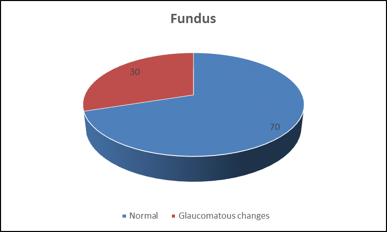
Figure 1. Fundus findings among study participants (n=40)
Table 5. Causes of poor visual outcome among affected patients (n=14)
|
Cause |
Number |
Percentage (%) |
|
Glaucomatous optic atrophy |
7 |
50.0 |
|
Delayed presentation |
4 |
28.6 |
|
No PL with irreversible damage |
3 |
21.4 |
|
Total |
14 |
100.0 |
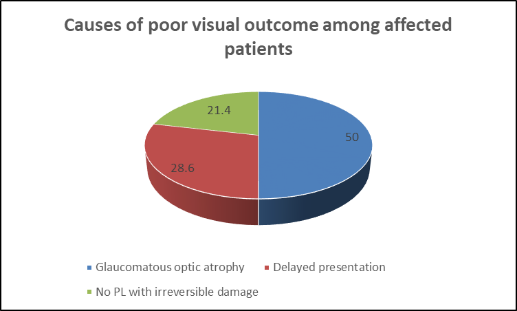
Figure 2. Causes of poor visual outcome among affected patients
Table 6. Comparison of preoperative and postoperative visual acuity (n=40)
|
Visual acuity |
Preoperative, n (%) |
Day 5, n (%) |
Day 30, n (%) |
|
6/6–6/12 |
0 (0.0) |
0 (0.0) |
5 (12.5) |
|
6/18–6/60 |
0 (0.0) |
13 (32.5) |
19 (47.5) |
|
<6/60–3/60 |
0 (0.0) |
15 (37.5) |
5 (12.5) |
|
<3/60–1/60 |
0 (0.0) |
2 (5.0) |
1 (2.5) |
|
<1/60–PL |
37 (92.5) |
7 (17.5) |
7 (17.5) |
|
No PL |
3 (7.5) |
3 (7.5) |
3 (7.5) |
|
Total |
40 (100.0) |
40 (100.0) |
40 (100.0) |
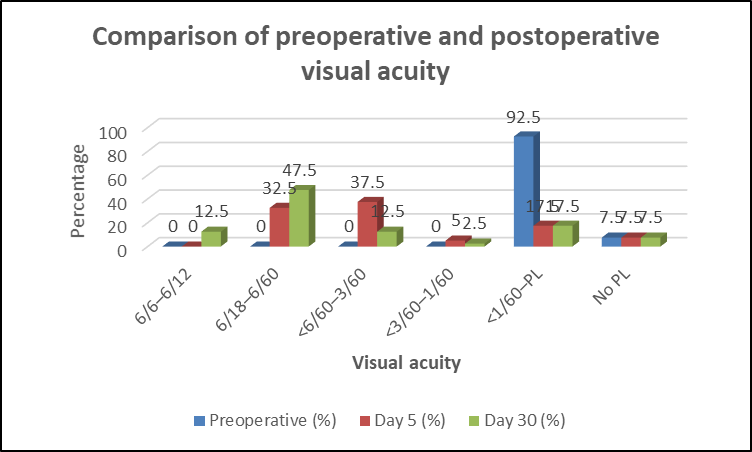
Figure 3. Postoperative visual acuity at day 5 and day 30
DISCUSSION:
The mean age in our study was 59.3 ± 11.2 years, with most patients aged 61–70 years (32.5%). Comparable mean ages were reported by Bhuyan and Baishyakh [11](61.54 years), Sharanabasamma et al. [15] (60.68 years), Shrestha et al. [16] (61.5 ± 8.9 years), and Ayub et al. [13] (63.8 years). Our study showed slight male predominance (52.5%), whereas Bhuyan and Baishyakh [11](58% females), Shrestha et al. [16] (56.6% females), and Ayub et al. [13] (58.8% females) reported female predominance. Rural patients constituted 90% in our study, similar to the rural predominance (62.5%) reported by Sinha et al. [10]Diminution of vision (100%), ocular pain (95%), and redness (90%) were the major presenting symptoms. Bhuyan and Baishyakh [11]similarly reported diminished vision and pain in 100% each and redness in 92%, while Shrestha et al. [16] reported diminished vision in 100% and pain in 96.2%.In our study, 62.5% presented within ≤7 days, with a mean symptom duration of 8.2 ± 5.3 days. Bhuyan and Baishyakh [11]reported presentation within 7 days in 56%, while Sinha et al. [10] observed better visual outcomes among patients presenting within 7 days.The mean preoperative IOP was 42.85 mmHg, with 60% having IOP >40 mmHg. Comparable findings were reported by Bhuyan and Baishyakh [11](43.84 ± 9.93 mmHg; 64% >40 mmHg) and Shrestha et al. [16] (64.2% >40 mmHg), while Rajkumari et al. [14] reported a lower mean IOP of 35.14 ± 6.35 mmHg.
In our study, phacomorphic glaucoma predominated (80%), while phacolytic glaucoma accounted for 20%. Phacomorphic predominance was also reported by Bhuyan and Baishyakh [11](58%), Shrestha et al. [16] (71.7%), and Sinha et al. [10] (68.3%). In contrast, Ayub et al. [13] reported phacolytic glaucoma in 70.3% and phacomorphic glaucoma in 29.7%.Glaucomatous fundus changes were present in 30% of our patients. Sharanabasamma et al. [15] reported optic disc damage in 42%, increasing to 80% among patients presenting after two weeks. Similarly, Shrestha et al. [16] reported that 8 of 11 patients with optic atrophy had presented with IOP >40 mmHg, highlighting the adverse effect of prolonged IOP elevation.Preoperatively, 92.5% of our patients had visual acuity <1/60–PL and 7.5% had no PL. Similarly, Sinha et al. [10] reported hand movements or worse in 92.5%, while Shrestha et al. [16] reported visual acuity <3/60 in all patients.At day 30, 12.5% achieved 6/6–6/12 and 47.5% achieved 6/18–6/60, giving an overall 60% with vision ≥6/60. Sinha et al. [10] reported vision ≥6/60 in 69.2%, Sharanabasamma et al. [15] reported ≥6/18 in 54%, and Ayub et al. [13] reported ≥6/18 in 75.6%. Rajkumari et al. [14] reported 6/6–6/12 in 70.8%, while Bhuyan and Baishyakh [11]reported 6/9 and 6/12 vision in 38% and 28%, respectively.Among 14 patients with poor visual outcome, glaucomatous optic atrophy was the commonest cause (50%), followed by delayed presentation (28.6%) and irreversible damage with no PL (21.4%). Sinha et al. [10], Shrestha et al. [16], and Ayub et al. [13] similarly identified optic atrophy as an important cause of poor postoperative vision. Sharanabasamma et al. [15] reported that 72% of patients presenting within two weeks achieved vision ≥6/12, whereas optic disc damage occurred in 80% of those presenting later.
CONCLUSION:
Lens-induced glaucoma commonly presented with severe visual impairment and markedly elevated IOP, with phacomorphic glaucoma being the predominant type. Surgical management resulted in substantial improvement in visual acuity in the majority of patients. Delayed presentation and glaucomatous optic atrophy were important causes of poor visual outcome, emphasizing the need for early diagnosis and timely cataract surgery.
LIMITATIONS
The study was limited by its small sample size, single-center design, and relatively short postoperative follow-up of 30 days. Longer multicentric studies with larger populations are required to evaluate long-term visual and IOP outcomes.
REFERENCES:
1. Shah SS, Meyer JJ. Lens-Induced Glaucoma. In: StatPearls [Internet]. Treasure Island (FL): StatPearls Publishing; 2024.
2. Chandrashekharan DK, Pawar N, Giri S, et al. Outcomes and favourable prognostic factors in patients of phacomorphic and phacolytic glaucoma managed by manual small-incision cataract surgery: a retrospective study. Indian J Ophthalmol. 2022;70(6):2078-2084.
3. Prajna NV, Ramakrishnan R, Krishnadas R, et al. Lens induced glaucomas--visual results and risk factors for final visual acuity. Indian J Ophthalmol. 1996;44(3):149-155.
4. Kothari M, Thomas R, Parikh R, et al. Lens-induced glaucoma: the need to spread awareness about early management of cataract among rural population. ISRN Ophthalmol. 2013;2013:581727.
5. Pascolini D, Mariotti SP. Global estimates of visual impairment: 2010. Br J Ophthalmol. 2012;96(5):614-618.
6. Singh N, Eeda SS, Gudapati BK, et al. Understanding the cataract treatment disparities among older adults in India: evidence from Longitudinal Aging Study in India. PLoS One. 2024;19(1):e0296040.
7. Rao SK, Padmanabhan P. Phacomorphic glaucoma. Ophthalmology. 1998;105(4):749-750.
8. Dhingra S, Khillan T, Sindal MD, et al. Phacolytic glaucoma: a nearly forgotten entity. Oman J Ophthalmol. 2019;12(1):46-48.
9. Epstein DL. Diagnosis and management of lens-induced glaucoma. Ophthalmology. 1982;89(3):227-230.
10. Sinha R, Das D, Karmakar A, et al. Analysis of clinical profile and outcome in cases of lens-induced glaucoma. Global J Cataract Surg Res Ophthalmol. 2024;3(1):56-62.
11. Bhuyan J, Baishyakh P. A clinical study on the incidence and visual outcome after surgical management of lens-induced glaucoma. Galore Int J Health Sci Res. 2021;6(4):42-53.
12. Vudayana D, Sativada L, Jerry P, et al. Clinical study of lens induced glaucoma at a tertiary eye care centre. J Evid Based Med Healthc. 2021;8(10):537-541.
13. Ayub R, Tom LM, Venkatesh R, et al. Outcomes and reasons for late presentation of lens induced glaucoma: a prospective study. Ophthalmol Glaucoma. 2021;4(5):504-511.
14. Rajkumari S, Phookun A, Deuri N, et al. Manual small incision cataract surgery in phacomorphic glaucoma: surgical technique and outcome in North-eastern India. Indian J Ophthalmol. 2013;61(6):279-284.
15. Sharanabasamma M, Vaibhav K, Divya N. Management and visual outcome in patients of lens-induced glaucomas at a tertiary eye care hospital in South India. J Clin Diagn Res. 2016;10(6):NC01-NC05.
16. Shrestha RK, Joshi SN, Shakya S, et al. Lens induced glaucoma in a tertiary eye care centre in Western Nepal. Nepal J Ophthalmol. 2019;11(22):159-165.