Tuberculous Pseudo aneurysm of the Ascending Aorta with Contained Rupture in a patient with Disseminated Tuberculosis: A Case Report.
- Dr. Zubin Parekh , Senior Resident TNMC & BYL Nair Ch hospital, Department of CVTS, Mumbai Maharashtra India.
- Dr. Atul Patil , Senior Resident TNMC & BYL Nair Ch hospital, Department of CVTS, Mumbai Maharashtra India.
- Dr. Husain Najmi , Senior Resident TNMC & BYL Nair Ch hospital, Department of CVTS, Mumbai Maharashtra India.
- Dr. Shweta Deshpande , Additional Professor TNMC & BYL Nair Ch hospital, Department of CVTS, Mumbai Maharashtra India.
- Dr. Kanak Nagle , Professor and Head TNMC & BYL Nair Ch hospital, Department of CVTS, Mumbai Maharashtra India.
Article Information:
Abstract:
Background: Mycotic aneurysms of the ascending aorta secondary to Mycobacterium tuberculosis are rare but life-threatening, and diagnosis is often delayed because their clinical and radiological features overlap with more common mediastinal diseases. Case Presentation: We report a 30-year-old man with microbiologically confirmed pulmonary tuberculosis receiving antitubercular therapy, in whom contrast-enhanced CT incidentally detected a saccular ascending aortic pseudoaneurysm. Serial imaging demonstrated rapid enlargement with a surrounding hematoma, consistent with contained rupture. The patient underwent successful excision of the pseudoaneurysm and ascending aortic reconstruction using a Dacron graft. Histopathology revealed necrotizing granulomatous inflammation, supporting tuberculous aortitis in the setting of disseminated tuberculosis despite negative AFB staining of the aortic tissue. Recovery was uneventful. Conclusion: Tuberculous pseudoaneurysm of the ascending aorta should be considered in patients with active tuberculosis and mediastinal abnormalities. Tuberculous pseudoaneurysm of the ascending aorta demands aggressive and prompt surgical intervention due to its extreme risk of catastrophic rupture. Despite formidable technical hurdles—including dense adhesions, intraoperative rupture necessitating deep hypothermic circulatory arrest, and active infection—timely surgical excision and graft reconstruction combined with antitubercular therapy successfully achieved anatomical restoration and long-term clinical stability.
Keywords:
Article :
INTRODUCTION:
Mycotic aneurysms constitute a small proportion of all aortic aneurysms, and tuberculous involvement of the aorta is an exceptionally rare manifestation of extra pulmonary tuberculosis. Tuberculous aortitis most commonly results from contiguous spread of infection from adjacent mediastinal lymph nodes, vertebral foci, or pleuropulmonary disease, although haematogenous dissemination or seeding via the vasa vasorum has also been described. The ensuing destruction of the aortic wall predisposes to pseudo aneurysm formation and rupture. Because the descending thoracic and abdominal aorta lies in closer proximity to mediastina and para-aortic lymph nodes, ascending aortic involvement is distinctly uncommon. Early recognition is essential, as delayed diagnosis carries a high risk of catastrophic rupture, whereas timely surgical intervention combined with ant tubercular therapy is associated with favourable outcomes.
We report a patient with disseminated tuberculosis in whom an incidentally detected ascending aortic psuedoaneurysm demonstrated rapid interval progression to contained rupture, necessitating urgent surgical repair. The case highlights the diagnostic challenges and the importance of integrating radiological, operative and histopathological findings in establishing the diagnosis of tuberculous aortitis.
MATERIALS AND METHODS:
History and Initial Presentation
A 30-year-old male, non-smoker, tobacco chewer, with no known comorbidities and a history of TB contact (father, pulmonary TB, treated), presented in October 2025 with dry cough, low-grade fever with chills, and loss of appetite/weight over several weeks. He had no dyspnea or chest pain at onset. Outside evaluation with chest imaging (14/10/2025) showed bilateral variable-sized cavitary consolidations suggestive of tuberculous etiology, and he was started on four-drug fixed-dose combination ATT (4FDC) under the National TB Elimination Programme on 15/10/2025.
Course and Incidental Discovery of Aortic Aneurysm
The patient was admitted to the Department of Pulmonary Medicine (24/12/2025–01/01/2026) for further evaluation of persistent consolidation and bilateral cervical lymphadenopathy. Sputum CBNAAT (27/12/2025) confirmed MTB detected (very low), rifampicin resistance not detected. During this admission, he was noted to have right-sided facial deviation (upper motor neuron pattern), prompting neurological evaluation.
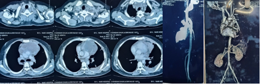
Contrast-enhanced CT thorax performed as part of TB workup (study date 27/12/2025) incidentally revealed a focal saccular aneurysm arising from the anterior wall of the ascending aorta, approximately 4.3 cm distal to the aortic annulus, measuring 23.4 mm with a neck of 14.7 mm, associated with a large perianeurysmal thrombus extending into the anterior mediastinum (7.3 × 8.9 × 6.2 cm), causing compression of the superior vena cava. No active contrast extravasation was noted at this stage.
MRI brain (11/01/2026) performed for the facial nerve findings showed an ill-defined lobulated T2-hyperintense, T1-hypointense lesion in the right pons (1.8 × 1.1 cm) without diffusion restriction, favored to be infective in etiology.
Repeat CT aortogram three weeks later (19/01/2026) showed interval enlargement of the aneurysm to 3.8 × 5.1 cm with a surrounding hematoma of approximately 250 cc and posterior displacement of mediastinal vessels — findings read as secondary to contained rupture of the aneurysm, with the possibility of mycotic aneurysm specifically raised by radiology. There was still no evidence of active contrast extravasation.
Contrast MRI brain (24/01/2026) showed the pontine lesion unchanged in appearance and reclassified as chronic insult sequelae with no features of active infection or neoplasm; a new incidental ring-enhancing granuloma was noted in the left parietal lobe (7 × 4 mm), read as suggestive of a TB granuloma in the context of the patient's known disease.
Given the enlarging aneurysm and perianeurysmal thrombus, the patient was started on anticoagulation (rivaroxaban 15 mg BD, later stepped down to 20 mg OD) and referred to the Department of Cardiovascular and Thoracic Surgery for further management.
Surgical Management
The patient was admitted under CVTS on 04/02/2026. Pre-operative 2D echocardiography showed normal biventricular function (LVEF 60%), mild tricuspid regurgitation, and no significant valvular pathology. He underwent excision of the ascending aortic pseudoaneurysm with ascending aortic augmentation using a Dacron graft on 11/02/2026.
Right infraclavicular incision was made at the deltopectoral groove, the right axillary artery was identified and looped, and a 10 mm straight graft was anastomosed to it in an end-to-side fashion for arterial inflow.A midline sternotomy was then performed.
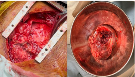
Operative findings included frank pus, a thick-walled pseudoaneurysmal sac, and intra-aneurysmal thrombus. Given the presence of dense interpericardial adhesions forming a mass-like structure over the aorta and the resulting risk of injury on direct sternotomy, arterial cannulation was established peripherally. Adhesiolysis was carried out to expose the right atrium, followed by right atrial purse-string placement and venous cannulation, establishing complete cardiopulmonary bypass. After completion of adhesiolysis, the pseudoaneurysmal sac was opened, releasing frank pus, which was sent for culture. During the process of adhesiolysis, the aneurysm inadvertently ruptured; consequently, the patient was placed on deep hypothermic circulatory arrest (DHCA).
After carefully defining the proximal and distal extent of the lesion, the aortic cross-clamp was applied and antegrade hyperkalemic sanguineous cardioplegia was administered to arrest the heart in diastole. The pseudoaneurysmal sac was opened, releasing frank pus which was sent for culture. Following the removal of intra-aneurysmal thrombus and the excision of the aneurysmal sac, the posterior wall was found to be intact and healthy; therefore, patch augmentation of the anterior wall of the aorta was performed using a Dacron graft with 5-0 Prolene. Thorough saline wash and antibiotic lavage were given prior to closure. The heart reverted to normal sinus rhythm after cross-clamp removal; the patient was weaned from bypass, protaminized, and de-cannulated with satisfactory hemostasis. No intra-aortic balloon pump was required. Cardiopulmonary bypass time was 100 minutes and aortic cross-clamp time was 48 minutes. Standard chest closure was performed over bilateral mediastinal drains, and the patient was shifted to the CVTS recovery room in stable hemodynamic condition for ventilatory support.
Histopathological examination (Surgical Pathology No. 832/26, reported 23/02/2026) of the excised aneurysmal/perianeurysmal tissue showed an ulcerated aortic wall with a large fibrin thrombus, wall disruption with extensive areas of necrosis, and epithelioid cell granulomas with giant cells — overall features of an aortic aneurysm with fresh fibrin thrombus and necrotising granulomatous inflammation. Special stains for AFB and fungi were both negative. The frank pus drained intraoperatively was also sent for routine (pyogenic) culture, which similarly showed no growth. Although microbiological confirmation could not be obtained from the vascular specimen , necrotizing granulomatous inflammation in the context of microbiologically confirmed pulmonary tuberculosis strongly supported tubercular aortitis.
Postoperative Course and Follow-up
The postoperative course was uneventful. Post-operative echocardiography showed preserved LV function (LVEF 60%) with trivial mitral regurgitation and mild tricuspid regurgitation. The patient was discharged on 20/02/2026 in stable condition on antitubercular therapy (ATT), antiplatelet therapy with aspirin (Ecosprin) 75 mg once daily, and a structured follow-up plan across CVTS, Cardiology, and Physiotherapy outpatient services. ATT was planned for a total duration of 6 months, comprising a 2-month intensive phase with isoniazid, rifampicin, pyrazinamide, and ethambutol (HRZE), followed by a 4-month continuation phase with isoniazid, rifampicin, and ethambutol (HRE).
Follow-up 2D echocardiography on 20/03/2026 showed mild aortic regurgitation with the remaining findings consistent with the immediate post-operative study, and no evidence of graft-related complications at that stage. CECT Thorax was done on 30/04/2026 suggestive of significant reduction in size of aneurysm and significantly reduced perianuerysmal thrombus. Focal out pouching of 1.4×0.7cm noted in ascending aorta . Pulmonary changes sequale of old infectious aetiology . At post operative follow up on 07 August 2026 , the patient remained clinically well , was hemodynamically stable and had gained weight and reported no new symptoms. He was advised to continue aspirin 75mg once daily. No evidence of recurrent cardiovascular symptoms was noted during follow up period.
Investigations Summary
Table! . Investigation
|
Investigation |
Date |
Key Finding |
|
Sputum CBNAAT |
27/12/2025 |
MTB detected (very low); rifampicin resistance not detected |
|
CECT Thorax |
27/12/2025 |
Saccular ascending aortic aneurysm (23.4 mm) with perianeurysmal thrombus/mediastinal mass (7.3×8.9×6.2 cm) compressing SVC |
|
USG Neck |
27/12/2025 |
Bilateral cervical lymphadenopathy, largest 1.4 cm, feasible for biopsy |
|
MRI Brain + Angio |
11/01/2026 |
Right pontine lesion 1.8×1.1 cm, T2 hyperintense, no restriction — favored infective |
|
USG Abdomen |
29/12/2025 |
Hepatosplenomegaly |
|
CT Aortogram |
19/01/2026 |
Aneurysm enlarged to 3.8×5.1 cm with surrounding hematoma (~250 cc) — contained rupture; mycotic aneurysm considered |
|
MRI Brain (contrast) |
24/01/2026 |
Pontine lesion unchanged — chronic sequelae; new left parietal ring-enhancing granuloma (7×4 mm), s/o TB granuloma |
|
2D Echo (pre-op) |
09/02/2026 |
LVEF 60%, mild TR, no RWMA, structurally normal valves |
|
Surgery |
11/02/2026 |
Excision of ascending aortic pseudoaneurysm with augmentation, Dacron graft |
|
Histopathology |
23/02/2026 |
Ulcerated aortic wall, necrosis, epithelioid granulomas with giant cells — necrotising granulomatous inflammation; AFB & fungal stains negative |
|
2D Echo (post-op, follow-up) |
20/03/2026 |
Mild AR; findings otherwise stable |
|
CECT Thorax |
30/04/2026 |
Significant reduction in size of aneurysm and significantly reduced perianuerysmal thrombus.Focal out pouching of 1.4×0.7cm noted in ascending aorta . Pulmonary changes sequale of old infectious etiology |
Pre operative
Intra operative
Post Operative
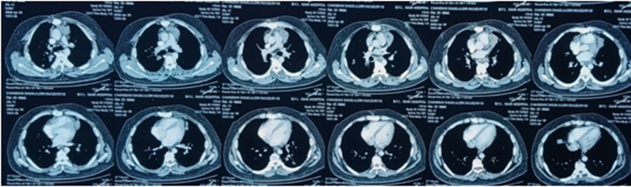
Table 2. Timeline of Clinical Events
|
Date |
Clinical Event |
|
October 2025 |
Patient presented with dry cough, low-grade fever, anorexia, and weight loss. |
|
14 October 2025 |
Chest imaging demonstrated bilateral cavitary pulmonary lesions suggestive of tuberculosis. |
|
15 October 2025 |
Four-drug antitubercular therapy (HRZE) initiated under the National TB Elimination Programme. |
|
24 December 2025 |
Admitted for evaluation of persistent pulmonary consolidation and bilateral cervical lymphadenopathy. |
|
27 December 2025 |
Sputum CBNAAT detected Mycobacterium tuberculosis (rifampicin sensitive). Contrast-enhanced CT thorax incidentally revealed a 23.4-mm saccular ascending aortic pseudoaneurysm with a large perianeurysmal thrombus causing superior vena cava compression. Ultrasonography confirmed bilateral cervical lymphadenopathy. |
|
29 December 2025 |
Ultrasonography of the abdomen demonstrated hepatosplenomegaly. |
|
11 January 2026 |
MRI brain revealed a right pontine lesion, initially considered infective in etiology. |
|
19 January 2026 |
CT aortogram demonstrated rapid enlargement of the pseudoaneurysm (3.8 × 5.1 cm) with a surrounding hematoma consistent with contained rupture. |
|
24 January 2026 |
Contrast-enhanced MRI brain showed a left parietal ring-enhancing granuloma suggestive of central nervous system tuberculosis. |
|
04 February 2026 |
Admitted under the Department of Cardiovascular and Thoracic Surgery for definitive surgical management. |
|
09 February 2026 |
Preoperative transthoracic echocardiography demonstrated preserved biventricular function (LVEF 60%) without significant valvular disease. |
|
11 February 2026 |
Excision of the ascending aortic pseudoaneurysm with ascending aortic augmentation using a Dacron graft. Intraoperative findings included a thick-walled pseudoaneurysm containing thrombus and frank pus. |
|
13 February 2026 |
Routine microbiological culture of intraoperative pus showed no bacterial growth. |
|
20 February 2026 |
Discharged in stable condition on antitubercular therapy, aspirin, and scheduled multidisciplinary follow-up. |
|
23 February 2026 |
Histopathological examination demonstrated necrotising granulomatous inflammation with epithelioid granulomas and giant cells; AFB and fungal stains were negative. |
|
20 March 2026 |
Follow-up echocardiography showed mild aortic regurgitation with preserved ventricular function and no graft-related complications. |
|
30 April 2026 |
Follow up CT suggests significant reduction in size of aneurysm and significantly reduced perianuerysmal thrombus. |
|
07 August 2026 |
Outpatient follow-up demonstrated a satisfactory clinical recovery. The patient was hemodynamically stable , had gained weight , remained asymptomatic , and was advised to continue tab aspirin 75mg once daily. |
DISCUSSION:
DISCUSSION
CONCLUSION:
Tuberculous pseudo aneurysm of the ascending aorta is a rare but serious complication of disseminated tuberculosis that can present incidentally and progress rapidly to contained rupture. Early recognition , prompt surgical repair and continued antitubercular therapy remains the cornerstone of successful management of tuberculous ascending aortic psueudoaneurysm. Granulomatous inflammation on histopathology, interpreted alongside confirmed systemic TB, is adequate support for a tuberculous etiology even in the absence of organism isolation from the aortic tissue itself.
Declarations
· Consent: Not applicable / Not required.
· Conflict of interest: None declared.
· Funding: None.
· Author contributions: All authors contributed to the clinical management, preparation, and review of the manuscript.
REFERENCES:
1. Ahmed A, Sheikh AM, Yusuf S, Al-Backr HA. Tuberculous ascending aortic pseudoaneurysm. Interact Cardiovasc Thorac Surg. 2013 Oct;17(4):742-744. doi: 10.1093/icvts/ivt277.
2. Bilfinger TV, Khan MA, DiMaio VJ, Meyer DB, Gore HF. Tuberculous pseudoaneurysm of the descending thoracic aorta: successful treatment by surgical excision and primary repair. Tex Heart Inst J. 1999;26(3):232-235.
3. Madabhavi I, Sarkar M, Chauhan C, Modi. Proximal Descending Thoracic Aortic Pseudoaneurysm Secondary to Pott’s Spine. AORTA. 2020;8. Doi: 10.1055/s-0040-1701212.
4. Choudhary SK, Bhan A, Talwar S, Goyal M, Sharma S, Venugopal P. Tubercular pseudoaneurysms of aorta. Ann Thorac Surg. 2001 Oct;72(4):1239-1244. Doi: 10.1016/s0003-4975(01)03002-8.
5. Kline JL, Durant J. Surgical resection of a tuberculous aneurysm of the ascending aorta. Report of a case. N Engl J Med. 1961 Dec 14;265(24):1185-1187. doi: 10.1056/NEJM196112142652404.
6. Jain AK, Chauhan RS, Dhammi IK, Maheshwari AV, Ray R. Tubercular pseudoaneurysm of aorta: a rare association with vertebral tuberculosis. Spine J. 2007 Mar-Apr;7(2):249-53. doi: 10.1016/j.spinee.2006.04.021. PMID: 17321978.
7. Méchaï F, Macaux L, Jeny F, Uzunhan Y, Cioni P, Martin A, Vignier N, Nunes H. Granulomatous lymphadenopathy and tuberculosis: accuracy of histology. Int J Infect Dis. 2025 Jul;156:107920. doi: 10.1016/j.ijid.2025.107920.
8. Jain D, Ghosh S, Teixeira L, Mukhopadhyay S. Pathology of pulmonary tuberculosis and non-tuberculous mycobacterial lung disease: Facts, misconceptions, and practical tips for pathologists. Semin Diagn Pathol. 2017 Nov;34(6):518-529. doi: 10.1053/j.semdp.2017.06.003.
9. Deipolyi AR, Czaplicki CD, Oklu R. Inflammatory and infectious aortic diseases. Cardiovasc Diagn Ther. 2018 Apr;8(Suppl 1):S61-S70. doi: 10.21037/cdt.2017.09.03.
10. Raavi L, Garg P, Hussain MWA, Wadiwala IJ, Mateen N, Elawady MS, Alomari MH, Alamouti-fard E, Pham SM, Jacob S. Mycotic Thoracic Aortic Aneurysm: Epidemiology, Pathophysiology, Diagnosis, and Management. Cureus. 2022 Nov 2;14(11):e31010. doi: 10.7759/cureus.31010.
11. Sörelius K, Mani K, Wanhainen A, Heinola I, Sörelius I, Wyss TR. Systematic review of the management of mycotic aortic aneurysms. Eur J Vasc Endovasc Surg. 2019 Sep;58(3):426-435. doi: 10.1016/j.ejves.2019.05.004.