A Prospective Study On Visual Outcome Of Toric Iol Implantation In Cataract Patients Having Regular Corneal Astigmatism.
- Dr. Yanee Tripathi , Senior Resident Manohar Das Netra Chikitsalaya, Motilal Nehru Medical College, Prayagraj, Uttar Pradesh, India
- Dr. Hari Shanker Nag , Senior Consultant Susrut Eye Foundation and Research Centre, Kolkata, West Bengal, India
- Dr. Suman Choudhary , Senior resident Maheshwara Medical College, Hyderabad, Telangana, India.
Article Information:
Abstract:
Background: Toric intraocular lenses (IOLs) enable simultaneous correction of cataract and pre-existing regular corneal astigmatism, improving postoperative visual outcomes. Aim: To evaluate the visual outcomes of toric IOL implantation in cataract patients with regular corneal astigmatism. Materials and Methods: This prospective observational study included 33 cataract patients with regular corneal astigmatism (1.5–4.0 D) undergoing phacoemulsification with toric IOL implantation at Susrut Eye Foundation and Research Centre, Kolkata. Uncorrected visual acuity (UCVA), best-corrected visual acuity (BCVA), and residual astigmatism were evaluated over three months. Results: Mean patient age was 64.27 ± 5.98 years. Mean UCVA improved from 0.8333 ± 0.1848 LogMAR preoperatively to 0.1455 ± 0.1563 at three months. Mean postoperative BCVA improved from 0.1000 ± 0.0901 LogMAR on day 1 to 0.0758 ± 0.0435 at three months. Complete astigmatic correction was achieved in 26 (78.8%) patients, while all patients had residual astigmatism ≤0.75 D. Conclusion: Toric IOL implantation provided substantial visual improvement and effective correction of regular corneal astigmatism, with favourable outcomes maintained at three months.
Keywords:
Article :
INTRODUCTION:
Cataract remains one of the leading causes of preventable blindness worldwide and is frequently associated with pre-existing corneal astigmatism, which may significantly affect postoperative visual outcomes. Studies have reported that approximately one-third of cataract patients have preoperative corneal astigmatism of 1.0 dioptre (D) or more, while a considerable proportion have astigmatism exceeding 1.5 D [1,2]. Therefore, correction of pre-existing astigmatism has become an important consideration in modern cataract surgery to achieve optimal visual rehabilitation and reduce spectacle dependence [3].Astigmatism is a refractive error resulting from variations in refractive power across different ocular meridians, primarily due to differences in corneal curvature or lenticular shape. It may be classified as regular or irregular. In regular astigmatism, the steepest and flattest principal meridians are perpendicular to each other, whereas this orthogonal relationship is absent in irregular astigmatism. Regular corneal astigmatism can be effectively corrected during cataract surgery through appropriate surgical techniques or implantation of specially designed intraocular lenses (IOLs) [3,4].Modern cataract surgery aims not only to remove the opacified crystalline lens but also to provide optimal refractive outcomes. Various techniques are available for correcting pre-existing astigmatism, including limbal relaxing incisions (LRI), astigmatic keratotomy (AK), opposite clear corneal incisions, laser-assisted in situ keratomileusis (LASIK), photorefractive keratectomy (PRK), and toric IOL implantation. However, the predictability of corneal relaxing incisions depends on surgical precision and individual wound-healing characteristics, while laser refractive procedures involve additional costs and surgical interventions [3,5]. In contrast, toric IOL implantation offers simultaneous correction of cataract and corneal astigmatism during a single procedure [4,5].Toric IOLs have gained increasing acceptance for correcting pre-existing regular corneal astigmatism, particularly in patients with astigmatism of 1.0 D or more [3,4]. These lenses incorporate cylindrical power along specific meridians to compensate for corneal astigmatism. Standard toric IOLs are available in different cylindrical powers to correct varying degrees of pre-existing corneal astigmatism. Accurate preoperative keratometry, appropriate IOL power calculation, and precise alignment along the intended axis are essential for satisfactory refractive outcomes [3,4,6].Despite these advantages, postoperative rotational stability remains an important determinant of toric IOL effectiveness [7,8]. Theoretical calculations suggest that approximately one-third of cylindrical correction is lost with 10° of misalignment and two-thirds with 20°, while rotation beyond 30° may increase astigmatism rather than correct it [3]. Therefore, accurate intraoperative positioning and postoperative assessment of lens alignment are crucial for achieving optimal visual outcomes [7,8].Considering the prevalence of pre-existing corneal astigmatism and increasing expectations for spectacle-independent vision, evaluating the effectiveness of toric IOL implantation is clinically relevant [6,9]. Hence, the present prospective study was designed to evaluate the visual outcomes of toric IOL implantation in cataract patients with regular corneal astigmatism, with particular emphasis on postoperative visual acuity, astigmatic correction, residual refractive error, and overall effectiveness of the procedure.
MATERIALS AND METHODS:
This single-centre, prospective observational study was conducted at Susrut Eye Foundation and Research Centre, Kolkata, a tertiary eye care centre. The study included patients with cataract and pre-existing regular corneal astigmatism undergoing phacoemulsification with toric intraocular lens (IOL) implantation.
Study Population
Patients aged 50–70 years with cataract and regular corneal astigmatism ranging from 1.5 to 4.0 dioptres (D) were included. Patients with uncontrolled hypertension, uncontrolled diabetes mellitus, bleeding disorders, corneal diseases causing irregular astigmatism, poor pupillary dilatation, traumatic or complicated cataract, subluxated cataract, or vitreoretinal pathology were excluded. Patients developing intraoperative complications, including zonular instability or posterior capsular dehiscence/rupture, were also excluded from the final analysis.Based on a previous study by Tiago B. Ferreira et al. (2016) [10], in which 98% of patients achieved uncorrected distance visual acuity of 0.3 LogMAR (6/12) or better following toric IOL implantation, the anticipated proportion was considered 0.98. Using a 95% confidence level (Z = 1.96) and 5% absolute precision, the calculated sample size was approximately 30 patients. Considering an anticipated 10% dropout rate, the planned minimum sample size was 33 patients.
Preoperative Evaluation
Written informed consent was obtained from all participants after explaining the study objectives, surgical procedure, and potential risks. Detailed demographic and clinical histories were recorded, including presenting complaints, systemic comorbidities, medication history, and previous ocular trauma or surgery. General examination included pulse rate, blood pressure, and relevant systemic assessment.A comprehensive ophthalmological examination was performed, including uncorrected visual acuity (UCVA) and best-corrected visual acuity (BCVA) using a LogMAR chart, slit-lamp biomicroscopy (Zeiss), cataract grading according to LOCS III, and intraocular pressure measurement using Goldmann applanation tonometry. Fundus examination was performed using +20 D and +90 D lenses, and lacrimal syringing was undertaken to exclude drainage obstruction or infection.Preoperative investigations included keratometry (Bausch & Lomb), axial length measurement and biometry (Alcon OcuScan), and IOL power calculation using the SRK/T formula. Corneal topography, aberrometry, and pachymetry were performed using Orbscan 3 and Zywave systems under mesopic lighting conditions. Toric IOL power and implantation axis were determined using Barrett Toric, Care Group, and Alcon Toric calculators. Routine investigations included fasting and postprandial blood glucose, HbA1c, and electrocardiography.
Surgical Procedure
All patients underwent standard phacoemulsification with toric IOL implantation. Following ocular preparation with 5% povidone-iodine, topical anaesthesia was administered, and reference axis marking was performed. Additional anaesthetic infiltration was provided when required. The planned incision site and implantation axis were marked according to preoperative calculations. Following routine phacoemulsification, the selected toric IOL was implanted into the capsular bag. After thorough removal of the ophthalmic viscosurgical device, final IOL alignment was performed along the predetermined axis to optimise astigmatic correction.
Postoperative Assessment
Patients were evaluated on postoperative days 1 and 7 and at one and three months. UCVA was assessed using a LogMAR chart at every follow-up visit, while BCVA was additionally recorded at one and three months. Postoperative visual improvement and astigmatic correction were assessed using the available clinical and refractive measurements.
Statistical Analysis
Data were entered into Microsoft Excel and analysed using GraphPad Prism version 5 and SPSS version 27.0 (SPSS Inc., Chicago, IL, USA). Continuous variables were expressed as mean ± standard deviation (SD) or median (interquartile range), as appropriate, while categorical variables were presented as frequencies and percentages. Preoperative and postoperative visual outcomes, including uncorrected visual acuity (UCVA), best-corrected visual acuity (BCVA), and refractive parameters, were compared using the paired Student's t-test for normally distributed data and the Wilcoxon signed-rank test for non-normally distributed data. Differences in proportions, wherever applicable, were assessed using the Z-test. All statistical tests were two-tailed, and a p-value ≤0.05 was considered statistically significant.
RESULTS:
A total of 33 patients with cataract and pre-existing regular corneal astigmatism underwent phacoemulsification with toric intraocular lens (IOL) implantation. Postoperative visual outcomes were evaluated in terms of uncorrected visual acuity (UCVA), best-corrected visual acuity (BCVA), and residual astigmatism over a three-month follow-up period.The majority of patients belonged to the 61–70-year age group (63.6%), followed by those aged below 60 years (24.2%) and above 70 years (12.1%). A slight male predominance was observed, with 18 (54.5%) males and 15 (45.5%) females. Regarding laterality, the right eye was operated on in 17 (51.5%) patients and the left eye in 16 (48.5%) patients (Table 1, Figure 1).Preoperative corneal astigmatism ranged from 1.50 D to 4.00 D in magnitude. The most frequently recorded cylindrical power was −3.00 D, observed in 6 (18.2%) patients, followed by −2.75 D in 4 (12.1%) patients. Astigmatism of −2.00 D and −2.50 D was observed in 3 (9.1%) patients each, while the remaining cylindrical powers were comparatively less frequent (Table 2).A substantial improvement in UCVA was observed following toric IOL implantation. Mean preoperative UCVA improved from 0.8333 ± 0.1848 LogMAR to 0.1750 ± 0.1685 LogMAR on postoperative day 1 and 0.1848 ± 0.1839 LogMAR at one week. Further improvement was observed at one month (0.1455 ± 0.1563 LogMAR), which remained unchanged at three months. Median UCVA improved from 0.80 LogMAR preoperatively to 0.10 LogMAR at all postoperative follow-up intervals, indicating considerable improvement in unaided distance vision (Table 3, Figure 2).Postoperative BCVA demonstrated progressive improvement during follow-up. Mean BCVA was 0.1000 ± 0.0901 LogMAR on postoperative day 1, improving to 0.0848 ± 0.0364 LogMAR at one week and 0.0758 ± 0.0435 LogMAR at one month. The mean BCVA remained unchanged at three months (0.0758 ± 0.0435 LogMAR), indicating maintenance of the visual improvement achieved during the early postoperative period (Table 4).BCVA remained better than UCVA throughout the postoperative follow-up period. The mean difference between UCVA and BCVA was 0.0750 LogMAR on day 1, 0.1000 LogMAR at one week, and 0.0697 LogMAR at both one and three months. Both parameters demonstrated improvement during the early postoperative period, with stable visual outcomes observed from one month onwards (Table 5, Figure 3).
Toric IOL implantation resulted in effective correction of pre-existing corneal astigmatism. Complete correction (0 D residual astigmatism) was achieved in 26 (78.8%) patients, while residual astigmatism of 0.25 D, 0.50 D, and 0.75 D was observed in 1 (3.0%), 3 (9.1%), and 3 (9.1%) patients, respectively. Overall, 30 (90.9%) patients achieved residual astigmatism of ≤0.50 D, while all 33 (100%) patients had residual astigmatism of ≤0.75 D. The reported statistical analysis demonstrated a significant result (Z=5.5435; p<0.00001) (Table 6).At three months, mean UCVA improved from 0.8333 LogMAR preoperatively to 0.1455 LogMAR, while mean postoperative BCVA reached 0.0758 LogMAR. Complete astigmatic correction was achieved in 78.8% of patients, with residual astigmatism not exceeding 0.75 D in any patient. The visual improvements achieved at one month were maintained throughout the three-month follow-up period, demonstrating favourable short-term visual and refractive outcomes following toric IOL implantation.
Table 1. Demographic and Clinical Characteristics of the Study Population (N=33)
|
Characteristics |
Frequency (n) |
Percentage (%) |
|
Age group (years) |
||
|
<60 |
8 |
24.2 |
|
61–70 |
21 |
63.6 |
|
>70 |
4 |
12.1 |
|
Sex |
||
|
Male |
18 |
54.5 |
|
Female |
15 |
45.5 |
|
Eye operated |
||
|
Right |
17 |
51.5 |
|
Left |
16 |
48.5 |
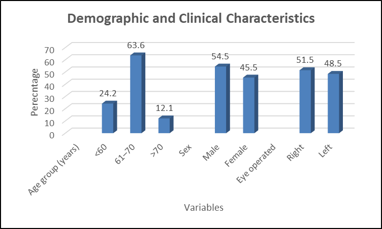
Figure 1. Demographic and Clinical Characteristics of the Study Population (N=33)
Table 2. Distribution of Preoperative Corneal Astigmatism (N=33)
|
Preoperative astigmatism (D) |
Frequency (n) |
Percentage (%) |
|
+1.75 |
2 |
6.1 |
|
+2.00 |
2 |
6.1 |
|
+2.75 |
1 |
3.0 |
|
+3.00 |
2 |
6.1 |
|
+3.25 |
1 |
3.0 |
|
−1.50 |
1 |
3.0 |
|
−1.75 |
2 |
6.1 |
|
−2.00 |
3 |
9.1 |
|
−2.50 |
3 |
9.1 |
|
−2.75 |
4 |
12.1 |
|
−3.00 |
6 |
18.2 |
|
−3.25 |
2 |
6.1 |
|
−3.50 |
2 |
6.1 |
|
−3.75 |
1 |
3.0 |
|
−4.00 |
1 |
3.0 |
|
Total |
33 |
100.0 |
Table 3. Comparison of Preoperative and Postoperative Uncorrected Visual Acuity (UCVA)
|
Follow-up interval |
N |
Mean ± SD (LogMAR) |
Minimum |
Maximum |
Median |
|
Preoperative |
33 |
0.8333 ± 0.1848 |
0.50 |
1.00 |
0.80 |
|
Postoperative day 1 |
33 |
0.1750 ± 0.1685 |
0.00 |
0.50 |
0.10 |
|
Postoperative week 1 |
33 |
0.1848 ± 0.1839 |
0.00 |
0.50 |
0.10 |
|
Postoperative month 1 |
33 |
0.1455 ± 0.1563 |
0.00 |
0.50 |
0.10 |
|
Postoperative month 3 |
33 |
0.1455 ± 0.1563 |
0.00 |
0.50 |
0.10 |
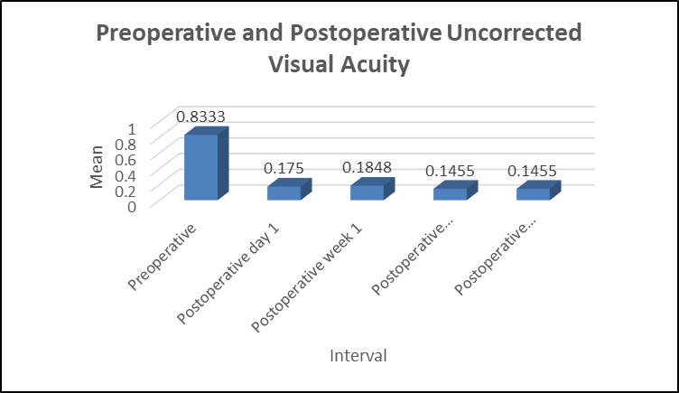
Figure 2. Comparison of Preoperative and Postoperative Uncorrected Visual Acuity (UCVA)
Table 4. Postoperative Best-Corrected Visual Acuity (BCVA) at Different Follow-up Intervals
|
Follow-up interval |
N |
Mean ± SD (LogMAR) |
Minimum |
Maximum |
Median |
|
Postoperative day 1 |
33 |
0.1000 ± 0.0901 |
0.00 |
0.50 |
0.10 |
|
Postoperative week 1 |
33 |
0.0848 ± 0.0364 |
0.00 |
0.10 |
0.10 |
|
Postoperative month 1 |
33 |
0.0758 ± 0.0435 |
0.00 |
0.10 |
0.10 |
|
Postoperative month 3 |
33 |
0.0758 ± 0.0435 |
0.00 |
0.10 |
0.10 |
BCVA: Best-corrected visual acuity.
Table 5. Comparison of Postoperative UCVA and BCVA Across Follow-up Intervals
|
Follow-up interval |
UCVA (Mean ± SD) |
BCVA (Mean ± SD) |
Mean difference (UCVA − BCVA) |
|
Day 1 |
0.1750 ± 0.1685 |
0.1000 ± 0.0901 |
0.0750 |
|
Week 1 |
0.1848 ± 0.1839 |
0.0848 ± 0.0364 |
0.1000 |
|
Month 1 |
0.1455 ± 0.1563 |
0.0758 ± 0.0435 |
0.0697 |
|
Month 3 |
0.1455 ± 0.1563 |
0.0758 ± 0.0435 |
0.0697 |
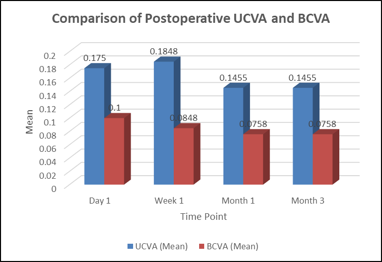
Figure 3. Comparison of Postoperative UCVA and BCVA Across Follow-up Intervals
Table 6. Distribution of Postoperative Residual Astigmatism Following Toric IOL Implantation (N=33)
|
Residual astigmatism (D) |
Frequency (n) |
Percentage (%) |
Cumulative percentage (%) |
|
0.00 |
26 |
78.8 |
78.8 |
|
0.25 |
1 |
3.0 |
81.8 |
|
0.50 |
3 |
9.1 |
90.9 |
|
0.75 |
3 |
9.1 |
100.0 |
|
Total |
33 |
100.0 |
100.0 |
DISCUSSION:
The present prospective observational study evaluated the visual outcomes of toric intraocular lens (IOL) implantation in 33 cataract patients with pre-existing regular corneal astigmatism. The findings demonstrated substantial improvement in postoperative visual acuity and effective astigmatic correction, with favourable outcomes maintained throughout the three-month follow-up period.The mean age of patients in our study was 64.27 ± 5.98 years, with the majority (63.6%) belonging to the 61–70-year age group. Males constituted 54.5% and females 45.5% of the study population. Ale et al. [11] reported a comparatively higher mean age of 75.56 ± 9.87 years among patients undergoing toric IOL implantation. Similarly, Zvorničanin et al. [12] investigated the prevalence of corneal astigmatism and its association with age in cataract patients, highlighting the importance of preoperative astigmatic assessment.In our study, preoperative astigmatism ranged from 1.5 to 4.0 D, with −3.0 D (18.2%) and −2.75 D (12.1%) being the most frequently observed values. Mendicute et al. [13] reported a mean preoperative refractive cylinder of −2.34 ± 1.28 D in 30 eyes undergoing toric IOL implantation. Similarly, Bandeira et al. [14] observed a mean preoperative keratometric cylinder of 1.92 ± 1.04 D (range: 0.75–6.78 D). These findings emphasise the clinical relevance of correcting pre-existing astigmatism during cataract surgery.A marked improvement in uncorrected visual acuity (UCVA) was observed in our study, with mean UCVA improving from 0.8333 ± 0.1848 LogMAR preoperatively to 0.1750 ± 0.1685 on postoperative day 1, 0.1848 ± 0.1839 at one week, and 0.1455 ± 0.1563 at both one and three months. Similarly, mean best-corrected visual acuity (BCVA) improved from 0.1000 ± 0.0901 LogMAR on day 1 to 0.0848 ± 0.0364 at one week and 0.0758 ± 0.0435 at one and three months.These findings are comparable with Mendicute et al. [13], who reported postoperative UCVA of 20/40 or better in 93.3% of eyes and 20/25 or better in 66.6%, while all eyes achieved BCVA of 20/25 or better. Bandeira et al. [14] also demonstrated favourable visual outcomes following Precizon toric IOL implantation, with no loss of corrected distance visual acuity at six months and 98% of eyes achieving spherical correction within ±1.00 D of the attempted target.
In the present study, complete astigmatic correction (0 D residual cylinder) was achieved in 26 (78.8%) patients, while 1 (3.0%) patient had 0.25 D and 3 (9.1%) patients each had 0.50 D and 0.75 D residual astigmatism. Thus, all patients achieved residual astigmatism below 1.0 D (reported Z=5.5435; p<0.00001).
Mendicute et al. [13] reported a significant reduction in mean refractive cylinder from −2.34 ± 1.28 D to −0.72 ± 0.43 D (p<0.01). Similarly, Bandeira et al. [14] observed a postoperative refractive cylinder of 0.77 ± 0.50 D, with 81% of eyes achieving residual cylinder ≤1.00 D. Patil et al. [15] demonstrated a reduction in refractive astigmatism from 3.55 ± 0.97 D preoperatively to 0.81 ± 0.28 D at one month and 0.79 ± 0.27 D at three months, with 92.5% of patients achieving residual astigmatism below 1.0 D. These findings are consistent with our results and support the effectiveness of toric IOLs in correcting regular corneal astigmatism.Although IOL rotation was not quantitatively evaluated in our study, postoperative visual outcomes remained unchanged between one and three months. Miyake et al. [16] reported effective astigmatic correction and good overall rotational stability with toric IOLs, although greater rotation was observed in eyes with longer axial lengths, particularly during the early postoperative period.Mendicute et al. [13] reported a mean IOL rotation of 3.63 ± 3.11°, with 96.7% of eyes showing rotation below 10°. Similarly, Patil et al. [15] observed rotation ≤5° in 72.5% of patients, 6–10° in 20%, and greater than 10° in 7.5%, requiring repositioning. Felipe et al. [17] demonstrated that toric IOL rotations below 10° generally produced refractive changes of less than 0.50 D. Furthermore, Singh et al. [18] emphasised the importance of appropriate patient selection, accurate preoperative measurements, precise surgical alignment, and early recognition of IOL misalignment for successful outcomes.
CONCLUSION:
Toric IOL implantation resulted in substantial improvement in postoperative visual acuity among cataract patients with regular corneal astigmatism. Complete astigmatic correction was achieved in 78.8% of patients, while all patients had residual astigmatism below 1.0 D. The favourable visual outcomes maintained at three months support toric IOL implantation as an effective approach for simultaneous correction of cataract and pre-existing regular corneal astigmatism.
LIMITATIONS
The study was limited by its small sample size (N=33), single-centre design, and relatively short follow-up period of three months. Additionally, the absence of a comparative control group and quantitative assessment of postoperative IOL rotation limited further evaluation of refractive outcomes and long-term rotational stability.
REFERENCES:
1. Hoffmann PC, Hütz WW. Analysis of biometry and prevalence data for corneal astigmatism in 23,239 eyes. J Cataract Refract Surg. 2010;36(9):1479-85.
2. Ferrer-Blasco T, Montés-Micó R, Peixoto-de-Matos SC, González-Méijome JM, Cerviño A. Prevalence of corneal astigmatism before cataract surgery. J Cataract Refract Surg. 2009;35(1):70-5.
3. Kaur M, Shaikh F, Falera R, Titiyal JS. Optimizing outcomes with toric intraocular lenses. Indian J Ophthalmol. 2017;65(12):1301-13.
4. Khan MI, Ch'ng SW, Muhtaseb M. The use of toric intraocular lens to correct astigmatism at the time of cataract surgery. Oman J Ophthalmol. 2015;8(1):38-43.
5. Lake JC, Victor G, Clare G, Porfírio GJ, Kernohan A, Evans JR. Toric intraocular lens versus limbal relaxing incisions for corneal astigmatism after phacoemulsification. Cochrane Database Syst Rev. 2019;12(12):CD012801.
6. Holland E, Lane S, Horn JD, Ernest P, Arleo R, Miller KM. The AcrySof Toric intraocular lens in subjects with cataracts and corneal astigmatism: a randomized, subject-masked, parallel-group, 1-year study. Ophthalmology. 2010;117(11):2104-11.
7. Bachernegg A, Rückl T, Riha W, Grabner G, Dexl AK. Rotational stability and visual outcome after implantation of a new toric intraocular lens for the correction of corneal astigmatism during cataract surgery. J Cataract Refract Surg. 2013;39(9):1390-8.
8. Zuberbuhler B, Signer T, Gale R, Haefliger E. Rotational stability of the AcrySof SA60TT toric intraocular lenses: a cohort study. BMC Ophthalmol. 2008;8:8.
9. Kim MH, Chung TY, Chung ES. Long-term efficacy and rotational stability of AcrySof toric intraocular lens implantation in cataract surgery. Korean J Ophthalmol. 2010;24(4):207-12.
10. Ferreira TB, Berendschot TT, Ribeiro FJ. Clinical outcomes after cataract surgery with a new transitional toric intraocular lens. J Refract Surg. 2016;32(7):452-9.
11. Ale JB, Power J, Zohs K, Cunningham F. Refractive and visual outcome of toric intraocular lens implantation following cataract surgery. Nepal J Ophthalmol. 2012;4(1):37-44.
12. Zvorničanin J. Corneal astigmatism in cataract surgery patients from Bosnia and Herzegovina. Int Ophthalmol. 2019;39(8):1753-60.
13. Mendicute J, Irigoyen C, Aramberri J, Ondarra A, Montés-Micó R. Foldable toric intraocular lens for astigmatism correction in cataract patients. J Cataract Refract Surg. 2008;34(4):601-7.
14. Bandeira F, Morral M, Elies D, Eguiza S, Souki S, Manero F, et al. Transitional conic toric intraocular lens for the management of corneal astigmatism in cataract surgery. Clin Ophthalmol. 2018;12:1071-9.
15. Patil MS, Nikose AS, Bharti S. Visual outcome and refractive status with monofocal toric intraocular lens implantation to correct astigmatism during cataract surgery. Indian J Ophthalmol. 2020;68(12):3016.
16. Miyake T, Kamiya K, Amano R, Iida Y, Tsunehiro S, Shimizu K. Long-term clinical outcomes of toric intraocular lens implantation in cataract cases with preexisting astigmatism. J Cataract Refract Surg. 2014;40(10):1654-60.
17. Felipe A, Artigas JM, Díez-Ajenjo A, García-Domene C, Alcocer P. Residual astigmatism produced by toric intraocular lens rotation. J Cataract Refract Surg. 2011;37(10):1895-901.
18. Singh VM, Ramappa M, Murthy SI, Rostov AT. Toric intraocular lenses: expanding indications and preoperative and surgical considerations to improve outcomes. Indian J Ophthalmol. 2022;70(1):10-23.