Clinicopathological Study Of Pediatric Lymphadenopathy With Fine Needle Aspiration Cytology (Fnac) Correlation.
- Dr. Richa Sisodia , Senior Resident Department of BMT, Superspeciality hospital & MGM medical college Indore.
- Dr. Rakesh Kumar Soni , Senior Resident, Department of Pediatrics, ESIC Medical College and Hospital, Indore, Madhya Pradesh
- Dr. Anil Malviya , Assistant Professor, Department of Pediatrics, ESIC Medical College and Hospital, Indore, Madhya Pradesh.
Article Information:
Abstract:
Background: Pediatric lymphadenopathy is a common clinical condition with causes ranging from reactive inflammatory lesions to infections and malignant disorders. Accurate diagnosis is essential for appropriate management. Fine Needle Aspiration Cytology (FNAC) is a minimally invasive, rapid, and cost-effective diagnostic technique that can aid in the evaluation of enlarged lymph nodes in children. Objective: To evaluate the clinicopathological spectrum of pediatric lymphadenopathy and determine the diagnostic utility of FNAC by correlating cytological findings with clinical and histopathological diagnoses wherever available. Materials and Methods: This hospital-based prospective observational study included pediatric patients (≤18 years) presenting with peripheral lymphadenopathy. Detailed demographic and clinical data were recorded, followed by FNAC of the affected lymph nodes under aseptic precautions. Cytological diagnoses were categorized, and histopathological examination was performed in selected cases. Statistical analysis was carried out using SPSS version 26.0, with a p-value <0.05 considered statistically significant. Results: Reactive lymphadenitis was the most frequent diagnosis, followed by granulomatous lymphadenitis, suppurative lesions, and malignant disorders. Cervical lymph nodes were the most commonly involved. FNAC demonstrated high diagnostic accuracy and showed good concordance with histopathological findings in cases where tissue diagnosis was available. Conclusion: FNAC is a reliable, safe, and effective first-line diagnostic modality for pediatric lymphadenopathy. It facilitates early diagnosis, guides clinical management, and reduces the need for unnecessary surgical biopsies.
Keywords:
Article :
INTRODUCTION:
Lymphadenopathy is a common clinical finding in the pediatric population and is frequently encountered in routine clinical practice. Although most enlarged lymph nodes are benign and self-limiting, they may also represent infections, granulomatous diseases, autoimmune disorders, or malignancies. Differentiating between these conditions is essential for timely diagnosis and appropriate management and avoidance of unnecessary invasive procedures [1,2].
The etiology of pediatric lymphadenopathy varies according to age, geographical region, nutritional status, immunological factors, and the prevalence of infectious diseases. Cervical lymphadenopathy is the most frequently affected site in children, mainly because of upper respiratory tract infections, odontogenic infections, and other localized inflammatory conditions. Careful clinical assessment, including the duration, size, consistency, tenderness, mobility and anatomical distribution of lymph nodes, helps guide the diagnostic approach but is often insufficient to establish a definitive diagnosis [3,4].
Fine Needle Aspiration Cytology (FNAC) is a simple, minimally invasive, rapid, safe, and cost-effective diagnostic procedure widely used for the evaluation of peripheral lymphadenopathy. It provides valuable cytomorphological information that helps differentiate reactive and infectious lesions from granulomatous diseases and malignant conditions, thereby reducing unnecessary surgical biopsies. Histopathological examination remains the gold standard in selected cases, particularly when malignancy is suspected or cytological findings are inconclusive. Correlation of FNAC findings further improves diagnostic confidence and clinical decision-making [5,6]. The present study was undertaken to evaluate the clinicopathological spectrum of pediatric lymphadenopathy and to assess the diagnostic utility of FNAC by correlating cytological findings with histopathological diagnoses wherever available.
MATERIALS AND METHODS:
Study design and settings:
This hospital-based prospective observational study was conducted in the Department of Pathology in collaboration with the Department of Pediatrics at a tertiary care teaching hospital over a period of 18 months.
Study populations
A total of 100 consecutive pediatrics patients aged 0-18 years presenting with clinically significant peripheral lymphadenopathy and referred for FNAC during the study period was included in the study. Detailed demographic and clinical information including age, sex, site of lymph node enlargement, duration of swelling, associated symptoms, and relevant clinical history, was obtained from the patients’ medical records and clinical examination
Inclusion Criteria
• Children aged 0–18 years presenting with clinically significant peripheral lymphadenopathy.
• Patients undergoing FNAC for diagnostic evaluation.
• Written informed consent obtained from parents or legal guardians.
Exclusion Criteria
• Patients older than 18 years.
• Inadequate or unsatisfactory FNAC smears.
• Patients whose parents or legal guardians declined to provide written informed consent.
FNAC procedure
Fine needle aspiration cytology was performed under strict aseptic precautions after obtaining informed consent from the parent or legal guardians. Aspiration was performed using a 22–23-gauge disposable needle attached to a 10 ml disposable syringe. Smear were prepared immediately following aspiration. Air dried smears were stained with may- Grunwald-Giemsa (MGG) stain, while alcohol fixed smears were stained using the Papanicolaou (Pap) stain. Ziehl- neelsen staining was performed in cases where granulomatous lymphadenitis or tubercular lymphadenitis was clinically or cytologically suspected.
Statistical Analysis
Data were analyzed using SPSS version 26.0. Continuous variables were expressed as mean ± standard deviation, and categorical variables as frequency and percentage. The Chi-square test or Fisher's exact test was used for comparison of categorical variables. A p-value <0.05 was considered statistically significant.
RESULTS:
A total of 100 pediatric patients with peripheral lymphadenopathy were included in the study. The majority were male 58 (58%), and 42 (42%) females, with a male to female ratio of approximately 1.4:1. The most commonly affected age group was 6–10 years. Cervical lymph nodes were the most frequently involved site. Reactive lymphadenitis was the predominant cytological diagnosis, followed by granulomatous lymphadenitis and suppurative lymphadenitis. Histopathological correlation was available in 30 cases and demonstrated a high concordance with FNAC findings.
Table 1: Age-wise Distribution of Study Participants (n=100)
|
Age Group (Years) |
Number |
Percentage (%) |
|
0-5 |
24 |
24% |
|
6-10 |
38 |
38% |
|
11-15 |
26 |
26% |
|
16-18 |
12 |
12% |
|
Total |
100 |
100% |
Interpretation: The highest proportion of children with lymphadenopathy belonged to the 6–10 years age group (38%), followed by the 11–15 years group (26%).
Cytological diagnosis on FNAC
Reactive lymphadenitis was the most common cytological diagnosis accounting for 54 (54%) cases. Granulomatous lymphadenitis was diagnosed in 22 (22%) cases, while 14 (14%) cases showed features of suppurative lymphadenitis. Malignant lesion included 6 (6%) cases of lymphoma and 2 (2%) cases of metastatic malignancy. Two cases (2%) were categorized as other benign lesion (table 2).
Table 2: Cytological Diagnosis on FNAC (n=100)
|
Cytological Diagnosis |
Number |
Percentage (%) |
|
Reactive lymphadenitis |
54 |
54% |
|
Granulomatous lymphadenitis |
22 |
22% |
|
Suppurative lymphadenitis |
14 |
14% |
|
Lymphoma |
6 |
6% |
|
Metastatic malignancy |
2 |
2% |
|
Others |
2 |
2% |
|
Total |
100 |
100% |
Interpretation: Reactive lymphadenitis was the most common cytological diagnosis (54%), whereas malignant lesions constituted 8% of all cases.
The cervical lymph node was the most frequently involved site accounting for 68 (68%). Other sites included the axillar (14%) inguinal (10%), and supraclavicular (5%) regions. Generalized lymphadenopathy was observed in 3(3%) patients [Table 3]
Table 3: Distribution according to Site of Lymph Node Involvement (n=100)
|
Site |
Number |
Percentage (%) |
|
Cervical |
68 |
68% |
|
Axillary |
14 |
14% |
|
Inguinal |
10 |
10% |
|
Supraclavicular |
5 |
5% |
|
Generalized |
3 |
3% |
|
Total |
100 |
100% |
Interpretation: The cervical region was the most common site of lymphadenopathy, accounting for 68% of the cases.
Histopathological examination was available in 30 patients who subsequently underwent lymph node biopsy. Among these cases, FNAC findings were concordant with the histopathological diagnosis in 28 (93.3%) cases, while 2 (6.7%) cases showed discordance [Table 4].
Table 4: FNAC–Histopathological Correlation (Biopsied Cases, n=30)
|
Correlation |
Number |
Percentage (%) |
|
Concordant |
28 |
93.3% |
|
Discordant |
2 |
6.7% |
|
Total |
30 |
100% |
Interpretation: FNAC findings showed 93.3% concordance with histopathological diagnosis, indicating excellent diagnostic reliability.
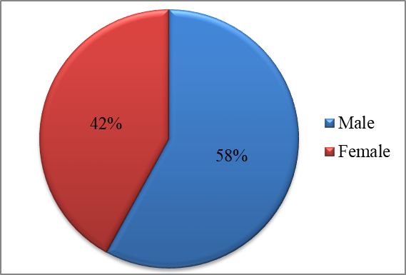
Figure 1: Gender Distribution of Study Participants
Figure legend: Male children were more frequently affected than female children, with a male-to-female ratio of approximately 1.4:1.
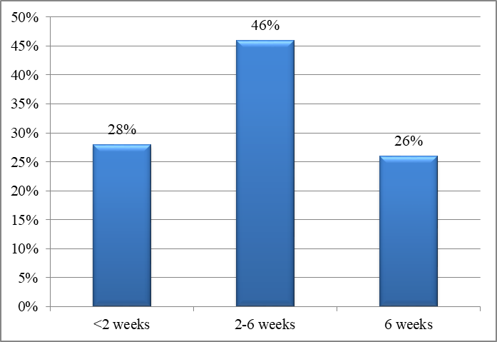
Figure 2: Duration of Lymphadenopathy at Presentation
Nearly half of the patients (46%) presented with duration of lymphadenopathy between 2 and 6 weeks, while 26% had persistent lymphadenopathy for more than six weeks.
DISCUSSION:
The present study showed that reactive lymphadenitis was the most common cause of pediatric lymphadenopathy, followed by granulomatous and suppurative lymphadenitis. Most patients belonged to the 6–10 years age group, and cervical lymph nodes were the most frequently involved. These findings are comparable with previous studies reporting that benign reactive lesions constitute the majority of pediatric lymph node enlargements [2,7]. The predominance of cervical lymphadenopathy observed in the present study is consistent with published literature, where cervical lymph nodes are commonly affected because of recurrent upper respiratory tract infections and other localized inflammatory conditions in children [4,8].
FNAC was found to be a reliable first-line diagnostic modality, accurately differentiating benign inflammatory lesions from malignant conditions. The high concordance between cytological and histopathological findings observed in our study supports previous reports demonstrating the excellent diagnostic accuracy of FNAC in evaluating pediatric lymphadenopathy [5,9]. Malignant lesions accounted for only a small proportion of cases, emphasizing that although most pediatric lymphadenopathy is benign, persistent or suspicious lymph node enlargement requires prompt evaluation to exclude serious underlying pathology. Early diagnosis using FNAC facilitates timely treatment and minimizes unnecessary surgical intervention [10].
Overall, the findings of the present study reaffirm that FNAC is a safe, rapid, minimally invasive, and cost-effective diagnostic technique for the evaluation of pediatric lymphadenopathy. When interpreted in conjunction with clinical findings and confirmed by histopathology in selected cases, FNAC significantly improves diagnostic accuracy and patient management [5, 9]. The present study was conducted at a single tertiary care center with a relatively small sample size. Therefore, multicenter studies involving larger pediatric populations are required to validate these findings.
CONCLUSION:
Pediatric lymphadenopathy is predominantly caused by benign conditions, with reactive lymphadenitis being the most common diagnosis. Fine Needle Aspiration Cytology (FNAC) is a safe, rapid, minimally invasive, and cost-effective diagnostic modality that provides reliable cytological evaluation of enlarged lymph nodes in children.
The high concordance between FNAC and histopathological findings observed in this study supports its usefulness as a first-line diagnostic tool. Early and accurate diagnosis through FNAC facilitates appropriate clinical management, reduces unnecessary surgical biopsies, and contributes to improved patient care.
REFERENCES:
1. Weinstock MS, Patel NA, Smith LP. Pediatric Cervical Lymphadenopathy. Pediatr Rev. 2018 Sep;39(9):433-443. doi: 10.1542/pir.2017-0249.
2. Deosthali A, Donches K, DelVecchio M, Aronoff S. Etiologies of Pediatric Cervical Lymphadenopathy: A Systematic Review of 2687 Subjects. Glob Pediatr Health. 2019 Jul 27;6:2333794X19865440. doi: 10.1177/2333794X19865440.
3. Pecora F, Abate L, Scavone S, Petrucci I, Costa F, Caminiti C, et al. Management of Infectious Lymphadenitis in Children. Children (Basel). 2021 Sep 27;8(10):860. doi: 10.3390/children8100860.
4. Park JE, Ryu YJ, Kim JY, Kim YH, Park JY, Lee H, et al. Cervical lymphadenopathy in children: a diagnostic tree analysis model based on ultrasonographic and clinical findings. Eur Radiol. 2020 Aug;30(8):4475-4485. doi: 10.1007/s00330-020-06794-w.
5. Khatri A, Mahajan N, Malik S, Rastogi K, Kumar P, Saikia D. Peripheral Lymphadenopathy in Children: Cytomorphological Spectrum and Interesting Diagnoses. Turk Patoloji Derg. 2021;37(3):219-225. doi: 10.5146/tjpath.2021.01537.
6. Jackson DL. Evaluation and Management of Pediatric Neck Masses: An Otolaryngology Perspective. Physician Assist Clin. 2018 Apr;3(2):245-269. doi: 10.1016/j.cpha.2017.12.003.
7. Cameron D, Stetson A, Blase J, Weldon CB. Pediatric Lymphadenopathy. Adv Pediatr. 2025 Aug;72(1):93-102. doi: 10.1016/j.yapd.2025.03.008.
8. Stanford EF, Levine HM, Cabana MD, Anosike BI. Lymphadenopathy: Differential Diagnosis and Indications for Evaluation. Pediatr Rev. 2024 Aug 1;45(8):429-439. doi: 10.1542/pir.2023-006291.
9. Vaughn JA. Imaging of Pediatric Cervical Lymphadenopathy. Neuroimaging Clin N Am. 2023 Nov;33(4):581-590. doi: 10.1016/j.nic.2023.05.008.
10. Zeppa P, Cozzolino I. Paediatric Lymphadenopathies. Monogr Clin Cytol. 2018;23:60-76. doi: 10.1159/000478882.