Isolation Of Candida Species From Various Clinical Samples, Their Socio-Demographic, Clinical Profile And Antifungal Susceptibility Pattern In A Tertiary Care Hospital.
- Dr. Joel Chelsea Rayen K , 2nd year Post graduate student, Department of Microbiology, Vydehi Institute of Medical Sciences and Research Center
- Dr. Supriya Dey , Associate Professor, Department of Microbiology, Vydehi Institute of Medical Sciences and Research Center
- Dr. Ranjini CY , Professor and HOD, Department of Microbiology.
Article Information:
Abstract:
Background: Candida species is one of the most frequent opportunistic fungal pathogens that lead to superficial mucosal infection as well as invasive candidemia. Recent change in the epidemiology of Candida albicans to non-albicans Candida (NAC) species that have been increasing in antifungal resistance has profound clinical consequences. Methodology: This retrospective, cross-sectional study evaluated clinical samples of adult patients in a tertiary care hospital. Candida isolates were determined to species level, and their socio-demographic, clinical profile, and antifungal susceptibility pattern were assessed with the help of standard laboratory techniques and CLSI guidelines. Results: Of the 233 positive fungal elements, 89 (38.2%) were Candida isolates. Candida albicans had a lesser proportion (n=38, 42.7%) than non-albicans Candida species. The most common isolate was C. tropicalis (n=23, 76.7%, which was mostly found in urine samples. Most of the patients were 60 years and above (44.4%), with a minor female preponderance (52.8%). The common comorbidities observed were diabetes mellitus and hypertension, whereas catheterization and extended hospital stay were the significant risk factors. The antifungal resistance was highest to fluconazole, especially with C. tropicalis and C. albicans. The isolates showed greater susceptibility to Echinocandins, accounting for only 7 resistant isolates. Conclusion: The predominance of NAC species and emergence of antifungal resistance necessitate the importance of routine species identification and susceptibility testing. Early detection.
Keywords:
Article :
INTRODUCTION:
Candidiasis is classified as one of the most frequent opportunistic fungal infections in clinical practice, which occurs due to the genus Candida that are normal components of the human microbiota.1 In cases where compromised host immunity or imbalance of normal flora, they may lead to superficial mucosal infections or invasive systemic disease.2
Candida albicans is traditionally the common species involved in human infections. Nevertheless, the past few decades have shown a tremendous epidemiological transition towards non-albicans Candida (NAC) species like C. tropicalis, C. parapsilosis, and C. krusei.3,4 The shift is clinically relevant, owing to differences in pathogenicity, virulence, and antifungal susceptibility between these species.5
Candida infections comprise a broad range of clinical manifestations such as oropharyngeal candidiasis, candiduria, vulvovaginal candidiasis, and invasive candidemia.6 Invasive candidiasis is one of them, and it is linked with considerable morbidity and mortality, especially among patients who are critically ill, and mortality rate has been reported between 30 and 50%.7
Candida infections are caused by several risk factors related to healthcare as well as the host. These encompass age extremes, malignancy, diabetes mellitus, immunosuppressive treatment, long-term administration of broad-spectrum antibiotics, indwelling catheters, and chronic hospitalization.8 Moreover, invasive surgeries and ICU hospitalization are also high-risk factors contributing to candidemia and other systemic infections.9
The incidence of Candida infection in India has been rising at a steady pace, especially among tertiary care facilities. Recent reports have indicated that NAC species have been reported to represent almost half of all Candida isolates and that C. tropicalis is often most common Candida species to appear.10 This epidemiological pattern has significant therapeutic consequences because NAC species tend to be less susceptible to frequently used antifungal agents.
The problem of Candida species antifungal resistance has been a major concern around the world. NAC species have been progressively reported to show azole resistance, especially fluconazole, and resistance to echinocandins and polyenes has also been reported.11 The extensive and frequent usage of empirical antifungal agents has also helped in selection of resistant strains, which has led to difficulties in treatment outcomes.
Proper clinical management of Candida isolates requires precise species-level identification, since various Candida species tend to exhibit different patterns of antifungal susceptibility.12 The development of advanced diagnostic tools, like automated systems and molecular methods, has enhanced the swift identification and susceptibility profile of Candida species.13
Although the clinical significance of Candida infections is increasingly becoming crucial, several studies on distribution of species, risk factors, and antifungal resistance profiles among tertiary care hospitals, especially in developing countries, are incomplete.14 Empirical therapy and antifungal stewardship programs depend on local epidemiological information to implement evidence-based interventions.
As the prevalence of NAC species infection and the emergence of antifungal resistance are constantly rising, the existing epidemiological trend and patterns of susceptibility to Candida infections in the tertiary care facility should be assessed to fill this research gap.
Aim
To determine the isolation of fungal species in various samples, clinical & socio-demographic profiles, and antifungal susceptibility of Candida albicans species and NAC species from various clinical samples in a tertiary care hospital.
Objectives
1. To isolate and identify Candida species from clinical samples acquired from adult patients.
2. To analyze socio-demographic (age, sex, comorbidities) and clinical profiles (comorbidities, underlying conditions, invasive procedures) associated with Candida infections.
3. To determine distribution of Candida albicans and non-albicans Candida species among the isolates.
4. To evaluate patterns of antifungal susceptibility of all isolates, with more emphasis on NAC species due to emerging resistance, using standardized methods.
METHODOLOGY:
Study design, setting and duration:
This retrospective, cross-sectional study was performed over 18 months (from August 2024 to January 2026) in the Department of Microbiology, Vydehi Institute of Medical Sciences and Research Centre, Karnataka, India.
Study population:
The targeted study population included adult patients who provided samples of blood, urine, pus, vaginal swabs, and bile fluid samples (for evaluation of candidemia, candiduria, vulvovaginal candidiasis, and wound infections). Paediatric patients (<18 years), improperly collected or poorly transported samples, sputum and tracheal aspirate samples (low diagnostic reliability), mixed bacterial-fungal cultures or contamination with normal flora (where Candida is not clinically significant), repeat isolates from the same patient within the same infection episode, and incomplete clinical/lab records were excluded from the study.
Sample size:
Based on a study done by Umamaheshwari et al.,15 prevalence of Candida infection was taken as 64%, Z = 1.96 (95% CI), p = 64%, q = 36%, absolute error d = 10%,
Sample size (n) = (Z)² pq
d2
n = 1.96 * 1.96 * 64 * 36
10*10
n ~ 89
Hence, the final sample size was calculated as 89.
Data collection procedure:
The study was conducted in the Department of Microbiology, Vydehi Institute of Medical Sciences and Research Institute, Bangalore. Data were collected from the Medical Records Department (MRD), Hospital Information System (HIS) and Laboratory Information System (LIS).
The variables collected:
1. Patient’s socio-demographic and clinical profile
2. Sample type
3. Organism isolated
4. Outcome details (Recovered/Discharged, etc.)
Clinical specimens like urine, blood, high vaginal swabs, and pus were collected and inoculated onto Sabouraud dextrose agar, followed by aerobic incubation at 37 °C for 24–48 hours. Blood samples (8–10 ml) were cultured in Brain Heart Infusion broth and incubated at 35-37 °C for up to 96 hours using the BacT/Alert system. Fungal isolates were identified based upon colony morphology, Gram staining, KOH/wet mount, and Lactophenol cotton blue mount. Candida species were confirmed by presence of Gram-positive budding yeast cells with pseudohyphae and further speciated using the germ tube test (C. albicans and C. dubliniensis). Germ tube test can be positive for both C. albicans and C. dubliniensis, further identification & antifungal susceptibility pattern for Fluconazole, Amphotericin B, Voriconazole, Caspofungin, and Micafungin was carried out by the broth microdilution method using the VITEK2 system (YST08 card) as per CLSI M27 guidelines.16,17
Operational Definition:
Multidrug resistance
“Acquired nonsusceptibility to at least 1 agent in 3 or more antimicrobial categories” 18
Data entry and analysis:
The collected data were entered in MS Excel and analyzed by IBM-SPSS software version 23.0. The qualitative variables were expressed in frequencies & percentages, and the quantitative variables were expressed as means & standard deviation.
Expected outcomes:
1. Identification of the most common sample source yielding Candida isolates
2. Determination of the proportion of Candida albicans versus Non-albicans Candida species.
Ethical consideration:
Data collection was initiated after obtaining approval from the Institutional Human Ethics Committee of Vydehi Institute of Medical Sciences and Research Institute (Ref no: VIEC/2026/APP/14). Strict confidentiality of patient information was maintained throughout the study period.
RESULTS:
Among the various samples of urine, blood, swab and bile screened, 233 samples showed fungal growth.
Table 1: Details of Fungal Growth
|
Category |
Number of Samples (n=233) |
Sub-details |
|
Candida species |
89 |
Positive Candida isolates |
|
Other fungi |
6 |
Aspergillus flavus (1) Aspergillus niger (1) Cryptococcus (2) Trichosporon (1) |
|
No Growth |
138 |
Among these, 15 samples showed fungal elements on microscopy; however, no fungal growth was obtained on culture. |
Table 1 exhibits the details regarding fungal growth. Out of 233 samples showing fungal growth, Candida species comprised the majority (89 isolates), followed by other fungi (6 isolates), such as Aspergillus, Cryptococcus, and Trichosporon. A significant number of samples (n = 138) showed no growth. However, 15 among no growth revealed fungal elements on microscopy, indicating possible non-viable organisms or prior antifungal exposure.
Table 2: Species Distribution of Candida Isolates
|
Setting & Sample Type |
C. albicans |
C. tropicalis |
C. guilliermondii |
C. parapsilosis |
C. glabrata |
C. lusitaniae |
C. auris |
C. krusei |
Total |
|
ICU |
|||||||||
|
Urine |
3 |
2 |
– |
– |
– |
– |
– |
– |
5 |
|
Blood |
– |
1 |
1 |
– |
– |
– |
– |
1 |
3 |
|
Swab |
1 |
– |
– |
– |
– |
– |
– |
– |
1 |
|
Bile |
1 |
– |
– |
– |
– |
– |
– |
– |
1 |
|
Ward |
|||||||||
|
Urine |
12 |
15 |
3 |
3 |
1 |
2 |
1 |
– |
37 |
|
Blood |
4 |
4 |
– |
1 |
– |
– |
– |
– |
9 |
|
Vaginal |
7 |
1 |
– |
– |
1 |
– |
– |
– |
9 |
|
Swab |
1 |
– |
– |
1 |
– |
– |
– |
– |
2 |
|
Bile |
– |
– |
– |
– |
– |
– |
– |
– |
0 |
|
OP |
|||||||||
|
Urine |
6 |
6 |
1 |
1 |
2 |
1 |
– |
– |
17 |
|
Blood |
– |
1 |
– |
– |
– |
– |
– |
– |
1 |
|
Vaginal |
2 |
– |
– |
– |
– |
– |
– |
– |
2 |
|
Swab |
1 |
– |
– |
– |
1 |
– |
– |
– |
2 |
Table 2 shows distribution of Candida species in various clinical settings (ICU, ward, OP) and sample types. Non-albicans Candida species exhibited predominance, with C. tropicalis being the most commonly isolated, particularly from urine samples. The ward comprised the largest number of isolates, and urine was the most commonly noted specimen. Rare species like C. krusei & C. auris were also isolated.
Table 2a: Distribution of Candida Species by Sample Type
|
Candida Species |
Urine n (%) |
Blood n (%) |
Vaginal n (%) |
Swab n (%) |
Bile n (%) |
Total n (%) |
|
C. albicans |
21 (55.3%) |
4 (10.5%) |
9 (23.7%) |
3 (7.9%) |
1 (2.6%) |
38 (42.7%) |
|
C. tropicalis |
23 (76.7%) |
6 (20.0%) |
1 (3.3%) |
0 |
0 |
30 (33.7%) |
|
C. guilliermondii |
4 (80.0%) |
1 (20.0%) |
0 |
0 |
0 |
5 (5.6%) |
|
C. parapsilosis |
4 (66.7%) |
1 (16.7%) |
0 |
1 (16.7%) |
0 |
6 (6.8%) |
|
C. glabrata |
3 (60.0%) |
0 |
1 (20.0%) |
1 (20.0%) |
0 |
5 (5.6%) |
|
C. lusitaniae |
3 (100%) |
0 |
0 |
0 |
0 |
3 (3.4%) |
|
C. auris |
1 (100%) |
0 |
0 |
0 |
0 |
1 (1.1%) |
|
C. krusei |
0 |
1 (100%) |
0 |
0 |
0 |
1 (1.1%) |
|
Total |
59 (66.3%) |
13 (14.6%) |
11 (12.4%) |
5 (5.6%) |
1 (1.1%) |
89 (100%) |
The distribution of the Candida species amongst various clinical specimens is shown in Table 2a. Candida albicans being the single most common species, had been found in various specimens like blood, urine, swab, and bile. Candida tropicalis and Candida guilliermondii have been seen in urine and blood specimens respectively. Candida parapsilosis was found in urine, blood, and swab specimens. Candida glabrata was present in urine and vaginal specimens. Some less frequent species, like Candida lusitaniae and Candida auris, were found only in urine specimens, while Candida krusei was found in blood specimens.
Table 3: Distribution of Socio-demographic and Clinical Profile of Candida Patients
|
Subgroup |
No. of Patients (%) |
|
Age Group |
|
|
18–39 yrs |
22 (24.4%) |
|
40–59 yrs |
28 (31.1%) |
|
≥60 yrs |
40 (44.4%) |
|
Mean ± SD age |
52.6 ± 14.8 yrs |
|
Median (Range) |
54 (18–84) yrs |
|
Gender |
|
|
Males |
42 (47.2%) |
|
Females |
47 (52.8%) |
Table 3 describes the distribution of demographic characteristics of patients with Candida infections. Most of the patients were aged ≥60 years (44.4%), with mean age of 52.6 ± 14.8 years. A minor female predominance (52.8%) was noted compared to males (47.2%).
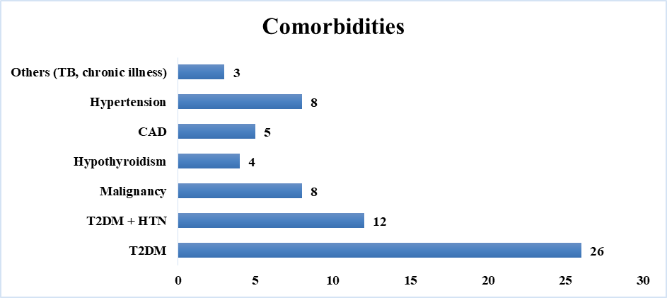
Figure 1: Distribution of Comorbidities
Figure 1 shows the distribution of comorbidities in patients infected with Candida species. Diabetes mellitus was the most commonly noted comorbidity, followed by hypertension. Other conditions, like coronary artery disease, malignancy, and hypothyroidism, were less frequently noted.
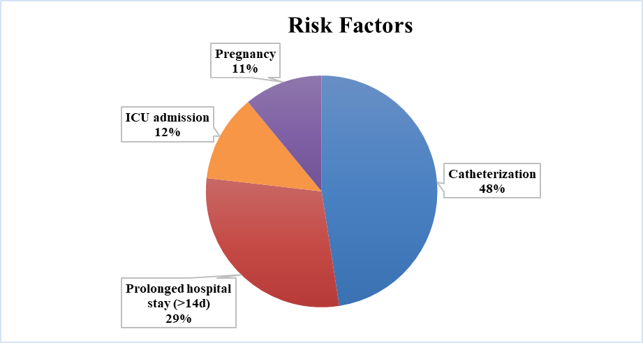
Figure 2: Distribution of Risk Factors
Figure 2 demonstrates the distribution of risk factors associated with Candida infections. Catheterization was the most common risk factor noted, followed by prolonged hospital stay (>14 days), ICU admission, and pregnancy, highlighting the profound association of Candida infections with interventions related to hospital care.
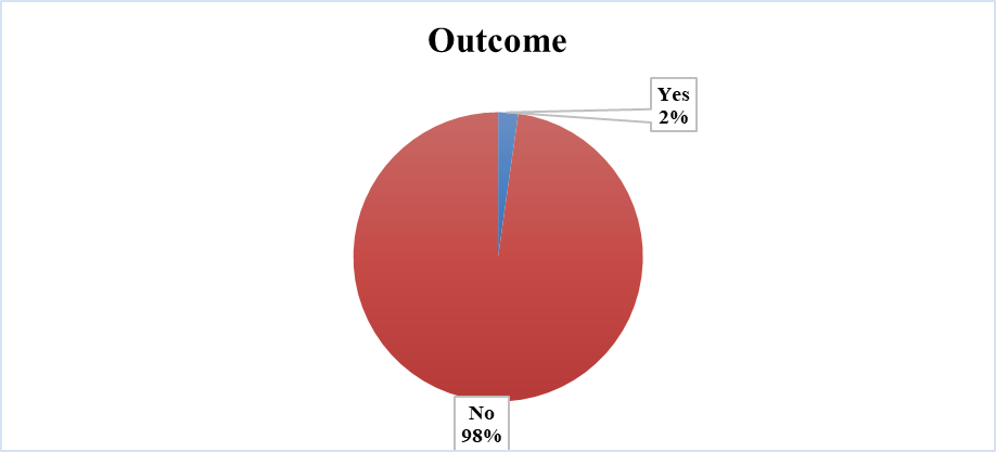
Figure 3: Distribution of Clinical Outcome
Figure 3 illustrates the distribution of patient outcomes infected with Candida. Mortality was lesser, with only two deaths reported, as the majority of patients (n=87) survived, indicating favourable outcomes in most cases. The two deaths were reported among participants infected with Candida krusei isolated from blood sample and Candida auris isolated from urine sample.
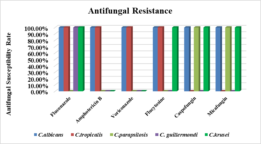
Figure 4: Distribution of Antifungal Resistance
Figure 4 demonstrates the distribution of antifungal resistance patterns among different Candida species. Resistance was mostly noted in C. albicans, where all antifungal drug resistance was noted. Candida tropicalis also demonstrated resistance to many antifungal drugs, especially to fluconazole. Resistance to echinocandins (caspofungin and micafungin) was minimal across species (n=7, 7.86%). Multidrug resistance was noted in Candida krusei, while several species, such as C. glabrata and C. lusitaniae, showed little to no resistance. Overall, azole resistance was more prominent compared to other antifungal classes (n=13, 14.61%. A noteworthy observation is the evolving antifungal resistance of the species C. tropicalis and C. krusei, indicating emerging resistance among non-Candida albicans species, whereas the resistance pattern is not significantly noted for other NAC species.
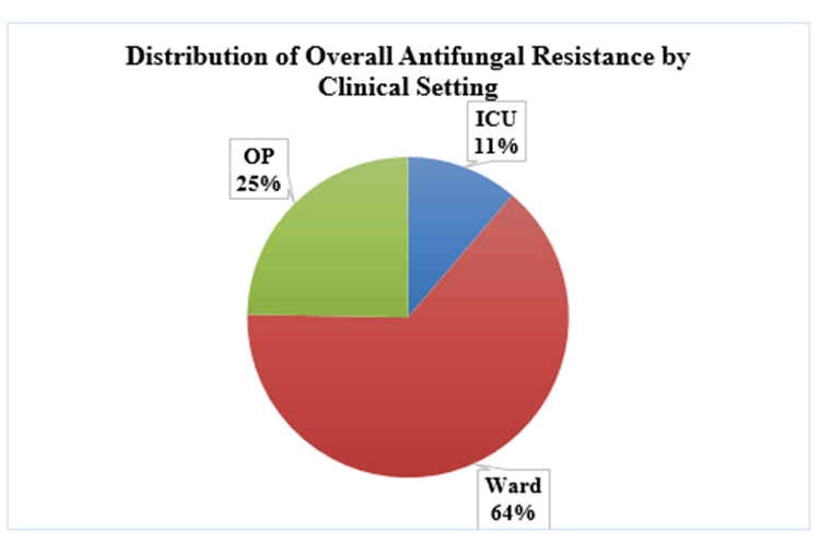
Figure 5: Distribution of Overall Antifungal Resistance by Clinical Setting
Figure 5 demonstrates the distribution of antifungal resistance across isolates from various clinical settings in the hospital. Samples isolated from ward accounts for the majority of resistance (64.0%), followed by those from OP (24.7%) and least resistance was noted in isolates taken from the ICU setting (11.2%). This finding suggests that patients admitted to hospital wards are more susceptible to antifungal resistance than in other clinical settings.
DISCUSSION:
The current study presents an outline of the distribution of Candida species, clinical features, and antifungal susceptibility profile in a tertiary care hospital. Candida species comprised a considerable proportion of total fungal isolates, depicting their significance as opportunistic pathogens among hospitalized patients.
One of the key results of the current study is the preponderance of Candida non-albicans species over C. albicans. Such observation can be explained by many recent studies that reported a similar epidemiological transition worldwide and in India.11,12 The emerging NAC species prevalence is especially due to the selective pressure created by intensive use of azoles and enhanced diagnostic possibilities that facilitate better identification of species.13
C. tropicalis was the most common organism of NAC species, especially in urine samples. This observation has been in concordance with other studies that have reported C. tropicalis to be a major cause of candiduria and candidemia in tropical countries.19 C. tropicalis has the potential to adhere to epithelial surfaces and create biofilms, explaining the reason why it remains pathogenic and persistent in both clinical and non-clinical environments.20
The most common clinical sample that yielded Candida isolates in the current study was urine. Comparable results were observed in existing studies in which candiduria was often linked to urinary catheterization and extended hospitalization.21 Candida colonization and infection due to catheter-associated infections remain a major problem in patients who are critically ill.22
The demographic profile of subjects in this study revealed that elderly patients were more prone to Candida infection, which could be attributed to a lack of immunosenescence with age and higher comorbidity rates. Similar research studies have recorded comparable findings where older age groups were reported to be at risk of invasive fungal infections.23
Diabetes mellitus was found to be the most prevalent comorbidity in the current study. This association is already known in the literature, since hyperglycemia disrupts neutrophil activity and encourages growth of fungi, thus predisposing people to Candida infections.24
Susceptibility patterns of antifungal agents in the current study revealed an increased fluconazole resistance, specifically towards C. albicans and C. tropicalis. Comparable patterns of resistance have been noted in recent surveillance studies, with increasing concern regarding the emerging trend of resistance to azole drugs.25,26 Resistance development has been frequently associated with long-term exposure to antifungal agents and misuse of antifungal drugs.
The present study and existing reports indicate that Echinocandins have better sensitivity indices compared to other medical agents, thus implying their efficacy in treating both C. albicans and NAC species.27 Amphotericin B was also active, but some cases of resistance in a few species have been reported.
The diagnosis of rarer species like C. auris and C. krusei in our study is noteworthy. C. auris has risen as a multidrug-resistant pathogen with high fatality rate and hospital outbreaks. Another interesting finding is the emerging antifungal resistance of the species C. tropicalis and C. krusei, which may increase further in future.28 Their existence, even in low numbers, highlights the necessity of vigilant surveillance and approaches to infection control.
The association between Candida infections and risk factors, including catheterization, prolonged hospital stay and ICU admission, is consistent with previous literature.29 These factors lead to higher exposure to invasive procedures and destruction of host defences supporting fungal invasion. Regarding clinical settings, highest antifungal resistance was noted among patients admitted to wards, which may be attributed to increased risk of exposure, suboptimal disinfection procedures and prolonged stay.
Candida score is a clinically proven tool intended to identify critically ill patients who are likely to have invasive candidiasis with the aim of recommending the use of antifungal drugs. This score takes into account variables such as total parenteral nutrition, surgery (especially abdominal), multifocal colonization with Candida and severe sepsis; scoring greater than or equal to 3 is an indicator of high risk of having candidemia. Early application of Candida score has proven to be effective in enabling early initiation of antifungal drugs, which leads to reduced mortality rate among ICU patients.30
The pathogenic potential and antifungal susceptibility of various Candida species vary considerably, making the distinction between species medically significant.3,4 C. tropicalis has been shown to be a causative agent of invasive disease with increased virulence and capacity for biofilm development.5 C. glabrata exhibits low susceptibility to azoles, which poses a clinical challenge.18 C. krusei is inherently resistant to fluconazole, while C. auris is regarded as a multidrug-resistant Candida species responsible for nosocomial infection outbreaks with high mortality rates.19 Hence, accurate species identification is vital for improved clinical outcomes and appropriate antifungal therapy.
Various clinical specimens contain important information about the anatomical location of Candida infections and their severity. For example, urine specimens can be a sign of candiduria, which is associated with catheterization and long-term hospitalizations. Blood samples indicate candidemia, a serious systemic infection with high morbidity and mortality rates.6 The presence of vaginal specimens implies the occurrence of vulvovaginal candidiasis, which usually affects women predisposed hormonally and immunologically.7 Swab samples, such as wound swabs, reflect localized infections, especially in immunocompromised or post-surgical patients8. Isolation of Candida albicans from bile, despite being low in this study (n=1), is clinically significant as it might indicate underlying infections such as hepatobiliary candidiasis. These conditions are frequently accompanied by biliary obstruction, suggesting a possible invasive infection. Therefore, the nature of clinical specimens is important when deciding on treatment and assessing disease severity.
In general, these research findings are similar to previous literature, confirming the pattern of emerging NAC prevalence and antifungal resistance worldwide. The disparities in results from a few similar studies can be attributed to patient population, geographical location, and healthcare practices. The prevalence of antifungal resistance in C. albicans species is relatively common and well-known in the field of healthcare. Hence, future studies should emphasize NAC resistance more.
Strengths and Limitations
The study provides in-depth information regarding the clinical profile, Candida species distribution, and antifungal drug susceptibility pattern within a tertiary care setting. The intricate analysis of risk factors and presence of multiple clinical samples also strengthen the clinical relevance further. Standardized susceptibility testing and identification techniques are used, which may contribute more to validity of the study. Also, due to the retrospective study design, causal relationship could not be proved, and limited clinical data were some of the unavoidable limitations of the study. The study was conducted only in a single tertiary care facility, which restricts the generalizability of the study findings.
CONCLUSION:
This study, despite the predominance of C. albicans growth, highlights a profound epidemiological transition toward non-albicans Candida species, with C. tropicalis emerging as the predominant isolate. The association of Candida infections with comorbidities, extremes of age, and risk factors related to hospital care indicates the need for vigilant clinical assessment.
The emerging antifungal resistance, especially to azoles, underscores the significance of routine species identification and antifungal susceptibility testing to facilitate proper therapy. Future studies should focus on multicentric surveillance and incorporation of molecular diagnostics to better understand resistance mechanisms and improve antifungal stewardship.
REFERENCES:
1. Calderone RA, Clancy CJ. Candida and candidiasis. 2nd ed. Washington DC: ASM Press; 2012.
2. Brown GD, Denning DW, Gow NAR, Levitz SM, Netea MG, White TC. Hidden killers: human fungal infections. Sci Transl Med. 2012;4(165):165rv13.
3. Pfaller MA, Diekema DJ. Epidemiology of invasive candidiasis: a persistent public health problem. Clin Microbiol Rev. 2007;20(1):133–63.
4. Guinea J. Global trends in the distribution of Candida species causing candidemia. J Antimicrob Chemother. 2014;69(1):1–11.
5. Mayer FL, Wilson D, Hube B. Candida albicans pathogenicity mechanisms. Virulence. 2013;4(2):119–28.
6. Kauffman CA. Clinical manifestations and diagnosis of candidiasis. Clin Infect Dis. 2005;41(Suppl 6):S371–6.
7. Pappas PG. Invasive candidiasis. N Engl J Med. 2006;354(14):1445–54.
8. Eggimann P, Garbino J, Pittet D. Epidemiology of Candida species infections in critically ill non-immunosuppressed patients. Lancet Infect Dis. 2003;3(11):685–702.
9. Vincent JL, Rello J, Marshall J, Silva E, Anzueto A, Martin CD, et al. International study of the prevalence and outcomes of infection in intensive care units. JAMA. 2009;302(21):2323–9.
10. Xess I, Jain N, Hasan F, Mandal P, Banerjee U. Epidemiology of candidemia in a tertiary care centre of North India: 5-year study. J Clin Microbiol. 2007;45(7):2155–60.
11. Colombo AL, Guimarães T. Epidemiology of hematogenous infections due to Candida spp. Braz J Infect Dis. 2003;7(3):173–82.
12. Kothavade RJ, Kura MM, Valand AG, Panthaki MH. Candida tropicalis: its prevalence, pathogenicity and increasing resistance to fluconazole. J Med Microbiol. 2010;59(8):873–80.
13. Arendrup MC. Epidemiology of invasive candidiasis. Curr Opin Crit Care. 2010;16(5):445–52.
14. Satoh K, Makimura K, Hasumi Y, Nishiyama Y, Uchida K, Yamaguchi H. Candida auris, a novel species causing invasive candidiasis in Japan. Microbiol Immunol. 2009;53(1):41–4.
15. Umamaheshwari S, Sumana MN. Retrospective analysis on distribution and antifungal susceptibility profile of Candida in clinical samples: a study from Southern India. Frontiers Publ Heal. 2023;11.1160841.
16. Lee H, Choi SH, Oh J, Koo J, Lee HJ, Cho S-I, et al. Comparison of six antifungal susceptibilities of 11 Candida species using the VITEK2 AST-YS08 card and broth microdilution method. Microbiol Spectr. 2022;10(2):e0125321.
17. Verma R, Pradhan D, Hasan Z, Singh H, Jain AK, Khan LA. A systematic review on distribution and antifungal resistance pattern of Candida species in the Indian population. Medical mycology. 2021;59(12):1145-65.
18. Arendrup MC, Patterson TF. Multidrug-Resistant Candida: Epidemiology, Molecular Mechanisms, and Treatment. The Journal of Infectious Diseases. 2017;216(3):445-51.
19. Playford EG, Marriott D, Nguyen Q, Chen S, Ellis D, Slavin M, et al. Candidemia in nonneutropenic critically ill patients: risk factors for non-albicans Candida spp. Crit Care Med. 2008;36(7):2034–9.
20. Chakrabarti A, Sood P, Rudramurthy SM, Chen S, Kaur H, Capoor MR, et al. Incidence, characteristics and outcome of ICU-acquired candidemia in India. Intensive Care Med. 2015;41(2):285–95.
21. Khan Z, Ahmad S, Joseph L, Hagen F, Meis JF. Candida auris: an emerging multidrug-resistant pathogen causing outbreaks of invasive infections. Clin Microbiol Rev. 2018;31(1):e00029-17.
22. Ben-Ami R. Treatment of invasive candidiasis: a narrative review. Clin Microbiol Infect. 2018;24(1):10–6.
23. Tumbarello M, Posteraro B, Trecarichi EM, Fiori B, Rossi M, Porta R, et al. Biofilm production by Candida species and inadequate antifungal therapy as predictors of mortality. Clin Infect Dis. 2007;44(3):387–94.
24. Falagas ME, Apostolou KE, Pappas VD. Attributable mortality of candidemia: a systematic review. Eur J Clin Microbiol Infect Dis. 2006;25(7):419–25.
25. Andes DR, Safdar N, Baddley JW, Playford G, Reboli AC, Rex JH, et al. Impact of treatment strategy on outcomes in patients with candidemia. Clin Infect Dis. 2012;54(8):1110–22.
26. Lamoth F, Lockhart SR, Berkow EL, Calandra T. Changes in the epidemiology of invasive candidiasis. Clin Infect Dis. 2018;66(6):947–53.
27. Bassetti M, Peghin M, Vena A. Challenges and solution of invasive candidiasis management. Drugs. 2019;79(1):1–14.
28. Pappas PG, Lionakis MS, Arendrup MC, Ostrosky-Zeichner L, Kullberg BJ. Invasive candidiasis. Nat Rev Dis Primers. 2018;4:18026.
29. Wisplinghoff H, Bischoff T, Tallent SM, Seifert H, Wenzel RP, Edmond MB. Nosocomial bloodstream infections in US hospitals. Clin Infect Dis. 2004;39(3):309–17.
30. León C, Ruiz-Santana S, Saavedra P, Almirante B, Nolla-Salas J, Alvarez-Lerma F, et al. A bedside scoring system ("Candida score") for early antifungal treatment in nonneutropenic critically ill patients with Candida colonization. Critical care medicine. 2006;34(3):730–37.