Immunohistochemical Expression Of P53 In Endometrial Hyperplasia And Its Clinicopathological Significance.
- Dr. Gayathri Elanchezhian , Assistant Professor, Department of Pathology, Madha Medical College and Research Institute, Chennai, India.
- Dr. J. Thanka , Director and Professor of Pathology, Sree Balaji Medical College and Hospital, Chennai-20, India.
- Dr. Sushma Nayar , Professor and Head, Department of Pathology, Madha Medical College and Research Institute, Chennai, India.
Article Information:
Abstract:
Background: Endometrial hyperplasia is a precursor lesion with variable risk of progression to endometrial carcinoma, particularly in atypical endometrial hyperplasia cases. Identification of reliable molecular markers that can identify high-risk lesions and assist in risk stratification and clinical management is essential. The tumor suppressor protein p53 has been implicated in early endometrial carcinogenesis and may serve as an adjunctive biomarker. Methods: This prospective study included 23 cases of endometrial hyperplasia (7 without atypia and 16 atypical endometrial hyperplasia) diagnosed on endometrial biopsy and hysterectomy specimens over a period from March 2020 to September 2021. Histopathological evaluation was performed using hematoxylin and eosin staining. Immunohistochemical analysis for p53 expression was carried out on paraffin-embedded tissue sections. Overexpression and null staining pattern of p53 was considered positive. The intensity of staining was assessed using a semi-quantitative H-score. Statistical analysis was performed using Pearson’s Chi-square test, with a p-value <0.05 considered statistically significant. Results: The majority of patients were in the age group of 41–50 years, with abnormal uterine bleeding being the most common clinical presentation. All cases of hyperplasia without atypia were negative for p53 expression. In contrast, 75% of atypical endometrial hyperplasia cases demonstrated positive p53 expression. A statistically significant association was observed between the type of endometrial hyperplasia and p53 expression (p = 0.001). Higher H-scores were predominantly observed in atypical hyperplasia, indicating increased intensity of p53 expression with increasing lesion severity. Conclusion: p53 expression correlates with the severity of endometrial hyperplasia and may represent an early molecular event in endometrial carcinogenesis. Immunohistochemical assessment of p53 can serve as a useful adjunct in identifying high-risk precursor lesions and may aid in clinical decision-making. However, larger studies with long-term follow-up are required to validate its prognostic significance.
Keywords:
Article :
INTRODUCTION:
Endometrial hyperplasia represents a spectrum of proliferative disorders of the endometrium, ranging from benign hormonal alterations to premalignant lesions with a significant risk of progression to endometrial carcinoma. (1) According to the World Health Organization (WHO) 2020 classification, endometrial hyperplasia is categorized into hyperplasia without atypia and atypical endometrial hyperplasia, also referred to as endometrioid intraepithelial neoplasia (EIN). (2) Hyperplasia is considered a true precursor lesion, with studies demonstrating a progression risk of approximately 25–40% to endometrial carcinoma if left untreated. (3) Early identification of high-risk lesions is essential, particularly in patients undergoing conservative management, where regular endometrial surveillance is recommended. (4) In this context, there is increasing interest in identifying reliable molecular markers that can identify lesions at higher risk of progression in endometrial hyperplasia and aid in clinical decision-making. (5) The tumor suppressor gene p53, located on chromosome 17, plays a critical role in regulating cell cycle progression and maintaining genomic stability by inducing cell cycle arrest and DNA repair. In normal cells, p53 protein levels are low due to rapid degradation; however, mutant forms of p53 accumulate within the nucleus and can be detected by immunohistochemistry. (6) Aberrant p53 expression has been associated with aggressive tumour behaviour and poor prognosis in various malignancies, including endometrial carcinoma. (7) Emerging evidence suggests that alterations in p53 may occur early in the carcinogenic pathway, particularly in atypical endometrial hyperplasia, indicating its potential role as an adjunctive biomarker for identifying higher-risk.(8) Therefore, the present study aims to evaluate the immunohistochemical expression of p53 in endometrial hyperplasia and to assess its association with histological severity and its potential role as an adjunctive biomarker for identifying high-risk lesions.
MATERIALS AND METHODS:
This Cross-sectional observational study was conducted on all consecutive eligible cases of histopathologically diagnosed endometrial hyperplasia received as endometrial biopsy or hysterectomy specimens in the Department of Obstetrics and Gynaecology, Sree Balaji Medical College and Hospitals, Chrompet, Chennai, between March 2020 and September 2021. A total of 23 eligible cases were included during the study period, comprising 7 cases of endometrial hyperplasia without atypia and 16 cases of atypical endometrial hyperplasia.
Inclusion criteria
All endometrial biopsy and hysterectomy specimens diagnosed histopathologically as endometrial hyperplasia, irrespective of patient age, were included in the study.
Exclusion criteria
Specimens with inadequate or unsatisfactory endometrial tissue (comprising only blood clots and fibrin without identifiable endometrial glands or stroma) and cases with associated cervical or vaginal pathology were excluded. The study was approved by the Institutional Ethics Committee of Sree Balaji Medical College and Hospital, Chennai (Approval No.: 1356/2020, dated 06/03/2020). Written informed consent was obtained from all participants before inclusion in the study
METHODOLOGY:
All endometrial biopsies and hysterectomy specimens received were examined grossly and fixed in 10% Formalin for about 16-18 hours. The clinical history of the patient including the age, clinical presentation and menstrual details were obtained. Proper sectioning from the representative site and the routine processing of dehydration, clearing and embedding was done. These sections were cut to 4µm thickness with Leica microtome (manual) and haematoxylin and eosin staining was done Immunohistochemistry was performed on 3–4 µm thick formalin-fixed, paraffin-embedded tissue sections using a mouse monoclonal anti-p53 antibody (Clone BP53-12). Antigen retrieval was carried out using Tris–EDTA buffer (pH 9.0), followed by incubation with the primary antibody. Detection was performed using the PolyExcel HRP/DAB Detection System according to the manufacturer's protocol. Appropriate positive and negative controls were included in each staining run.
INTERPRETATION OF IMMUNOHISTOCHEMISTRY:
The percent of p53 immunoreactive tumor cells was scored as 0 to 3+ in p53positive regions. Nuclear staining in >=10% of tumor cells was scored as aberrant expression; the evaluations of the immunohistochemistry were done utilizing H-SCORE. Stained slides were examined under a light microscope. Five distinct fields were examined for immunohistochemical staining of the antibodies. The staining was graded semi quantitatively, taking into account the intensity of particular staining in different fields.
Interpretation of p53 immunohistochemistry:
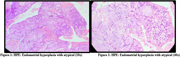
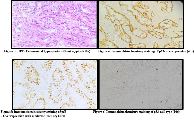
p53 immunostaining was interpreted according to the observed staining pattern. A wild-type (scattered nuclear) staining pattern was considered normal, whereas overexpression (strong diffuse nuclear staining) and complete absence of nuclear staining (null pattern) were considered aberrant (mutant-type) expression. The percentage of positively stained nuclei and staining intensity were assessed using the H-score method. Five representative high-power fields were evaluated for each case.
H-score was calculated using the formula:
H-score = ΣPi × (i + 1)
where Pi is the percentage of cells stained at each intensity and i represents the staining intensity (0 = none, 1 = weak, 2 = moderate, 3 = strong).
STATISTICAL ANALYSIS
Statistical analysis was performed using IBM SPSS Statistics for Windows, Version 16.0 (IBM Corp., Armonk, NY, USA). Continuous variables were expressed as mean ± standard deviation (SD), while categorical variables were presented as frequencies and percentages. The association between categorical variables was assessed using Pearson's Chi-square test or Fisher's exact test, where appropriate. A two-tailed p-value of <0.05 was considered statistically significant.
RESULTS:
A total of 23 cases of endometrial hyperplasia were included in the study, comprising 7 cases of hyperplasia without atypia and 16 cases of endometrial atypical hyperplasia. The majority of patients belonged to the age group of 41–50 years, followed by 31–40 years, with a mean age of 43.61 ± 5.63 years (range: 28–56 years). Regarding clinical presentation, the most common symptom was abnormal uterine bleeding (69.5%), followed by postmenopausal bleeding (21.7%), vaginal discharge (4.3%), and abdominal pain (4.3%).
Distribution of endometrial hyperplasia
Out of 23 cases:
● Hyperplasia without atypia: 7 cases (30.4%)
● Endometrial atypical hyperplasia: 16 cases (69.6%)
P53 expression in endometrial hyperplasia
All cases of hyperplasia without atypia (100%) were negative for p53 expression.
Among cases of atypical endometrial hyperplasia, 75% showed positive p53 expression, while 29.4% were negative.
There was a statistically significant association between the type of endometrial hyperplasia and p53 expression (p < 0.05).
Immunohistochemical (IHC) staining pattern
Overall, 52.2 % of cases showed positive p53 expression, while 47.8% were negative.
Among the 12 p53-positive cases, 6 (50.0%) had an H-score of 101–200, 5 (41.7%) had an H-score of 201–300, and 1 (8.3%) had an H-score of 40–100. Higher H-scores were predominantly observed in atypical endometrial hyperplasia.
Table 1: Distribution of age of patients (N=23):
|
Age (years) |
N |
% |
|
≤ 30 |
1 |
4.3 |
|
31 – 40 |
5 |
21.7 |
|
41 – 50 |
15 |
65.2 |
|
51 – 60 |
2 |
8.7 |
|
Total |
23 |
100.0 |
|
Mean ± SD |
43.61 ± 5.63 years |
|
|
Range |
28 – 56 years |
|
Table 2: Distribution of clinical presentation of hyperplasia (N=23):
|
Clinical presentation |
N |
% |
|
Post-menopausal bleeding |
5 |
21.7 |
|
Vaginal discharge |
1 |
4.3 |
|
Abnormal uterine bleeding |
16 |
69.5 |
|
Abdomen pain |
1 |
4.3 |
|
Total |
23 |
100.0 |
Table 3: Distribution of precursor lesions (N=23):
|
Precursor lesions |
N |
% |
|
Hyperplasia without atypia |
7 |
30.4% |
|
Atypical endometrial hyperplasia |
16 |
69.6 % |
Table 4: Comparison of p53 Expression Between Hyperplasia Without Atypia and Atypical Endometrial Hyperplasia (N=23):
|
Precursor lesions |
P53 positive |
P53 negative |
Total |
|||
|
N |
% |
N |
% |
N |
% |
|
|
Hyperplasia without atypia |
0 |
0 |
7 |
100.0 |
7 |
100.0 |
|
Atypical endometrial hyperplasia |
12 |
75 |
4 |
25 |
16 |
100.0 |
|
Total |
12 |
52.2 |
11 |
47.8 |
23 |
100.0 |
|
Chi square |
15.78 |
|||||
|
P value |
0.001 (Statistically significant) |
|||||
Table 5: Distribution of H score of P53 staining (N=12):
|
H score |
N |
% |
|
40 – 100 |
1 |
8.33% |
|
101 – 200 |
6 |
50% |
|
201 – 300 |
5 |
41.7% |
|
Total |
12 |
100% |
DISCUSSION:
Endometrial hyperplasia represents a continuum of proliferative disorders with variable potential for progression to endometrial carcinoma. The distinction between hyperplasia without atypia and endometrial atypical hyperplasia (endometrioid intraepithelial neoplasia) is clinically significant, as the latter is associated with a substantially higher risk of malignant transformation (1,9). The majority of patients in the present study belonged to the 41–50-year age group, with a mean age of 43.61 ± 5.63 years. This finding is consistent with previous reports demonstrating that endometrial hyperplasia predominantly affects perimenopausal women, likely due to prolonged unopposed estrogen exposure and anovulatory cycles. Abnormal uterine bleeding was the most common presenting symptom, emphasizing its importance as the principal clinical manifestation warranting endometrial evaluation. (10) The principal finding of the present study was the significantly higher expression of p53 in atypical endometrial hyperplasia compared with hyperplasia without atypia. The absence of p53 expression in all non-atypical lesions and its positivity in the majority of atypical lesions suggests that aberrant p53 expression is associated with increasing histological severity and may reflect underlying molecular alterations occurring during endometrial carcinogenesis (11). The observed increase in p53 expression with increasing histological is consistent with previous evidence suggesting that molecular alterations may occur during the early stages of endometrial carcinogenesis. Although TP53 mutations are classically associated with serous endometrial carcinoma, aberrant p53 expression in atypical endometrial hyperplasia may reflect early molecular dysregulation in lesions with greater biological aggressiveness.(12)
The present findings are in agreement with previous studies by Masjeed et al. and Ilie et al., both of which demonstrated progressively increased p53 expression from endometrial hyperplasia to carcinoma. Minor differences in reported expression rates among studies are likely attributable to variations in patient populations, antibody clones, staining protocols, and interpretation criteria rather than fundamental biological differences. (8,13) In the present study, a statistically significant association was observed between the type of endometrial hyperplasia and p53 expression (p < 0.05), indicating that p53 positivity is associated with increasing histological severity of endometrial hyperplasia. These findings suggest that p53 immunohistochemistry may serve as a useful adjunct in risk stratification, particularly in patients with atypical endometrial hyperplasia where treatment decisions may be individualized..(14) The H-score analysis further demonstrated that higher intensity of p53 expression was predominantly observed in atypical hyperplasia cases, supporting the association between increased p53 accumulation and severity of the lesion.
This semi-quantitative assessment adds additional value by not only identifying positivity but also reflecting the degree of protein expression, which may correlate with underlying genetic alterations (15). The biological basis of these findings lies in the role of p53 as a tumor suppressor gene that regulates cell cycle progression and genomic stability. Mutation of p53 leads to accumulation of dysfunctional protein within the nucleus, which can be detected by immunohistochemistry. The presence of such mutations in endometrial atypical hyperplasia suggests that these lesions may already harbor early molecular changes associated with malignant transformation.(6) From a clinical perspective, the identification of p53 expression in endometrial atypical hyperplasia has important implications. Patients demonstrating aberrant p53 expression may warrant closer clinical surveillance; however, the present study was not designed to determine whether p53 independently predicts malignant progression. Prospective longitudinal studies are therefore required before p53 can be recommended as an independent prognostic biomarker.(4,16). The present study has several limitations. First, the relatively small sample size and single-centre design may limit the generalizability of the findings. Second, molecular confirmation of TP53 mutations was not performed; therefore, immunohistochemical expression could not be correlated with mutation status. Finally, the absence of long-term follow-up precluded assessment of the relationship between p53 expression and subsequent progression to endometrial carcinoma.. Despite these limitations, the present study provides valuable insight into the immunohistochemical expression of p53 in endometrial hyperplasia using standardized histopathological evaluation and semi-quantitative H-score assessment. The findings contribute to the limited available data on p53 expression in endometrial hyperplasia from the Indian population and provide a basis for future multicenter studies.
CONCLUSION:
The present study demonstrated a statistically significant association between p53 immunohistochemical expression and atypical endometrial hyperplasia. Aberrant p53 expression was observed predominantly in atypical endometrial hyperplasia, while hyperplasia without atypia showed no aberrant expression, suggesting that p53 immunohistochemistry may serve as a useful adjunctive marker in identifying high-risk lesions. These findings support the potential role of p53 immunohistochemistry as an ancillary tool in the histopathological evaluation and risk stratification of endometrial hyperplasia. However, larger multicenter studies incorporating molecular correlation and long-term follow-up are required to validate its prognostic significance and establish its clinical utility.
REFERENCE:
1. Montgomery BE, Daum GS, Dunton CJ. Endometrial hyperplasia: a review. Obstet Gynecol Surv. 2004 May;59(5):368-78. doi: 10.1097/00006254-200405000-00025. PMID: 15097798.
2. Baak JP, Mutter GL, Robboy S, van Diest PJ, Uyterlinde AM, Ørbo A, et al. The molecular genetics and morphometry‐based endometrial intraepithelial neoplasia classification system predicts disease progression in endometrial hyperplasia more accurately than the 2020 World Health Organization classification system. Cancer. 2005;103(11):2304–12
3. Bourdel N, Chauvet P, Tognazza E, Pereira B, Botchorishvili R, Canis M. Sampling in atypical endometrial hyperplasia: which method results in the lowest underestimation of endometrial cancer? A systematic review and meta-analysis. Journal of minimally invasive gynaecology. 2016 Jul 1;23(5):692-701.
4. Gallos ID, Alazzam M, Clark T, Faraj R, Rosenthal A, Smith P GJ. RCOG Green-top Guideline: Management of Endometrial Hyperplasia. Royal College of Obstetricians &Gynaecologists. 2016.
5. Sanderson PA, Critchley HO, Williams AR. Endometrial hyperplasia and cancer: molecular mechanisms. J Pathol. 2017;241(2):242–56.
6. Finlay CA, Hinds PW, Levine AJ. The p53 proto-oncogene can act as a suppressor of transformation. Cell. 1989 Jun 30;57(7):1083-93.
7. Lax SF, Kendall B, Tashiro H, Slebos RJC, Ellenson LH. The frequency of p53, k‐ras mutations, and microsatellite instability differs in uterine endometrioid and serous carcinoma. Cancer. 2000;88(4):814–24.
8. Masjeed NMA, Khandeparker SGS, Joshi AR, Kulkarni MM, Pandya N. Immunohistochemical study of ER, PR, KI67 and p53 in endometrial hyperplasia and endometrial carcinomas. J ClinDiagn Res. 2017; 11(8):31-34.
9. Bourdel N, Chauvet P, Tognazza E, Pereira B, Botchorishvili R, Canis M. Sampling in atypical endometrial hyperplasia. J Minim Invasive Gynecol. 2016;23(5):692–701.
10. Yano M, Ito K, Yabuno A, et al. Impact of TP53 immunohistochemistry on histological grading. Mod Pathol. 2019;32(7):1023–31.
11. Hachisuga T, Fukuda K, Uchiyama M, et al.Immunohistochemical study of p53 expression in endometrial carcinomas: correlation with markers of proliferating cells and clinicopathologic features. Int J Gynecol Cancer. 1993;3(6):363-368
12. Ilie D, Georgescu CV, Simionescu C, et al. Immunohistochemical aspects of endometrium hyperplasias. Curr Health Sci J. 2011;37(2).
13. Allison KH, Tenpenny E, Reed SD, Swisher EM, Garica RL. Immunohistochemical markers in endometrial hyperplasia: is there a panel with promise? A review. Appl Immunohistochem Mol Morphol. 2008 Jul;16(4):329-43. doi: 10.1097/PAI.0b013e318159b88e. PMID: 18528284.
14. Ahmed NY, Isaac RH. P53 expression in endometrial hyperplasia and carcinoma. Zanco J Med Sci. 2010;14(2):28–34.
15. Swami RM, Lakhe R, Doshi P, et al. Immunohistochemical study of ER, PR, p53 and Ki67. IP Arch CytolHistopathol Res. 2020;5(4):274–279.
16. Jastania R, Nageeti T. Is p53 overexpression a predictor of worse outcomes. Int J PatholClin Res. 2018;4(4):081.