CBCT-Based Assessment of Mandibular Bone Parameters and Fracture Risk in Patients with Deeply Impacted Mandibular Third Molars.
- Muhammad Shoaib , Assistant Professor, Oral and Maxillofacial Surgery, Multan Medical and Dental College / Ibn-e-Siena Hospital, Multan, Pakistan
- Neeta Kumari , Assistant Professor, Department of Oral Medicine and Diagnosis, Fatima Jinnah Dental College, Karachi, Pakistan
- Syed Muhammad Abdullah Salman , Assistant Professor, Department of Oral and Maxillofacial Surgery, Fatima Jinnah Dental College, Karachi, Pakistan
- Ejaz Ahmad , Classified Medical Specialist, Combined Military Hospital (CMH), Rawalpindi, Pakistan
- Mehak Anis , Assistant Professor, Department of Oral and Maxillofacial Surgery, Baqai Dental College, Karachi, Pakistan
- Faisal Asghar , Assistant Professor Oral Medicine, Multan Medical and Dental College, Multan, Pakistan
Article Information:
Abstract:
Background: Deeply impacted mandibular third molars may reduce the amount of supporting bone at the mandibular angle and alter cortical morphology, potentially increasing susceptibility to fracture. Objective: To assess CBCT-based mandibular bone parameters and identify anatomical factors associated with increased fracture risk in patients with deeply impacted mandibular third molars. Methods: This prospective observational CBCT-based study was conducted at Fatima Jinnah Dental College, from March 2025 to November 2025, included 140 patients with deeply impacted mandibular third molars. Demographic characteristics, third-molar angulation, Pell and Gregory classification, buccal and lingual cortical thickness, total mandibular height, residual bone height, buccolingual width, and root relationship with the inferior alveolar canal were recorded. Results: Of 140 patients, 46 (32.9%) were categorized as high fracture risk. Compared with lower-risk patients, the high-risk group had significantly lower buccal cortical thickness (2.52±0.71 vs. 3.18±0.84 mm), lingual cortical thickness (0.79±0.49 vs. 1.39±0.66 mm), residual bone height (9.4±1.8 vs. 16.5±3.0 mm), and B/A ratio (0.33±0.05 vs. 0.55±0.08) (all p<0.001). Horizontal impaction showed the lowest B/A ratio (0.40±0.10) and the highest frequency of high fracture risk, 18 (54.5%). Independent predictors included horizontal impaction (aOR 3.21; 95% CI 1.39–7.42), Pell and Gregory Class III (aOR 2.54; 95% CI 1.18–5.47), lingual cortical thickness <1 mm (aOR 2.83; 95% CI 1.29–6.21), lingual cortical perforation (aOR 3.64; 95% CI 1.43–9.27), and age ≥30 years (aOR 2.21; 95% CI 1.11–4.42). Conclusion: It is concluded that CBCT-based assessment of residual bone and cortical morphology can help identify patients with deeply impacted mandibular third molars who have increased radiographic fracture susceptibility. Reduced residual bone height, cortical thinning, horizontal impaction, Class III position, and lingual cortical perforation were important high-risk features.
Keywords:
Article :
INTRODUCTION:
Aimed mandated third molars are a common occurrence in oral and maxillofacial surgery and can be partially or completely impacted in the mandibular bone due to insufficient eruption space, abnormal angulation or poor anatomical relationships [1]. Third molars are clinically significant due to their large size and deep involvement in the mandibular angle, and the possibility that they can distort the surrounding cortical and cancellous bone structure [2]. The third molar is already a relatively weak part of the skull, because all the bone volume in the angle area could be lost even more by the presence of an impacted third molar [3]. Past meta-analytic studies have demonstrated that there is a correlation between presence of a third molar and the risk of fracture of the mandibular angle, and this risk is related to the level and position of impaction [4][5]. Several factors are important in creating fracture susceptibility, such as impaction depth, angulation, buccolingual position, thickness of the cortical plates, residual height of the mandible, and the percentage of the mandibular angle that is taken up by the tooth [6]. Three-dimensional studies have found that an impacted third molar can affect the distribution of stress along the mandibular angle, and that with loading, the stress tends to focus in the region of the root and spread towards the angle [7]. More recent evidence from computed tomography studies has also indicated that the position of the third molar in the third dimension, and the percent of the mandibular angle volume occupied by the third molar, are significant factors as far as the severity and the pattern of mandibular angle fracture [8].
Cone-beam computed tomography (CBCT) allows for a 3D visualization of the mandible area of the third molars and for detailed evaluation of the bone thickness, the angulation of the tooth, the depth of impaction and the spatial relationship, which cannot be completely assessed on conventional 2D radiographs [9]. Previous studies using CBCT have shown significant differences in the thickness of buccal and lingual cortical bone depending on the position and inclination of impacted third molars [10,11]. Deeply involved teeth can be very near the outer cortical plates and the inferior alveolar canal, which makes the evaluation of the remaining surrounding bone critical prior to surgery, especially [12]. Residual bone mass seems particularly important with regard to fracture risk. Recent study has reported that in patients with iatrogenic mandibular angle fractures, the ratio between the residual bone height at the region of the mandibular third molar and the total height of the mandible was significantly lower, and a ratio below 0.4 was found to have a high risk of fracture [13]. Similarly, CBCT has been shown to correlate with other anatomical risk factors, such as the thickness of the lingual cortex, bone density, impaction, and angulation [14]. The present results suggest that the mechanical strength of the mandibular angle cannot be reliably assessed by the traditional impaction classification [15].
Objective
To assess CBCT-based mandibular bone parameters and identify anatomical factors associated with increased fracture risk in patients with deeply impacted mandibular third molars.
MATERIALS AND METHODS:
This prospective observational CBCT-based study was conducted at Fatima Jinnah Dental College, from March 2025 to November 2025. A total of 140 patients with deeply impacted mandibular third molars were included. The sample size was selected in accordance with a previous prospective CBCT study that evaluated mandibular cortical bone characteristics in 140 patients with impacted mandibular third molars. Patients aged ≥18 years with a completely bony impacted mandibular third molar were included. Deep impaction was defined as Pell and Gregory Level C, with the tooth preferably belonging to Class II or III according to its relationship with the anterior border of the ramus. Patients with mandibular cysts or tumors, previous mandibular fractures or surgery, craniofacial anomalies, metabolic bone disorders, incomplete third-molar root formation, or CBCT images of inadequate diagnostic quality were excluded.
Data Collection
After approval from the institutional ethical review committee and informed consent, demographic information including age and gender was recorded. All patients underwent standardized CBCT examination of the mandibular third-molar region. The position of the impacted tooth was classified according to Winter’s classification as vertical, mesioangular, distoangular, or horizontal, while the depth and ramus relationship were recorded using the Pell and Gregory classification. CBCT images were evaluated in axial, coronal, and sagittal planes.
Mandibular parameters included buccal cortical thickness, lingual cortical thickness, total mandibular bone height, residual bone height inferior to the impacted third molar, buccolingual mandibular width, and the relationship of the roots with the inferior alveolar canal. Cortical thickness was measured at the cervical, middle-root, and apical levels where appropriate. For fracture-risk assessment, total mandibular height at the third-molar region was recorded as A, while the residual bone height between the lowest point of the impacted tooth and the inferior mandibular border was recorded as B. The residual bone ratio was calculated as B/A. A B/A ratio <0.40 was considered indicative of high radiographic mandibular fracture risk, based on previous research showing substantially increased fracture susceptibility below this threshold. Lingual cortical perforation, marked cortical thinning, deep horizontal impaction, and reduced residual bone height were additionally documented as anatomical risk characteristics.
Statistical Analysis
Data were analyzed using SPSS version 26.0. Continuous variables including age, cortical bone thickness, mandibular height, residual bone height, buccolingual width, and B/A ratio were presented as mean±standard deviation. Categorical variables including gender, impaction type, Pell and Gregory classification, cortical perforation, and fracture-risk category were expressed as frequencies and percentages. Independent-samples t-test or one-way ANOVA was used to compare quantitative bone parameters between impaction categories, while chi-square or Fisher’s exact test was used for categorical comparisons. A p-value <0.05 was considered statistically significant.
RESULTS:
The study included 140 patients with a mean age of 26.44±6.03 years. Females accounted for 72 (51.4%) and males 68 (48.6%). Mesioangular impaction was most common, 54 (38.6%), followed by vertical 38 (27.1%), horizontal 33 (23.6%), and distoangular 15 (10.7%). Pell and Gregory Class II was present in 92 (65.7%) and Class III in 48 (34.3%) patients. Mean mesiodistal angulation was 35.68±35.19°, while 46 (32.9%) patients were categorized as high fracture risk based on a B/A ratio <0.40.
Table 1. Demographic and impaction characteristics of the study participants (n=140)
|
Characteristic |
Result |
|
Age (years), mean±SD |
26.44±6.03 |
|
Male |
68 (48.6%) |
|
Female |
72 (51.4%) |
|
Mesioangular impaction |
54 (38.6%) |
|
Vertical impaction |
38 (27.1%) |
|
Horizontal impaction |
33 (23.6%) |
|
Distoangular impaction |
15 (10.7%) |
|
Pell & Gregory Class II |
92 (65.7%) |
|
Pell & Gregory Class III |
48 (34.3%) |
|
Level C impaction |
140 (100%) |
|
Mesiodistal angulation (degrees) |
35.68±35.19 |
|
High fracture risk (B/A <0.40) |
46 (32.9%) |
|
Lower fracture risk (B/A ≥0.40) |
94 (67.1%) |
Patients in the high-risk group were older than those in the lower-risk group, with mean ages of 29.4±6.7 versus 25.0±5.2 years (p<0.001). High-risk patients also had significantly lower buccal cortical thickness (2.52±0.71 vs. 3.18±0.84 mm), lingual cortical thickness (0.79±0.49 vs. 1.39±0.66 mm), residual bone height (9.4±1.8 vs. 16.5±3.0 mm), buccolingual width (12.8±1.6 vs. 13.7±1.7 mm), and B/A ratio (0.33±0.05 vs. 0.55±0.08), all showing statistically significant differences.
Table 2. Comparison of CBCT mandibular bone parameters according to fracture-risk category
|
CBCT Parameter |
High Risk (n=46) |
Lower Risk (n=94) |
p-value |
|
Age (years) |
29.4±6.7 |
25.0±5.2 |
<0.001 |
|
Buccal cortical thickness (mm) |
2.52±0.71 |
3.18±0.84 |
<0.001 |
|
Lingual cortical thickness (mm) |
0.79±0.49 |
1.39±0.66 |
<0.001 |
|
Total mandibular height (mm) |
28.7±3.1 |
30.2±3.4 |
0.011 |
|
Residual bone height (mm) |
9.4±1.8 |
16.5±3.0 |
<0.001 |
|
Buccolingual mandibular width (mm) |
12.8±1.6 |
13.7±1.7 |
0.003 |
|
Residual bone ratio (B/A) |
0.33±0.05 |
0.55±0.08 |
<0.001 |
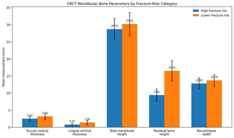 Figure 1. Comparison of CBCT-Based Mandibular Bone Parameters between High- and
Lower-Fracture-Risk Groups
Horizontal impaction showed the least favorable bone profile, with a mean B/A ratio of 0.40±0.10 and lingual cortical thickness of 0.74±0.50 mm, while 18 (54.5%) patients were classified as high fracture risk. In comparison, vertical impaction had a higher B/A ratio of 0.54±0.10, greater lingual cortical thickness of 1.42±0.65 mm, and only 7 (18.4%) high-risk cases. Differences in B/A ratio, cortical thickness, and fracture-risk frequency were statistically significant (p<0.05).
Table 3. CBCT bone characteristics according to third-molar angulation
|
Impaction Type |
n (%) |
B/A Ratio, mean±SD |
Lingual Cortical Thickness (mm), mean±SD |
High Fracture Risk n (%) |
|
Mesioangular |
54 (38.6%) |
0.49±0.11 |
1.08±0.63 |
17 (31.5%) |
|
Vertical |
38 (27.1%) |
0.54±0.10 |
1.42±0.65 |
7 (18.4%) |
|
Horizontal |
33 (23.6%) |
0.40±0.10 |
0.74±0.50 |
18 (54.5%) |
|
Distoangular |
15 (10.7%) |
0.51±0.12 |
1.35±0.61 |
4 (26.7%) |
|
p-value |
<0.001 |
<0.001 |
0.012 |
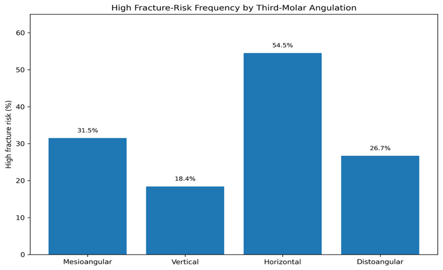
Figure 2. Distribution of High Fracture Risk According to Mandibular Third-Molar Impaction Type
High fracture risk was significantly associated with several anatomical characteristics. Pell and Gregory Class III was present in 24 (52.2%) high-risk versus 24 (25.5%) lower-risk patients (p=0.003). Lingual cortical thickness <1 mm was found in 32 (69.6%) versus 36 (38.3%) (p=0.001), while cortical perforation occurred in 16 (34.8%) versus 9 (9.6%) patients (p<0.001). Root contact with the inferior alveolar canal was also more frequent in the high-risk group, 27 (58.7%) versus 34 (36.2%) (p=0.019), and age ≥30 years was observed in 23 (50.0%) versus 25 (26.6%) patients (p=0.011).
Table 4. Association of anatomical characteristics with high radiographic fracture risk
|
Characteristic |
High Risk (n=46) |
Lower Risk (n=94) |
p-value |
|
Pell & Gregory Class II |
22 (47.8%) |
70 (74.5%) |
0.003 |
|
Pell & Gregory Class III |
24 (52.2%) |
24 (25.5%) |
|
|
Lingual cortex <1 mm |
32 (69.6%) |
36 (38.3%) |
0.001 |
|
Lingual cortex ≥1 mm |
14 (30.4%) |
58 (61.7%) |
|
|
Lingual cortical perforation |
16 (34.8%) |
9 (9.6%) |
<0.001 |
|
No cortical perforation |
30 (65.2%) |
85 (90.4%) |
|
|
Root–inferior alveolar canal contact |
27 (58.7%) |
34 (36.2%) |
0.019 |
|
No canal contact |
19 (41.3%) |
60 (63.8%) |
|
|
Age ≥30 years |
23 (50.0%) |
25 (26.6%) |
0.011 |
|
Age <30 years |
23 (50.0%) |
69 (73.4%) |
On multivariable analysis, horizontal impaction was associated with 3.21 times greater odds of high fracture risk (95% CI: 1.39–7.42; p=0.006). Other significant predictors included Pell and Gregory Class III (aOR 2.54; 95% CI: 1.18–5.47), lingual cortical thickness <1 mm (aOR 2.83; 95% CI: 1.29–6.21), cortical perforation (aOR 3.64; 95% CI: 1.43–9.27), and age ≥30 years (aOR 2.21; 95% CI: 1.11–4.42). Root–inferior alveolar canal contact was not independently significant (aOR 1.76; p=0.148).
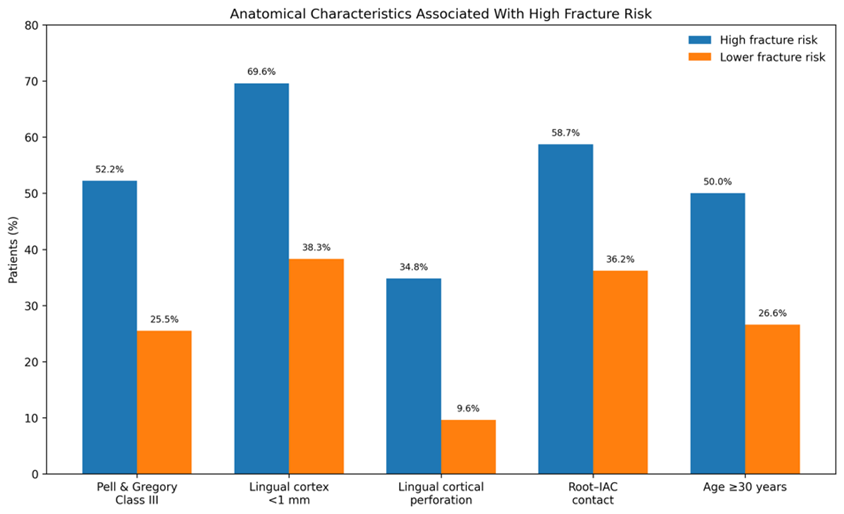
Figure 3. Association of Anatomical and Clinical Characteristics With High Mandibular Fracture Risk
Table 5. Multivariable analysis of factors associated with high mandibular fracture risk
|
Predictor |
Adjusted OR |
95% CI |
p-value |
|
Horizontal impaction |
3.21 |
1.39–7.42 |
0.006 |
|
Pell & Gregory Class III |
2.54 |
1.18–5.47 |
0.017 |
|
Lingual cortical thickness <1 mm |
2.83 |
1.29–6.21 |
0.009 |
|
Lingual cortical perforation |
3.64 |
1.43–9.27 |
0.007 |
|
Root–inferior alveolar canal contact |
1.76 |
0.82–3.79 |
0.148 |
|
Age ≥30 years |
2.21 |
1.11–4.42 |
0.024 |
DISCUSSION:
In the present study it was shown that these mandibles containing deeply impacted TM3s can significantly change the structural features of the mandibular angle. A study of 140 patients showed that 46 (32.9%) had a high radiographic fracture risk (residual bone ratio (B/A) <0.40). A similar study has previously shown that there is a strong link between B/A ratio with fracture occurrence, with a mean B/A ratio of 0.28±0.08 for patients with iatrogenic mandibular fracture and 0.62±0.09 for patients without iatrogenic fracture who had an impacted tooth extracted. A B/A ratio of less than 0.40 was deemed as an important threshold for risk of fracture [16]. High risk patients in this study had significantly lower supporting bone dimensions. The mean buccal cortical thickness was 2.52±0.71 mm in the medium-risk group as compared to 3.18±0.84 mm in the lower-risk group (p<0.001), and the mean lingual cortical thickness was 0.79±0.49 mm in the medium-risk group versus 1.39±0.66 mm in the lower-risk group (p<0.001). Residual bone height was also markedly lower at 9.4±1.8 versus 16.5±3.0 mm, and the corresponding B/A ratios were 0.33±0.05 and 0.55±0.08 (p<0.001). Previous studies have also documented the significant loss of residual bone height in patients with mandibular angle fractures and that the amount of residual bone may be a good predictor of the fractures [17]. Angle of the third molars also had effect on the bone profile. The horizontal impactions had the lowest mean B/A ratio (0.40±0.10) and lingual cortical thickness (0.74±0.50 mm) of which 18 (54.5%) were high risk. In comparison, vertical impactions had a B/A ratio of 0.54±0.10, lingual cortical thickness of 1.42±0.65 mm and only 7 (18.4%) high-risk cases. Past studies also reported that there are significant differences in lingual cortical thickness based on third molar angulation using CBCT [18]. Other previous studies, which were more recent, have also found that lingual cortical perforation was most often found on horizontal and deeply impacted third molars [19]. High-risk patients had significantly higher rates of Pell and Gregory Class III impaction (24 (52.2%) vs. 24 (25.5%) lower-risk patients (p=0.003)). Multivariable analysis revealed that Class III impaction was an independent risk factor for increased fracture risk with an aOR of 2.54 (95% CI 1.18–5.47; p=0.017). Previous studies have reported this correlation as well, with meta-analytic data indicating an even more significant increase for the mandibular angle fracture risk of Class III and Position C third molars, with a relative risk of about 2.99 and 2.72, respectively [20]. A previous study also found that patients with position C of third molars with angle Class III were more prone to angle fracture than those with other positions [21]. There were a number of limitations to this study. The fracture-risk classification was based on radiographic CBCT parameters and did not actually indicate fracture incidence over a period of time, but rather on anatomical susceptibility. The study was performed in one center and the number of patients is relatively small, so that the results might not be generalizable. Additionally, the interpretation of the CBCT measurements (cortical thickness and residual bone height) can be impacted by resolution, slice orientation and observer variability. Other surgical factors like the experience of the operator, osteotomy extent, sectioning technique and postoperative trauma were also not taken into consideration, but these could have an effect on the occurrence of mandibular fracture.
CONCLUSION:
It is concluded that CBCT can provide useful three-dimensional information for identifying mandibular configurations associated with increased fracture susceptibility in patients with deeply impacted mandibular third molars. High-risk patients showed significantly reduced buccal and lingual cortical thickness, lower residual bone height, and a lower B/A ratio. Horizontal impaction, Pell and Gregory Class III position, lingual cortical thickness <1 mm, cortical perforation, and age ≥30 years were significant independent predictors of high radiographic fracture risk. These findings support the use of CBCT-based assessment of residual bone and cortical morphology for preoperative risk stratification and surgical planning in deeply impacted mandibular third molars.
REFERENCES:
1. Mu J, Wu C, Ouyang L, Yang Y, Wu Y, Jin B. Evaluation of residual bone mass of the mandibular third molar and the risk of mandibular fracture. Med Oral Patol Oral Cir Bucal. 2024;29(4):e537-e544. doi:10.4317/medoral.26520.
2. Gumber TK, Kandiara P, Bhullar RS, Dhawan A, Kapila S, Singh B. Assessment and correlation of variation in lingual cortical plate thickness with different angulations of impacted mandibular third molar using cone beam computed tomography in North Indian population. J Maxillofac Oral Surg. 2023;22(3):590-602. doi:10.1007/s12663-022-01835-x.
3. Halder M, Chhaparwal Y, Patil V, Smriti K, Chhaparwal S, Pentapati KC. Quantitative and qualitative correlation of mandibular lingual bone with risk factors for third molar using cone beam computed tomography. Clin Cosmet Investig Dent. 2023; 15:267-277. doi:10.2147/CCIDE.S428908.
4. Safi Y, Moshfeghi M, Ghazizadeh Ahsaie M, Zameni M, Sahafi SA. Relationship between impacted mandibular third molars and the mandibular canal on CBCT scans. J Long Term Eff Med Implants. 2024;34(3):65-74. doi:10.1615/JLongTermEffMedImplants.2022042569.
5. Parhiz SA, Bakhtiary P, Mosavat F, Kharazifard MJ. Thickness of buccal and lingual alveolar bone plates according to the position of impacted mandibular third molars on cone-beam computed tomography scans. Front Dent. 2019;16(4):279-289. doi:10.18502/fid.v16i4.2087.
6. Ge J, Zheng JW, Yang C, Qian WT. Variations in the buccal-lingual alveolar bone thickness of impacted mandibular third molar: our classification and treatment perspectives. Sci Rep. 2016; 6:16375. doi:10.1038/srep16375.
7. Amin SA, Elsheikh AK, Meyer RA, Bagheri SC. Incidence of pre-existing lingual cortex perforation before removal of mandibular third molars. J Oral Maxillofac Surg. 2020;78(12):2129-2137. doi: 10.1016/j.joms.2020.08.010.
8. Yüceer-Çetiner E, Sakarya Ö, Vural A, Kazan D. Does the position of the mandibular third molar have an effect on the lingual bone morphology? A cone beam computed tomography evaluation. Diagnostics (Basel). 2025;15(18):2401. doi:10.3390/diagnostics15182401.
9. Mohanty R, Rout P, Singh V. Preoperative anatomic evaluation of the relationship between inferior alveolar nerve canal and impacted mandibular third molar in a population of Bhubaneswar, Odisha, using CBCT: a hospital-based study. J Maxillofac Oral Surg. 2020;19(2):257-262. doi:10.1007/s12663-019-01193-1.
10. Mu J, Wu Y, Wu C, Piao H, Jin B. Relationship between mandibular third molars and mandibular angle and condylar fractures. Med Oral Patol Oral Cir Bucal. 2024;29(5):e634-e643. doi:10.4317/medoral.26604.
11. Kandel L, Mishra R, Yadav D, Tripathi S, Shubham S, Chhetri P. Impact of mandibular third molars on angle fractures: a retrospective study. Dent Traumatol. 2021;37(1):103-107. doi:10.1111/edt.12608.
12. Samieirad S, Eshghpour M, Dashti R, Tohidi E, Javan AR, Mianbandi V. Correlation between lower third molar impaction types and mandibular angle and condylar fractures: a retrospective study. J Oral Maxillofac Surg. 2019;77(3):556-564. doi: 10.1016/j.joms.2018.09.028.
13. Brucoli M, Romeo I, Pezzana A, Boffano P, Benech A. The relationship between the status and position of third molars and the presence of mandibular angle and condylar fractures. Oral Maxillofac Surg. 2020;24(1):31-36. doi:10.1007/s10006-019-00811-z.
14. Lee JT, Dodson TB. The effect of mandibular third molar presence and position on the risk of an angle fracture. J Oral Maxillofac Surg. 2000;58(4):394-398.
15. Halmos DR, Ellis E, Dodson TB. Mandibular third molars and angle fractures. J Oral Maxillofac Surg. 2004;62(9):1076-1081.
16. Meisami T, Sojat A, Sàndor GKB, Lawrence HP, Clokie CML. Impacted third molars and risk of angle fracture. Int J Oral Maxillofac Surg. 2002;31(2):140-144. doi:10.1054/ijom.2001.0215.
17. Ma'aita J, Alwrikat A. Is the mandibular third molar a risk factor for mandibular angle fracture? Oral Surg Oral Med Oral Pathol Oral Radiol Endod. 2000;89(2):143-146.
18. Subhashraj K. A study on the impact of mandibular third molars on angle fractures. J Oral Maxillofac Surg. 2009;67(5):968-972. doi: 10.1016/j.joms.2008.09.005.
19. Duan DH, Zhang Y. Does the presence of mandibular third molars increase the risk of angle fracture and simultaneously decrease the risk of condylar fracture? Int J Oral Maxillofac Surg. 2008;37(1):25-28. doi: 10.1016/j.ijom.2007.07.010.
20. Takada H, Abe S, Tamatsu Y, Mitarashi S, Saka H, Ide Y. Three-dimensional bone microstructures of the mandibular angle using micro-CT and finite element analysis: relationship between partially impacted mandibular third molars and angle fractures. Dent Traumatol. 2006;22(1):18-24. doi:10.1111/j.1600-9657.2006.00368. x.
21. Panneerselvam E, Prasad PJ, Balasubramaniam S, Somasundaram S, Raja KVB, Srinivasan D. The influence of the mandibular gonial angle on the incidence of mandibular angle fracture: a radiomorphometric study. J Oral Maxillofac Surg. 2017;75(1):153-159. doi: 10.1016/j.joms.2016.08.016.