Impact of Dexmedetomidine Dose on Hemodynamics and Surgical Field Quality During Middle Ear Surgery.
- Dr. Neha kargutkar , Clinical associate Department of Anaesthesiology, Jaslok Hospital, Mumbai.
- Dr. Shilpa Gurav , Professor and HOD Department of Anaesthesiology, MIMER Medical College, Talegaon Dabhade, Pune.
- Dr. Manisha Anilkumar Khamkar , Associate professor Department of Anaesthesiology, MIMER Medical College, Talegaon Dabhade, Pune.
Article Information:
Abstract:
Background: Middle ear microsurgeries demand a clear, oligemic field for optimal visualization. This can be achieved through controlled hypotension. Dexmedetomidine, a selective α2-adrenergic agonist, has been widely used as an adjuvant to general anaesthesia for controlled hypotension, which improves surgical field visibility and volatile anaesthetic sparing effects. However, the optimal infusion dose with minimal side effects remains unclear. The study aimed to evaluate the impact of two Dexmedetomidine infusion doses (0.5 μg/kg/hr and 0.7 μg/kg/hr) on achieving a bloodless surgical field and maintaining hemodynamic stability in patients undergoing middle ear surgery under general anesthesia. Methods: A prospective observational study was carried out over a period of 18 months at a tertiary healthcare facility. The study included 46 ASA I–II participants aged 18-60 years undergoing middle ear surgery under general anesthesia. Participants were randomly distributed into two groups: Group I (0.5 μg/kg/h) and Group II (0.7 μg/kg/h) dexmedetomidine infusion post-induction. Hemodynamic parameters, Boezaart surgical field score, sevoflurane requirements, and adverse effects were recorded and analysed. Results: Both the groups showed similar surgical field optimization and comparable stable hemodynamics throughout the surgery, with no statistically significant differences in heart rate or blood pressure at any time point (p>0.05). Sevoflurane requirements were significantly lower (p<0.05) in Group II at multiple time points (30-150 minutes). No significant adverse effects were reported in either group. Conclusion: Both doses of dexmedetomidine infusion are effective and safe for achieving controlled hypotension, stable hemodynamics, and optimal surgical field during middle ear surgeries. For higher doses, careful monitoring is advisable.
Keywords:
Article :
INTRODUCTION:
Middle ear surgery poses unique anaesthetic challenges, requiring a near-bloodless field for optimal visualization under the microscope. Achieving such an oligemic field has traditionally relied on techniques such as controlled hypotension, topical vasoconstrictors, and head elevation to reduce intraoperative bleeding. [1,2] The advent of α2-adrenergic agonists like injection dexmedetomidine has transformed perioperative anaesthetic strategies by offering sedation, analgesia, and controlled hypotension, with minimal respiratory depression and stable hemodynamics. [3,4] Dexmedetomidine achieves these effects via central and peripheral α2-adrenoceptor stimulation. Its mechanisms involve presynaptic inhibition of norepinephrine release and postsynaptic attenuation of sympathetic activity, resulting in reduced heart rate and blood pressure. [5] It has been shown to decrease the requirements for inhalational anaesthetics and opioid analgesics, thereby reducing side effects like postoperative nausea and vomiting (PONV) and respiratory depression. [6]
However, optimal dosing remains controversial. While higher doses (>0.7 μg/kg/hour) improve field visibility and reduce anaesthetic needs, they are also associated with adverse effects such as bradycardia and hypotension. [7] Given these considerations, this research was designed to evaluate and contrast the impact of two separate infusion dosages of dexmedetomidine (0.5 μg/kg/hour and 0.7 μg/kg/hour) on intraoperative hemodynamic stability, the quality of the surgical field, and volatile anaesthetic requirements during middle ear surgeries under general anesthesia. The study aims to identify the most appropriate dosing strategy to optimize surgical conditions while minimizing adverse effects.
MATERIALS AND METHODS:
A prospective observational comparative study was conducted after institutional ethical committee approval from July 2023 to December 2024 at the Department of Anaesthesiology, a tertiary healthcare centre in western Maharashtra. Participants aged 18–60 years belonging to ASA physical status I or II, posted for elective middle ear surgery under general anesthesia lasting for 2-3 hours were included in study. Participants with bleeding disorders, hepatic/renal impairment, on sedatives, anti-hypertensives, or β-blockers, pregnant individuals, or those with heart blocks or anaphylactic reaction to study drug were excluded.
Considering mean and standard deviation, as reported by previous studies as 79.47, 4.65 and 75.03, 5.8, [8] a minimum sample size of 46 was calculated at 95% confidence Interval.
Primary outcome variable was to note the surgical field for bleeding with Boezaart surgical field score which was rated by surgeon. Secondary outcome variables were Heart rate (HR), Mean arterial pressure (MAP), Systolic blood pressure (SBP), Diastolic blood pressure (DBP) and Sevoflurane requirement (Dial concentration).
Data collection procedure:
Individuals between 18 and 60 years old, classified as ASA physical status I or II, were included in the study following institutional ethics committee approval. Patients scheduled for middle ear surgery were considered. A comprehensive preoperative assessment was conducted to identify any notable medical or clinical history. Systemic and general examinations were performed along with standard laboratory investigations. Informed consent was obtained a day prior to surgery by either the primary investigator or co-investigator, after providing a detailed explanation of the procedure. The patient was given an information sheet (English and local language) for an explanation, and any query was answered by the investigator.
Subsequently, participants were assigned to two groups of 23 each using a computer-generated randomization table.
Group I: Dexmedetomidine infusion at 0.5 μg/kg/hr
Group II: Dexmedetomidine infusion at 0.7 μg/kg/hr
The drug was prepared by the investigator, who did not participate in the surgical procedure. An independent assessor carried out anesthesia administration following institutional guidelines and documented all the necessary parameters.
In recovery room, pre-operative vitals of the patients were noted, Intravenous access established and then the participants were shifted to Operation theatre. Participants were premedicated as per standard institutional protocol. Following three minutes of preoxygenation with 100% oxygen, anesthesia induction was performed using intravenous propofol until patients no longer responded to verbal stimuli. Inj. Atracurium at a dose of 0.5 mg/kg was administered to enable tracheal intubation. Dexmedetomidine Infusion was started post-induction according to the group allotted by investigator. Hemodynamic parameters (HR, SBP, DBP, MAP) and sevoflurane requirement (dial concentration) were recorded by assessor every 15 minutes intraoperatively. The concentration of sevoflurane on the vaporizer was modified to manage hypotension exceeding the desired blood pressure range (MAP< 30% of baseline). Dexmedetomidine infusion was discontinued 20 minutes before surgery completion. Postoperatively, any remaining neuromuscular blockade was reversed using Inj. Neostigmine (0.05 mg/kg) and Inj. glycopyrrolate (8 μg/kg). Once sufficient motor function returned and spontaneous respiration resumed of the participants, extubation was done. Post-operative pain was tackled by injection Paracetamol 1gm IV.
At the end of the procedure, surgeon was asked to assess the bleeding at the surgical site according to Boezaart surgical field grading scale (Table 1). Any adverse effects like bradycardia, hypotension were noted. At the end of 15 minutes after extubation, postoperative HR, SBP, DBP, MAP, oxygen saturation were noted in OT. Following surgery, the participants were transferred to the postoperative recovery area where they were observed for 30 minutes. They were sent to the ward upon fulfilling the established discharge criteria.
Statistical Analysis:
All information was recorded using a pre-structured study proforma. Categorical data were expressed as frequencies and percentages, with relationships analyzed using the Chi-Square test. Numerical data were reported as Mean ± SD and compared between groups with the unpaired t-test. Statistical significance was assigned to a p-value less than 0.05. Graphs were utilized for results visualization as appropriate. Most data analyses were conducted with SPSS Version 26.0, while graphical depiction was done using Microsoft Excel 2021.
RESULTS:
A total of 46 patients were enrolled in the study over 18 months period. The demographic data of the subjects are presented in Table-2. There was no statistically significant difference in age, gender, weight, ASA grade or duration of surgery between the study groups (p > 0.05).
Surgical Field Visibility (Boezaart Score):
All participants in both groups achieved a Boezaart score of 2, indicating optimal surgical field visibility with minimal bleeding, supporting equal efficacy in surgical field optimization for both groups. (Figure 1)
Hemodynamic Parameters:
Hemodynamic values were assessed and compared at multiple time points between both groups. Heart rate showed a gradual decline post-induction in the two groups, and at most intervals there was no statistically significant differences. (p>0.05) While both groups demonstrated a declining trend in SBP post-induction, no time point showed a significant inter-group difference (p-values > 0.05). Both dosages effectively maintained SBP within target hypotensive range. Diastolic Blood Pressure showed a similar declining pattern, with Group II trending slightly lower than Group I. However, differences were not statistically significant at any time point (all p-values > 0.05), supporting dose-equivalence in diastolic control. MAP reductions followed expected hypotensive patterns in both groups. Group II consistently exhibited slightly lower MAP values, though without statistical significance (p > 0.05), reflecting clinical comparability in hypotensive response. (Table 3&4) (Figure 2-5)
Sevoflurane Requirement:
Sevoflurane requirement (dial concentration) values were significantly lower in Group II from 30 to 150 minutes (p < 0.05), indicating a clear dose-dependent inhalational agent-sparing effect with 0.7 μg/kg/hr. (Table 5, Figure 6)
Adverse effects:
Hypotension and bradycardia occurred exclusively in Group II (each in 3 participants, 13%). Group I had no reported bradycardia or hypotensive episodes. The differences were not statistically significant (p = 0.233 for both).
Table 1: Boezaart surgical field grading scale
|
Grade |
Assessment |
|
0 |
No bleeding (cadaveric conditions) |
|
1 |
Slight bleeding- no suctioning required |
|
2 |
Slight bleeding- occasional suctioning required |
|
3 |
Slight bleeding- frequent suctioning required; bleeding threatens surgical field a few seconds after suction is removed |
|
4 |
Moderate bleeding- frequent suctioning required and bleeding threatens surgical field directly after suction is removed |
|
5 |
Severe bleeding- constant suctioning required; bleeding appears faster than can be removed by suction; surgical field severely threatened and surgery usually not possible |
Table 2: Comparison of Demographic parameters between study groups
|
Parameter |
Group I |
Group II |
p-value |
|
Mean Age (years) |
41.1 ± 14.1 |
37.6 ± 14.9 |
0.42 |
|
Female/Male (%) |
73.9/26.1 |
43.5/56.5 |
0.07 |
|
ASA I /II (%) |
43.5/ 56.5 |
52.2/ 47.8 |
0.76 |
|
Weight (Kg) |
52.8 + 9.3 |
54.4 + 9.2 |
0.55 |
|
Duration of Surgery (mins) |
178.7 + 39.2 |
165.0 + 37.0 |
0.23 |
Table 3: Comparison of Mean Heart Rate between study groups
|
Heart Rate (/min) |
Group I |
Group II |
P value |
|
0 min |
90.65 + 8.04 |
90.96 + 12.26 |
0.92 |
|
15 min |
88.52 + 10.20 |
84.83 + 12.09 |
0.27 |
|
30 min |
83.43 + 11.90 |
82.74 + 13.28 |
0.85 |
|
45 min |
78.52 + 11.95 |
77.39 + 13.04 |
0.76 |
|
60 min |
75.74 + 10.79 |
77.22 + 12.93 |
0.67 |
|
75 min |
71.61 + 9.51 |
73.57 + 11.95 |
0.54 |
|
90 min |
69.78 + 10.28 |
72.00 + 11.60 |
0.51 |
|
105 min |
70.50 + 10.45 |
71.05 + 11.17 |
0.87 |
|
120 min |
69.10 + 10.83 |
71.89 + 8.62 |
0.38 |
|
135 min |
69.61 + 10.53 |
70.67 + 7.20 |
0.73 |
|
150 min |
69.24 + 9.11 |
70.56 + 6.09 |
0.62 |
|
165 min |
69.18 + 9.26 |
69.00 + 8.36 |
0.96 |
|
180 min |
68.59 + 9.55 |
68.67 + 9.42 |
0.98 |
Table 4: Comparison of Mean arterial Pressure between study groups
|
Heart Rate (/min) |
Group I |
Group II |
P value |
|
0 min |
90.65 + 8.04 |
90.96 + 12.26 |
0.92 |
|
15 min |
88.52 + 10.20 |
84.83 + 12.09 |
0.27 |
|
30 min |
83.43 + 11.90 |
82.74 + 13.28 |
0.85 |
|
45 min |
78.52 + 11.95 |
77.39 + 13.04 |
0.76 |
|
60 min |
75.74 + 10.79 |
77.22 + 12.93 |
0.67 |
|
75 min |
71.61 + 9.51 |
73.57 + 11.95 |
0.54 |
|
90 min |
69.78 + 10.28 |
72.00 + 11.60 |
0.51 |
|
105 min |
70.50 + 10.45 |
71.05 + 11.17 |
0.87 |
|
120 min |
69.10 + 10.83 |
71.89 + 8.62 |
0.38 |
|
135 min |
69.61 + 10.53 |
70.67 + 7.20 |
0.73 |
|
150 min |
69.24 + 9.11 |
70.56 + 6.09 |
0.62 |
|
165 min |
69.18 + 9.26 |
69.00 + 8.36 |
0.96 |
|
180 min |
68.59 + 9.55 |
68.67 + 9.42 |
0.98 |
Table 5: Comparison of Sevoflurane requirement between study groups
|
Sevoflurane requirement(Dial concentration) |
Group I |
Group II |
p- value |
|
0 min |
1.37 + 0.34 |
1.33 + 0.39 |
0.689 |
|
15 min |
1.34 + 0.36 |
1.19 + 0.36 |
0.170 |
|
30 min |
1.32 + 0.37 |
1.04 + 0.19 |
0.002 |
|
45 min |
1.23 + 0.37 |
0.99 + 0.18 |
0.008 |
|
60 min |
1.23 + 0.37 |
0.97 + 0.19 |
0.004 |
|
75 min |
1.19 + 0.39 |
0.94 + 0.16 |
0.001 |
|
90 min |
1.15 + 0.34 |
0.92 + 0.10 |
0.004 |
|
105 min |
1.15 + 0.36 |
0.90 + 0.10 |
0.006 |
|
120 min |
1.12 + 0.35 |
0.88 + 0.10 |
0.008 |
|
135 min |
1.12 + 0.38 |
0.89 + 0.10 |
0.016 |
|
150 min |
1.11 + 0.40 |
0.89 + 0.10 |
0.033 |
|
165 min |
1.05 + 0.37 |
0.91 + 0.10 |
0.243 |
|
180 min |
1.04 + 0.38 |
0.89 + 0.11 |
0.267 |
Figures Legends
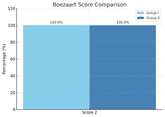
Figure 1: Bar diagram showing mean Boezaart score comparison between study groups
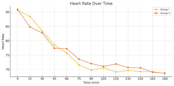
Figure 2: Line diagram showing Heart Rate variation over time in both study groups
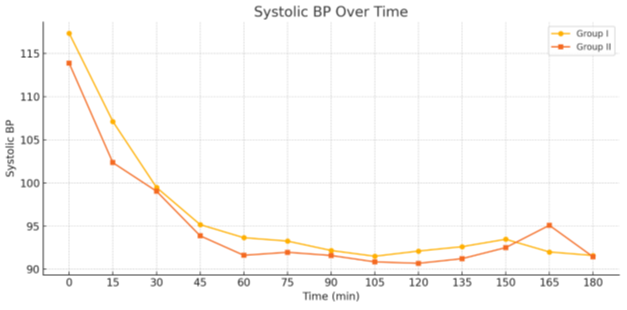
Figure 3: Line diagram showing Systolic Blood Pressure variation over time in study groups
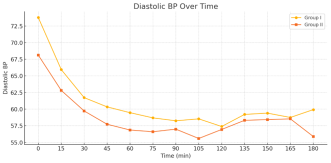
Figure 4: Line diagram showing Diastolic Blood Pressure variation over time in both study groups
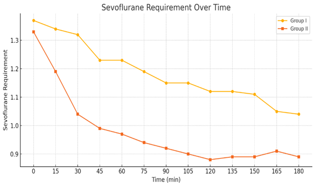
Figure 5: Line diagram showing Mean Arterial Pressure variation over time in study groups
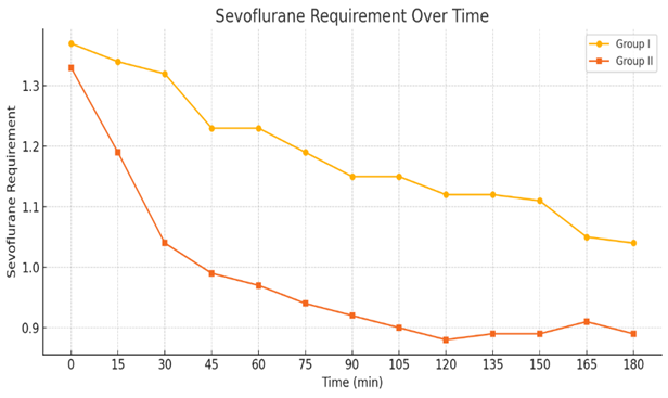
Figure 6: Line diagram showing Sevoflurane requirement (dial concentration) over time in both study groups
DISCUSSION:
The present study compared the effects of two different doses of dexmedetomidine infusion 0.5 μg/kg/hr (Group I) and 0.7 μg/kg/hr (Group II) on intraoperative hemodynamic stability, surgical field quality, and Sevoflurane requirements during middle ear surgeries. The Boezaart surgical field grading scores were optimal and identical in both groups, with all patients achieving a score of 2, signifying adequate visibility and minimal bleeding. This supports the notion that even the lower dose of dexmedetomidine (0.5 μg/kg/hr) suffices to maintain a satisfactory surgical field, corroborating findings from Gupta et al. and Lakshmi et al. [2,9]
Both doses effectively maintained heart rate, systolic, diastolic, and mean arterial pressures within the target hypotensive range, without statistically significant differences between the groups. This finding suggests that both doses are similarly effective in achieving controlled hypotension during these procedures, aligning with prior studies. [2,9-14]. Notably, the requirement of sevoflurane was significantly lower in Group II compared to Group I from 30 to 150 minutes intraoperatively. This indicates a dose-dependent volatile anaesthetic-sparing effect of dexmedetomidine at higher infusion rates, consistent with observations from Gupta et al., Bijay et al., Fernandes et al. and Aho et al [2,11,15,16]
In terms of safety, no significant adverse hemodynamic events were recorded in either group, though Group II exhibited a non-significant trend toward slightly lower MAP and HR at few time points but was managed promptly. Overall, the results of this study reinforce the clinical utility of dexmedetomidine as an adjunct for middle ear surgeries, with both tested doses providing comparable hemodynamic stability and surgical field conditions. The higher dose (0.7 μg/kg/hr) demonstrated a more pronounced reduction in sevoflurane requirements, suggesting it may be preferred in situations where minimizing inhalational agent exposure is desirable, provided close monitoring is ensured.
Limitations:
Although statistically powered, the study was conducted on a relatively small cohort of 46 participants. This study took place at a single tertiary healthcare institution located in Western Maharashtra. This may limit the generalizability of the findings. As surgical expertise, anaesthesia practices, and patient characteristics vary across institutions, a multicentric approach could provide more robust data.
CONCLUSION:
Present study concluded that 0.5 μg/kg/hr and 0.7 μg/kg/hr infusion of Dexmedetomidine in middle ear surgery during general anaesthesia were comparable in terms of hemodynamic stability contributing to bloodless intraoperative surgical field with better visibility. We further conclude that though hemodynamic stability was good in both groups, Dexmedetomidine infusion in dose of 0.7 μg/kg/hr significantly reduced requirement of intraoperative Sevoflurane with minimal but manageable adverse effects. So, with real time hemodynamic monitoring this dose may be adopted as a preferred protocol for middle ear surgeries.
REFERENCES:
1. De C Deacock AR. Anæsthesia for Middle Ear Surgery: Aspects of Anesthesia for Middle Ear Surgery an Blood Loss during Stapedectomy.
2. Gupta K, Bansal M, Gupta PK, Pandey MN, Agarwal S. Dexmedetomidine infusion during middle ear surgery under general anaesthesia to provide oligaemic surgical field: A prospective study. Indian J Anaesth. 2015 Jan 1;59(1):26–30.
3. Nasreen F, Bano S, Khan RM, Hasan SA. Dexmedetomidine used to provide hypotensive anesthesia during middle ear surgery. Indian J Otolaryngol Head Neck Surg. 2009;61:205–7.
4. Turan G, Ozgultekin A, Turan C, Dincer E, Yuksel G. Advantageous effects of dexmedetomidine on haemodynamic and recovery responses during extubation for intracranial surgery. Eur J Anaesthesiol. 2008;25:816–20.
5. Petroianu GA, Aloum L, Adem A. Neuropathic pain: Mechanisms and therapeutic strategies. Frontiers in Cell and Developmental Biology. 2023 Jan 16;11:1072629.
6. Khan ZP, Munday IT, Jones RM, Thornton C, Mant TG, Amin D. Effects of dexmedetomidine on isoflurane requirements in healthy volunteers. 1: Pharmacodynamic and pharmacokinetic interactions. Br J Anaesth 1999; 83: 372-380.
7. Keniya VM, Ladi S, Naphade R. Dexmedetomidine attenuates sympathoadrenal response to tracheal intubation and reduces perioperative anaesthetic requirement. Indian J Anaesth. 2011 Jul;55(4):352–7.
8. Sebastian B, Talikoti AT, Krishnamurthy D. Attenuation of haemodynamic responses to laryngoscopy and endotracheal intubation with intravenous dexmedetomidine: A comparison between two doses. Indian J Anaesth. 2017 Jan 1;61(1):48–54.
9. Lakshmi K Swamy, Ravi Madhusudhana. Effects of Lower Doses of Dexmedetomidine on Controlled Hypotension During Middle Ear Surgery. Indian Journal of Anesthesia and Analgesia 2019; 6(3) (Part - I): 721-726.
10. Nuzhat Anjum Abdul Hafeez, Kirti Arvind Kundalwal, Prajakta Madhukar Tayade, Nanasaheb Khandu Bhangare. A Comparative Study of IV Bolus versus IV Infusion of Dexmedetomidine on Intraoperative Haemodynamics in ENT Surgeries under General Anaesthesia: A Randomized Double-Blind Study. International Journal of Pharmaceutical and Clinical Research 2023; 16(5); 940-945.
11. Bijay M, Ganapathi P, Sharma NGA, et al. A comparison of two different doses of dexmedetomidine infusion during maintenance of general anaesthesia in patients undergoing spine surgeries, functional endoscopic sinus surgery and middle ear surgeries. J. Evid. Based Med. Healthc. 2016; 3(21), 872-875.
12. Kamibayashi T, Maze M. Clinical use of alpha-2adrenergic agonists. Anesthesiology 2000; 93: 1345-1349
13. Govil N, Dadu S, Singh VP, Rai V, Kumar P. Dexmedetomidine improves surgical field in cochlear implant surgery: a randomized, double-blind, placebo-controlled trial. Anaesthesia, Pain & Intensive Care. 2019 Jan 19:147-53
14. Eremenko AA, Chemova EV. Comparison of dexmedetomidine and propofol for short-term sedation in early postoperative period after cardiac surgery. Anesteziologiya i Reanimatologiya. 2014.
15. FERNANDES S, RAMCHANDANI P, HARDE M. Impact of Dexmedetomidine Infusion during Functional Endoscopic Sinus Surgery: A Randomised Controlled Trial. Journal of Clinical & Diagnostic Research. 2023 Aug 1;17(8).
16. Aho M, Lehtinen AM, Erkola O, Kallio A, Korttila K. The effect of intravenously administered dexmedetomidine on perioperative hemodynamics and isoflurane requirements in patients undergoing abdominal hysterectomy. Anesthesiology 1991; 74: 997-1002.